 E-submission
 TOTA
TOTA TOTS
TOTS
Search
- Page Path
- HOME > Search
Review Article
- Combined acetabular and pelvic ring injuries: a reference-frame algorithm for definitive fixation sequencing
- Jeong-Hyun Koh, Seungyeob Sakong
- J Musculoskelet Trauma 2026;39(2):83-92. Published online April 9, 2026
- DOI: https://doi.org/10.12671/jmt.2026.00031
-
 Abstract
 PDF
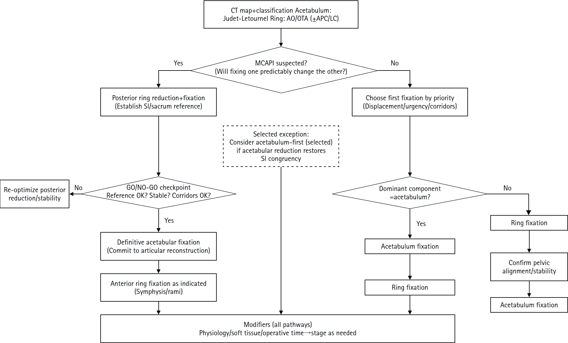
PDF - Combined acetabular and pelvic ring injuries are not simply “two fractures in one patient.” Reduction and fixation of one component can alter the alignment and reducibility of the other, rendering operative sequencing a primary decision variable rather than a secondary consideration. These injuries typically result from high-energy trauma, frequently occur in patients with polytrauma, and are further influenced by physiological tolerance and the feasibility of available operative corridors. The existing evidence base remains constrained by retrospective study designs, inconsistent definitions, variable classification systems, and heterogeneous outcome reporting, all of which limit the strength of comparative recommendations. This state-of-the-art review presents a surgeon-facing, algorithmic approach grounded in a reference-frame mindset. We emphasize computed tomography (CT)-based mapping and the use of consistent terminology to characterize acetabular morphology, pelvic ring instability, deformity vectors, suspicion of mechanical coupling, and feasible operative corridors. Mechanically connected acetabular and pelvic ring injuries (MCAPI) are introduced as a working framework for identifying patterns in which reduction or fixation of one injury predictably influences the other. In cases of suspected MCAPI, a posterior ring-based sequence is generally preferred, typically consisting of posterior ring reduction and fixation, definitive acetabular reconstruction, and subsequent anterior ring fixation. We propose an explicit intraoperative “GO/NO-GO” checkpoint (reference acceptable, stable, corridors feasible) to prevent acetabular reconstruction on a moving target. Acetabulum-first strategies may be appropriate only in selected anteroposterior compression- type configurations in which acetabular fixation plausibly restores sacroiliac congruency and posterior stabilization remains technically feasible. We summarize key outcome domains and complication patterns, highlighting hip dislocation as an important risk factor associated with both neurologic deficits and overall complications. Standardized CTbased definitions and outcome instruments, together with multicenter cohorts employing predefined decision pathways, are required to test sequencing strategies and to determine whether improved radiographic reduction translates into durable functional benefit.
- 697 View
- 20 Download

Original Articles
- Percutaneous anterior leverage technique for anteromedial cortical support in intertrochanteric femur fractures: a computed tomography-based validation study
- Whee Sung Son, Bum Jin Shim, Oog-jin Shon
- J Musculoskelet Trauma 2026;39(2):117-129. Published online March 27, 2026
- DOI: https://doi.org/10.12671/jmt.2025.00311
-
Abstract
PDF
- Background
Anteromedial cortical support (AMCS) enhances stability in intertrochanteric femur fractures. However, reproducible, validated methods of achieving AMCS have not previously been reported. This study introduces a percutaneous anterior leverage technique and validates its AMCS effects using computed tomography (CT).
Methods
We retrospectively reviewed patients treated by a single surgeon between March 2022 and December 2024. The inclusion criteria were an AO/OTA classification of A1–A3, application of the percutaneous anterior leverage technique, available pre- and postoperative CT, and ≥6 months follow-up. Outcomes included CT-based AMCS (anterior on axial and medial on coronal images, classified as positive, neutral, or negative), time to union, union rate, changes in neck-shaft angle, and treatment failure (varus collapse, blade cut-through, or nonunion without the former two). The risk factors for failure were analyzed.
Results
Of 273 patients reviewed, 53 met the inclusion criteria. Follow-up was at least 6 months in all cases. Positive anterior support was achieved in 37 patients (69.8%) and positive medial support in 42 (79.25%). No patient demonstrated negative anterior support; one (1.9%) had negative medial support. Cortical support improved significantly after surgery. CT images demonstrated significant postoperative improvements (anterior P=0.026; medial P<0.001). Bone union was achieved in 50 patients (94.34%) at a mean of 3.93±1.48 months. The mean change in the neck-shaft angle at last follow-up was 1.75°±2.34° varus. Three patients (5.66%) experienced treatment failure. Anteromedial cortical breakage during follow-up differed between failure and nonfailure groups (P=0.002), but regression identified no independent predictors. No technique-related complications were observed.
Conclusions
Our percutaneous anterior leverage technique produced favorable CT-confirmed AMCS and high union with low failure, supporting its safety and effectiveness in intertrochanteric femur fractures. Level of evidence: IV.
- 668 View
- 32 Download
- Sex-specific bottlenecks and risk zones in the retrograde superior pubic ramus screw corridor: a 3D CT-based morphometric cadaver study
- Ji Won Jeong, Jung Tae Ahn, Gu Hee Jung, Kun Tae Kim
- J Musculoskelet Trauma 2026;39(2):103-116. Published online March 26, 2026
- DOI: https://doi.org/10.12671/jmt.2026.00066
-
Abstract
PDF
 Supplementary Material
 - Background
Superior ramus screw fixation is commonly used to stabilize anterior pelvic ring injuries but is constrained by a narrow, irregular, and curved intraosseous corridor. Trajectory-based morphometric analysis may assist in screw diameter selection and enable identification of reproducible anatomic constriction zones.
Methods
We conducted a cross-sectional computed tomography (CT)-based morphometric study of 82 cadaveric pelvises (42 males, 40 females). Bottleneck diameter was defined as the diameter of the largest fully contained virtual cylinder along the planned trajectory, and cylinder length was recorded. Orthogonal cross-sections at 9.5-mm intervals (up to 12 segments) were generated to measure segment-wise effective diameter (defined as twice the minimum centerline-to-cortex distance) and cortical clearance, which was used as a diameter-based safety margin. Segments were realigned to the acetabular start segment to define relative segment positions (Δ seg). Feasibility was assessed for prespecified screw diameters ranging from 3.5 to 7.3 mm.
Results
Mean bottleneck diameter was larger in males than in females (7.34±1.10 vs. 5.93±0.98 mm), whereas trajectory length was similar between sexes (127.85±8.54 vs. 128.85±8.20 mm). Δ seg realignment localized corridor constriction to two discrete zones: a preacetabular zone (Δ seg −6 to −4) and a periacetabular zone (Δ seg 1 to 2), where effective diameter and cortical clearance were most limited. Feasibility rates were 100% at 3.5–4.5 mm, 95.2% vs. 82.5% at 5.0 mm, 81.0% vs. 27.5% at 6.5 mm, and 59.5% vs. 10.0% at 7.3 mm in males and females, respectively.
Conclusions
Female models demonstrated smaller trajectory-wide bottleneck diameters and segment-wise effective diameters than male models. Acetabular-referenced Δ seg realignment identified two reproducible anatomic risk zones: a preacetabular zone adjacent to the obturator neurovascular bundle and a periacetabular zone near the external iliac vessels. At diameters ≥6.5 mm, cortical proximity increased more prominently in females than in males. Level of evidence: III.
- 724 View
- 24 Download
Review Article
- Definitive fixation for traumatic pelvic ring injuries: a dynamically informed, posterior-referenced framework
- Jeong-Hyun Koh, Seungyeob Sakong
- J Musculoskelet Trauma 2026;39(2):73-82. Published online March 24, 2026
- DOI: https://doi.org/10.12671/jmt.2026.00045
-
Abstract
PDF
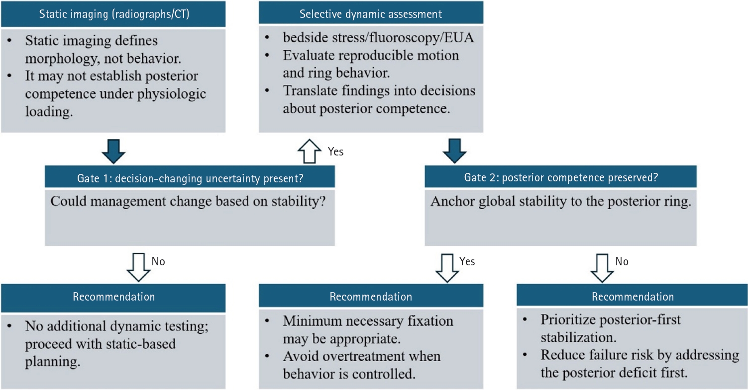
- Optimal definitive fixation for traumatic pelvic ring injuries remains challenging because static radiographs and computed tomography, although essential for defining morphology, do not consistently predict load-dependent behavior during early mobilization. This uncertainty contributes to substantial practice variation and continued reliance on simplified displacement thresholds, such as the 2.5 cm rule. Such rules can misclassify instability by underrepresenting posterior competence and by privileging static measurements over functional behavior. In this narrative review, we propose a dynamically informed, posterior- referenced framework composed of three linked elements: (1) decision-linked terminology that explicitly distinguishes dynamic instability, radiographic change, and clinical failure; (2) selective stress-based assessment when uncertainty is likely to alter management; and (3) escalation along a fixation continuum that weighs incremental stability against operative burden. When static imaging cannot establish posterior competence with confidence, we outline selective stress-based approaches to assess pelvic ring behavior and to translate demonstrated instability into fixation selection along a defined continuum. Across all steps, the framework emphasizes minimum necessary fixation and explicitly incorporates the cost of selection as a primary decision variable. The operative question, therefore, shifts from gap width alone to clinically relevant motion and preservation of posterior competence. In doing so, this approach aims to reduce both undertreatment and overtreatment and to improve the consistency and defensibility of definitive fixation strategies across diverse practice environments.
- 813 View
- 32 Download
Original Articles
- Comparative results of the femoral neck system versus the dynamic hip screw for stable femoral neck fractures in older adults in Korea: a retrospective cohort study
- Byung-Chan Choi, Byung-Woo Min, Kyung-Jae Lee, Jun-Sik Hong
- J Musculoskelet Trauma 2025;38(4):203-211. Published online October 24, 2025
- DOI: https://doi.org/10.12671/jmt.2025.00276
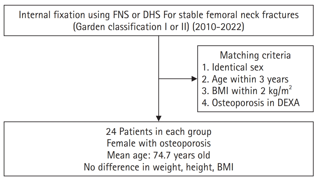
-
Abstract
PDF
- Background
This study aimed to compare the clinical and radiological outcomes of the femoral neck system (FNS) and the dynamic hip screw (DHS) for the internal fixation of stable femoral neck fractures in older adults.
Methods
This retrospective cohort study included 48 matched older adult patients based on sex, age, BMI, and osteoporosis status, who had undergone internal fixation with either FNS or DHS for stable femoral neck fractures between January 2010 and December 2022. To minimize selection bias, a 1:1 case-control matching was performed based on sex, age, body mass index (BMI), and the presence of osteoporosis. A total of 48 patients (24 in each group) were included. We compared perioperative data (operation time, hemoglobin change, transfusion rate), functional outcomes using the Koval score, and radiological outcomes, including union rate, femoral neck shortening, and complication rates.
Results
The mean operation time was significantly shorter in the FNS group than in the DHS group (60.9 minutes vs. 70.8 minutes; P=0.007). There were no statistically significant differences between the two groups in the union rate (87.5% in FNS vs. 95.8% in DHS), femoral neck shortening, final Koval score distribution, or overall complication rates (12.5% in both groups).
Conclusions
For treating stable femoral neck fractures in older adults, the FNS demonstrated comparable clinical and radiological outcomes to the DHS, with the distinct advantage of a shorter operation time. While these findings suggest that the FNS is a promising and safe alternative that may reduce the surgical burden, definitive conclusions are precluded by the small sample size, warranting further research to corroborate these results. Level of evidence: IV.
- 2,547 View
- 37 Download
- Hook plate versus periarticular-type volar locking plate for distal radius fractures involving the volar lunate facet in Korea: a retrospective cohort study
- Hyun-Jae Park, Joo-Hak Kim
- J Musculoskelet Trauma 2025;38(4):221-228. Published online October 24, 2025
- DOI: https://doi.org/10.12671/jmt.2025.00241
-
Abstract
PDF
- Background
This study investigated the clinical and radiographic outcomes of hook plate (HP) fixation for volar lunate facet fractures, comparing them with periarticular-type volar locking plates (PVLPs).
Methods
A retrospective review was conducted on 24 patients with distal radius fractures involving volar lunate facet fragments who underwent surgery between January 2016 and April 2021. Patients were divided into two groups: HP (n=12) and PVLP (n=12). Radiographic union, wrist range of motion, Disabilities of the Arm, Shoulder and Hand (DASH) scores, and implant-related complications were compared. Statistical analyses included the Mann-Whitney U test and Fisher exact test.
Results
Radiographic union was achieved in all patients (100%), without secondary displacement or hardware failure. No significant differences were observed between the two groups in wrist flexion (P=0.152), extension (P=0.832), pronation (P=0.792), or supination (P=0.328). The mean DASH scores were 12.8±5.5 in the HP group and 14.6±6.0 in the volar plate group (P=0.449). One patient in the HP group experienced mild flexor tendinopathy that resolved with conservative management. No cases of tendon rupture or early reoperation were reported.
Conclusions
Fixation of volar lunate facet fractures using a HP yielded clinical and radiographic outcomes comparable to those of PVLPs, with a low rate of complications and reliable bony union. Due to its mechanical stability, compatibility with standard surgical approaches, and low risk of flexor tendon irritation, the HP may serve as a valuable alternative for managing volar lunate facet fractures. Level of evidence: IV.
- 919 View
- 25 Download
Review Article
- Atypical ulnar fractures: a narrative review of current concepts and a case of bilateral surgical management
- Chi-Hoon Oh, Hyun Tak Kang, Jun-Ku Lee
- J Musculoskelet Trauma 2025;38(3):124-132. Published online July 24, 2025
- DOI: https://doi.org/10.12671/jmt.2025.00227
-
Abstract
PDF
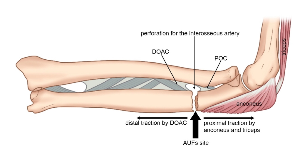
- Atypical ulnar fractures (AUFs) are rare complications that are often linked to long-term antiresorptive therapy. Although atypical femoral fractures are well-studied, AUFs lack standardized diagnostic and treatment protocols. This review summarizes current knowledge on AUFs, including their pathophysiology, diagnostic criteria, and management. A case of bilateral AUFs treated with two distinct osteosynthesis methods is presented, emphasizing the principles of biological healing and mechanical stabilization.
- 2,222 View
- 64 Download
Original Articles
- Comparison of outcomes of reinforced tension band wiring and precontoured plate and screw fixation in the management of Mayo type IIIB olecranon fractures
- Hyun Goo Kang, Tong Joo Lee, Samuel Jaeyoon Won
- J Musculoskelet Trauma 2025;38(2):96-101. Published online February 28, 2025
- DOI: https://doi.org/10.12671/jmt.2025.00059
- Correction in: J Musculoskelet Trauma 2025;38(3):168
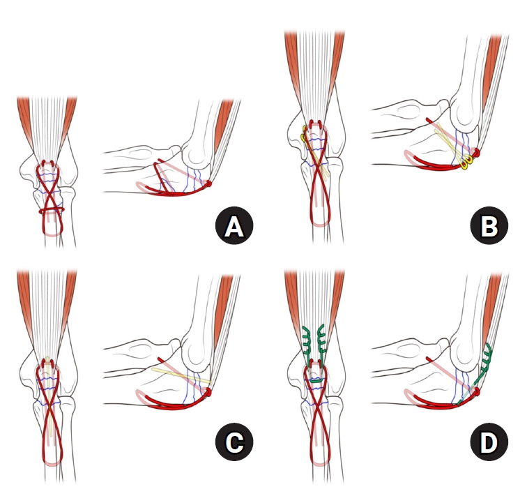
-
Abstract
PDF
- Background
Mayo type IIIB olecranon fractures are characterized by significant displacement and comminution, presenting a challenge in selecting the appropriate fixation technique. This study compared the clinical and radiographic outcomes, complications, and reoperation rates of reinforced tension band wiring (TBW) and precontoured plate and screw fixation (PF) in the surgical treatment of Mayo type IIIB olecranon fractures.
Methods
This retrospective review analyzed 24 patients diagnosed with Mayo type IIIB olecranon fractures, who were treated between 2005 and 2023. Of these, 11 patients underwent reinforced TBW, and 13 received precontoured PF. Clinical outcomes were assessed using Disabilities of the Arm, Shoulder, and Hand (DASH) scores and the Mayo Elbow Performance Score (MEPS). Radiographic outcomes focused on fracture union. Operative times, complication rates, and reoperation rates were compared between the groups.
Results
Both the reinforced TBW and PF groups achieved satisfactory clinical outcomes, with no significant between-group differences in DASH and MEPS scores (P>0.05). Radiographic union was achieved in all patients. The reinforced TBW group demonstrated a significantly shorter operative time than the PF group (93.6±7.4 min vs. 132.3±13.7 min; P<0.001). Complication rates were similar between the two groups (reinforced TBW, 38.4%; PF, 36.3%), but hardware-related irritation occurred more frequently in the reinforced TBW group. Reoperations were required in 15.8% of the reinforced TBW group due to hardware irritation, whereas no reoperations were necessary in the PF group.
Conclusions
Reinforced TBW and PF are both effective surgical options for managing Mayo type IIIB olecranon fractures, yielding comparable clinical and radiographic outcomes. While reinforced TBW offers shorter operative times and lower costs, PF is associated with fewer hardware-related complications. Further prospective studies are needed to optimize treatment strategies for these complex fractures. Level of Evidence: Level III. -
Citations
Citations to this article as recorded by
- Suture tension band fixation of olecranon fractures: description and early outcomes of a novel technique
Joseph G. Monir, Frank L. Vazquez, Musab Gulzar, Kevin Cuneo, Thomas McQuillan, Michael B. Gottschalk, Eric R. Wagner
JSES Reviews, Reports, and Techniques.2026; 6(2): 100707. CrossRef - Are posterior olecranon locking plates a problem for patients after fracture healing because of prominence?
Reva Qiu, Mallika Makkar, Richard Buckley
Injury.2025; 56(11): 112769. CrossRef
- Suture tension band fixation of olecranon fractures: description and early outcomes of a novel technique
- 3,358 View
- 60 Download
- 2 Crossref
- Outcomes of open reduction and internal fixation using 2.0/2.4 mm locking compression plate in isolated greater tuberosity fractures of humerus
- Sung Choi, Dongju Shin, Sangwoo Kim, Byung Hoon Kwack
- J Musculoskelet Trauma 2025;38(1):32-39. Published online January 24, 2025
- DOI: https://doi.org/10.12671/jmt.2025.00005

-
Abstract
PDF
- Background
The purpose of this study was to retrospectively evaluate the radiographic and clinical results of a small single or double low-profile plate fixation of 2.0/2.4 mm locking compression plate (LCP) in treating isolated greater tuberosity (GT) fractures of the humerus. Methods: From June 2015 to October 2022, patients who underwent LCP in treating isolated GT fractures of the humerus were included in this study. The radiological and clinical results were analyzed in 15 patients who underwent open reduction and internal fixation used 2.0/2.4 mm LCP. Results: Bone union was achieved in 14 patients (93.3%) and one failed case was treated with a 2.4 mm single LCP fixation. Radiological union was achieved within 10–20 weeks. Complications occurred in two patients (13.3%), including the reduction failure and shoulder stiffness. At the final follow-up, the average clinical scores were as follows: a visual analog scale for pain of 2.1 (range, 0–5) and a University of California, Los Angeles score of 27.2 (range, 18–31). Regarding range of motion (ROM), the average active ROMs were 142° for forward flexion (range, 120°–150°), 147.1° for abduction (range, 120°– 180°), and 59.3° for external rotation (range, 45°–80°). For internal rotation, the average was observed to reach the 10th thoracic vertebra (range, 1st lumbar vertebra–7th thoracic vertebra). Conclusions: The clinical and radiologic outcomes of treating isolated GT fracture using 2.0/2.4 mm LCP were favorable, and double low-profile plate fixation may be beneficial for sufficient fracture stability if possible. Level of evidence: Level IV, case series.
- 2,943 View
- 72 Download
- Restoration of Lateral Tibial Plateau Widening and Articular Depression Is Necessary to Prevent Valgus Deformities after Arthroscopic Reduction and Internal Fixation in AO/OTA 41.B2 or B3 Fractures
- Jun-Ho Kim, Kang-Il Kim, Sang-Hak Lee, Gwankyu Son, Myung-Seo Kim
- J Korean Fract Soc 2024;37(3):125-136. Published online July 31, 2024
- DOI: https://doi.org/10.12671/jkfs.2024.37.3.125
-
Abstract
PDF
- Purpose
This study examined the factors affecting valgus deformities after arthroscopic reduction and internal fixation (ARIF) in lateral joint-depression tibial plateau fractures.
Materials and Methods
Patients with lateral joint-depression tibial plateau fractures treated with ARIF were assessed retrospectively. The radiological evaluations included the articular depression distance (ADD) and the lateral plateau widening distance (LPWD) on preoperative and postoperative computed tomography. A postoperative valgus deformity was defined as valgus malalignment (mechanical axis ≥3°) and valgus deviation (Δmechanical axis of the operated knee from the healthy knee of ≥5°). Subgroup analyses based on a postoperative valgus deformity were performed to compare the clinical outcomes, including the range of motion, patient-reported outcomes measures, and failure and osteoarthritis progression. Furthermore, factors affecting the postoperative mechanical and Δmechanical axes were assessed.
Results
Thirty-nine patients were included with a mean follow-up of 44.6 months (range, 24-106 months). Valgus malalignment and valgus deviation were observed after ARIF in 10 patients (25.6%) and five patients (12.8%), respectively. The clinical outcomes were similar in patients with and without a postoperative valgus deformity. On the other hand, lateral compartment osteoarthritis progression was significantly higher in the valgus deformity group than in the non-valgus deformity group (valgus malalignment group: 50.0% vs 6.9%, p=0.007; valgus deviation group: 60.0% vs 11.8%, p=0.032). One patient with valgus deformity underwent realignment surgery at postoperative five years. The preoperative ADD and postoperative LPWD were significantly associated with the postoperative mechanical (both, p<0.001) and Δmechanical (ADD, p=0.001; LPWD, p=0.025) axes. Moreover, the lateral meniscectomized status during ARIF was significantly associated with the Δmechanical axis (p=0.019).
Conclusion
Osteoarthritis progression was highly prevalent in patients with postoperative valgus deformity. Thus, the restoration of lateral plateau widening and articular depression and preservation of the meniscus are necessary to prevent a valgus deformity after ARIF in lateral joint-depression tibial plateau fractures.
- 3,725 View
- 51 Download
- Risk Factors of Fixation Failure in Femoral Neck Fractures
- Sung Hyun Yoon, Kyu Beom Kim, Hyung Jun Lee, Kyung Wook Kim
- J Korean Fract Soc 2023;36(4):118-124. Published online October 31, 2023
- DOI: https://doi.org/10.12671/jkfs.2023.36.4.118
-
Abstract
PDF
- Purpose
Internal fixation after a femoral neck fracture (FNF) is one of the conventional treatment options for the young and active elderly patients. However, fixation failure of internal fixation is a probable complication. The treatment of fixation failure after a primary internal fixation of the FNF remains a challenge.
Materials and Methods
Between July 2002 and March 2017, 83 patients who underwent internal fixation after FNF were retrospectively analyzed. Radiological assessments, including Pauwels’ angle, fracture level, reduction quality, and bone union, were measured, preoperatively and postoperatively. Moreover, intraoperative variables such as time to surgery, surgical time, and estimated blood loss were also evaluated.
Results
The patients were divided into the fixation failure and the non-failure groups. Among the 83 patients, 17 cases (20.5%) of fixation failure after the primary internal fixation of the FNF were identi-fied. When comparing the two groups according to the radiographic data, Pauwels’ angle and the reduction quality based on Garden’s angle showed significant differences (p<0.001). Moreover, when comparing the intraoperative variables, unlike the surgical time and estimated blood loss, significant differences were noted in the time interval from injury to surgery and specifically in whether the surgery was performed within 12 hours after injury (p<0.001).
Conclusion
Pauwels’ angle, reduction quality, and time to surgery are the major factors that can predict the possibility of internal fixation failure of the FNF. Early and accurate anatomical reduction is needed to decrease complications after the internal fixation of the FNF. -
Citations
Citations to this article as recorded by- Factors associated with implant- and bone-related complications and fixation-failure–related readmissions after internal fixation for femoral neck fractures in young and middle-aged adults: a population-based study
Jiong Mei
International Orthopaedics.2026;[Epub] CrossRef
- Factors associated with implant- and bone-related complications and fixation-failure–related readmissions after internal fixation for femoral neck fractures in young and middle-aged adults: a population-based study
- 3,423 View
- 48 Download
- 1 Crossref
- Primary Open Reduction and Plate Fixation in Open Comminuted Intra-Articular Distal Radius Fracture
- Jun-Ku Lee, Soonchul Lee, Weon Min Cho, Minkyu Kil, Soo-Hong Han
- J Korean Fract Soc 2021;34(1):16-22. Published online January 31, 2021
- DOI: https://doi.org/10.12671/jkfs.2021.34.1.16
-
Abstract
PDF
- Purpose
There are no standard surgical treatments for open distal radius fractures (DRFs), and the fracture fixator is chosen by the surgeon’s own experience. This study compared the outcomes of open reduction and volar locking plating (OR VLP) between closed and open AO-OTA type C3 DRFs. Materials and Methods: Patient data were retrospectively collected between January 2010 and December 2018. Only patients aged >18 years with AO-OTA C3 DRFs were included. After further exclusion, the patients with DRFs were divided into two groups: 13 patients with open DRFs in Group 1 and 203 patients with closed DRFs in Group 2. Data on the patient characteristics and treatment-related factors were further investigated. For the radiological evaluation, the radial height, volar height, and volar titling were measured based on the final plain radiography, and the union time was measured. The wrist range of motion (ROM), pain visual analogue scale score, and modified Mayo wrist score for function were measured at the final outpatient follow-up. Finally, the complications associated with OR VLP fixa-tion were investigated. Results: In the demographic comparison, the patients with open fractures were older (mean age, 62years) than those with closed fractures (mean age, 57 years), without a statistically significant differ-ence. The patients with open DRFs had longer antibiotic therapy and hospital stay durations. Although they presented a higher radial inclination, with statistical significance, the clinical implication was low with a mean difference of 3°. No significant differences were observed for the remaining radiological parameters, wrist ROM, and functional scores. An open DRF did not increase the complication rates,including deep infection. Conclusion: Depending on the expertise of the operating surgeon, the primary OR VLP fixation in open intra-articular comminuted DRF did not increase the incidence of deep infections and yielded similar outcomes to a closed intra-articular comminuted DRF.
- 1,585 View
- 12 Download
- Does the Use of a Silicone Ring Tourniquet Help Reduce Bleeding in the Minimally Invasive Internal Fixation with Locking Plate for Distal Femoral Fractures?
- Ki-Bong Park, Hong-Ki Jin, Il-Yeong Hwang, Sung-Who Chang, Sung-Cheon Na
- J Korean Fract Soc 2020;33(3):148-153. Published online July 31, 2020
- DOI: https://doi.org/10.12671/jkfs.2020.33.3.148
-
Abstract
PDF
- Purpose
This study evaluated the usefulness of a silicone ring tourniquet by analyzing the changes in the perioperative hemoglobin (Hb) levels or amount of perioperative bleeding compared to those of a pneumatic tourniquet or no usage during minimally invasive plate fixation for distal femoral fractures.
Materials and Methods
From January 2017 to December 2019, 30 patients who underwent minimally invasive plate fixation using a locking compression plate for distal femoral fractures were evaluated and classified as a silicone ring tourniquet (Group 1), a pneumatic tourniquet (Group 2), and no usage (Group 3). The variables for analysis were age, sex, preoperative Hb (preHb), postoperative 72-hour Hb (postHb), differences between preHb and postHb (preHb-postHb), amount of intraoperative and overall transfusion, estimated unit of transfusion corrected by preHb-postHb and total transfusion (Hb-lost), amount of intraoperative and postoperative and total bleeding. One-way ANOVA was used to identify the differences between the groups.
Results
The age, sex, operation time, preHb, preHb-postHb, amount of intraoperative and overall transfusion and Hb-lost were similar in the two groups. The amount of intraoperative bleeding was significantly lower in Group 1 than Group 3 (p=0.004), but there was no difference in the amount of postoperative and total bleeding between the two groups.
Conclusion
The use of a silicone ring tourniquet in the minimally invasive plate fixation for distal femoral fractures decreased the amount of intraoperative bleeding compared to no use of a tourniquet. -
Citations
Citations to this article as recorded by- Silicone ring tourniquet could be a substitute for a conventional tourniquet in total knee arthroplasty with a longer surgical field: a prospective comparative study in simultaneous total knee arthroplasty
Tae sung Lee, Kwan Kyu Park, Byung Woo Cho, Woo-Suk Lee, Hyuck Min Kwon
BMC Musculoskeletal Disorders.2023;[Epub] CrossRef
- Silicone ring tourniquet could be a substitute for a conventional tourniquet in total knee arthroplasty with a longer surgical field: a prospective comparative study in simultaneous total knee arthroplasty
- 1,421 View
- 6 Download
- 1 Crossref
- Percutaneous Iliosacral Screw Fixation with Cement Augmentation in Osteoporotic Sacral Fracture
- Cheol hwan Kim, Young yool Chung, Seung woo Shim, Sung nyun Baek, Choong young Kim
- J Korean Fract Soc 2019;32(4):165-172. Published online October 31, 2019
- DOI: https://doi.org/10.12671/jkfs.2019.32.4.165
-
Abstract
PDF
- PURPOSE
The prevalence of osteoporotic sacral fractures is increasing. Traditionally, conservative treatment is the 1st option, but it can increase the risk of comorbidity in the elderly. To reduce the complications and allow early mobility, iliosacral screw fixation with cement augmentation will be one of the treatment options for patients with osteoporotic sacral fractures.
MATERIALS AND METHODS
This study reviewed 25 patients (30 cases) who had undergone percutaneous iliosacral screw fixation with cement augmentation for osteoporotic sacral fractures from July 2012 to December 2018 with a minimum follow up of six months. The clinical outcomes were assessed using the measures of pain (visual analogue scale [VAS] score), hospital stay and the date when weight-bearing started. All patients were evaluated radiologically for pull-out of screw, bone-union, and cement-leakage.
RESULTS
Bone union was achieved in 30 cases (100%). The mean duration of the hospital stay was 24 days (4–66 days); weight-bearing was performed on an average nine days after surgery. The VAS scores immediately (3.16) and three months after surgery (2.63) were lower than that of the preoperative VAS score (8.3) (p<0.05). No cases of cement-leakage or neurologic symptoms were encountered. Two patients (6.7%) experienced a pulling-out of the screw, but bone-union was accomplished without any additional procedures.
CONCLUSION
Percutaneous iliosacral fixation with cement augmentation will be an appropriate and safe surgical option for osteoporotic sacral fractures in the elderly in terms of early weight-bearing, pain reduction, and bone-union. -
Citations
Citations to this article as recorded by- Role of Augmentation in the Fixation of Osteoporotic Fractures
Chinmoy Das, Partha Pratim Das
Indian Journal of Orthopaedics.2025; 59(3): 294. CrossRef
- Role of Augmentation in the Fixation of Osteoporotic Fractures
- 1,548 View
- 4 Download
- 1 Crossref
Case Report
- Arthroscopic Assisted Bioabsorbable Screw Fixation for Radial Head Fractures: A Report of Two Cases
- Bong Ju Park, Ki Yong An, Yong Suk Choi
- J Korean Fract Soc 2017;30(1):35-39. Published online January 31, 2017
- DOI: https://doi.org/10.12671/jkfs.2017.30.1.35
-
Abstract
PDF
- Most radial head fractures occur as the result of low-energy mechanisms, such as a trip or fall on the outstretched hand. These fractures typically occur when an axial load is applied to the forearm, causing the radial head to hit the capitellum of the humerus. Good results are shown with nonsurgical treatments for Mason type 2 fractures. However, if there is a limitation of elbow joint exercise or displacement of more than 2 mm, an operative treatment should be considered. We treated two patients with arthroscopic assisted bioabsorbable screw (K-METâ„¢; U&I Corporation, Uijeongbu, Korea) fixation for radial head fractures to prevent complications of open reduction and minimize radiation exposure.
-
Citations
Citations to this article as recorded by- Bioabsorbable Screws Used in Hallux Valgus Treatment Using Proximal Chevron Osteotomy
Woo-Jin Shin, Young-Woo Chung, Ki-Yong An, Jae-Woong Seo
Journal of Korean Foot and Ankle Society.2018; 22(4): 181. CrossRef
- Bioabsorbable Screws Used in Hallux Valgus Treatment Using Proximal Chevron Osteotomy
- 999 View
- 3 Download
- 1 Crossref
Original Articles
- Treatment of Olecranon Fractures with Proximal Ulna Comminution Using Locking Compression Plates
- Ki Do Hong, Tae Ho Kim, Jae Cheon Sim, Sung Sik Ha, Min Chul Sung, Jong Hyun Jeon
- J Korean Fract Soc 2015;28(1):59-64. Published online January 31, 2015
- DOI: https://doi.org/10.12671/jkfs.2015.28.1.59
-
Abstract
PDF
- PURPOSE
The purpose of this study is to evaluate the clinical results of locking compression plate (LCP) fixation for olecranon fractures with proximal ulna comminution.
MATERIALS AND METHODS
We review 10 cases of olecranon fractures with proximal ulna comminution treated with LCPs from August 2011 to August 2013. Follow-up period was from 12 months to 18 months. Mean age was 63.1 years (35-84 years). According to the Mayo classification, there were eight type IIB, and two type IIIB fractures. We used Mayo classification. Clinical evaluation was performed based on radiographic union of olecranon and measurements of range of motion at last follow-up. Disability of the arm, shoulder and hand (DASH) score and Mayo elbow performance score (MEPS) were used for evaluation of functional recovery.
RESULTS
All patients had bone union. According to the MEPS, nine of ten patients had a good or excellent outcome. The mean DASH score was 18.6. All cases started postoperative range of motion (ROM) within 14 days. Elbow ROM was more than 110degrees in all cases except one. Mean radiological bony union time was 4.2 months (2.5-6.0 months) postoperatively. Complication was hardware irritation in three patients.
CONCLUSION
Internal fixation using LCP for olecranon fractures with proximal ulna comminution can be a good treatment option which obtains good clinical results and enables early ROM.
- 919 View
- 7 Download
- A Retrospective Comparative Study of Internal Fixation with Reconstruction Plate Versus Anatomical Locking Compression Plate in Displaced Intercondylar Fractures of Humerus
- Tong Joo Lee, Young Tae Kim, Dae Gyu Kwon, Ju Yong Park
- J Korean Fract Soc 2014;27(4):294-300. Published online October 31, 2014
- DOI: https://doi.org/10.12671/jkfs.2014.27.4.294
-
Abstract
PDF
- PURPOSE
To analyze the clinical result of a conventional reconstruction plate (CRP) fixation and locking compressive plate (LCP) fixation on the surgical treatment of an adult's displaced intercondylar fracture of humerus.
MATERIALS AND METHODS
A total of 40 patients enrolled in the study were treated between August 2002 and May 2012. Fixation with a CRP was performed in 20 patients (group A) and anatomical locking compression plate fixation was performed in 20 patients (group B). The clinical and functional evaluation was performed according to the Mayo elbow performance score and Cassebaum classification of elbow range of motion (ROM), disabilities of the arm, shoulder and hand score.
RESULTS
The Mayo elbow functional evaluation scores, eight cases were excellent, 10 cases were good, and two cases were fair in group A, and 12 cases were excellent, seven cases good, and one case fair in group B; both groups showed satisfactory results. The durations of attaining 90 to 120 degrees of the ROM of joints postoperatively were 8.3 days on average (6 to 15 days) in group A and 5.5 days on average (5 to 9 days) in group B, demonstrating a significant difference between the two groups (p=0.04). Although the correlations of clinical results according to the difference of bone mineral densities (BMDs) were not statistically significant between the two groups (p=0.35), loss of fixation occurred due to loosening of screws in two patients with low BMDs in whose operations reconstruction plates were used.
CONCLUSION
The use of locking compressive plate on the surgical treatment of an diaplaced intercondylar fracture of humerus have a good clinical results because that permits early rehabilitation through good fixation and reduces the complications such as loosening of screws.
- 603 View
- 0 Download
- Result of Surgical Treatment for the Femoral Head Fracture
- Joon Soon Kang, Kyoung Ho Moon, Tong Joo Lee, Jong Hyuck Yang
- J Korean Fract Soc 2014;27(3):198-205. Published online July 31, 2014
- DOI: https://doi.org/10.12671/jkfs.2014.27.3.198
-
Abstract
PDF
- PURPOSE
This study analyzed the clinical and radiological long-term follow-up results of patients with femoral head fracture who received surgical treatments.
MATERIALS AND METHODS
Retrospective evaluation was performed for 20 patients with femoral head fracture who received surgical treatments between December 1997 and May 2010. According to Pipkin's classification, there were five type I, six type II, one type III, and eight type IV fractures.
RESULTS
The average Merle d'Aubigne'-Postel score was 12.8 (12.80+/-3.53). According to surgical method, the score for the bony fragment excision group was 9.8 (9.83+/-2.79), and that for the open reduction and internal fixation group was 13.9 (13.92+/-3.07). Depending on Thompson-Epstein criteria, two patients were good, two were fair, and two were poor in the bony fragment excision group. Four patients were excellent, six were good, and three were poor in the open reduction and internal fixation group.
CONCLUSION
Bony fragment excision should be performed with caution in patients with femoral head fracture. Considering fragment size, location, and presence of acetabular fracture, better outcome can be expected using the open reduction and internal fixation method in comparison with excision.
- 747 View
- 3 Download
- Results of Intramedullary Nailing of Femoral Shaft Fracture: Trochanteric Entry Portal (Sirus Nail) versus Piriformis Entry Portal (M/DN Nail)
- Sang Ho Ha, Woong Hee Kim, Gwang Chul Lee
- J Korean Fract Soc 2014;27(1):50-57. Published online January 31, 2014
- DOI: https://doi.org/10.12671/jkfs.2014.27.1.50
-
Abstract
PDF
- PURPOSE
To compare treatment results obtained using the trochanteric (Sirus nail) entry portal with those obtained using the Piriformis fossa (M/DN) entry portal during intramedullary (IM) nailing of femur shaft fractures.
MATERIALS AND METHODS
Four hundreds and thirty-two patients treated for femur shaft fracture using IM nails from February, 2001 to May, 2010 were divided into two groups. group 1 was composed of 180 patients treated through the trochanteric (Sirus nail; n=180) entry portal, while group 2 contained 170 patients treated through the piriformis fossa (M/DN nail; n=170) entry portal. We compared the clinical and radiographic findings of both groups to evaluate the treatment results.
RESULTS
Functional result, range of motion and union time (18, 20 weeks) were similar in both groups. The operation time of patients in the over-weighted group was 90 minutes in group 1 and 120 minutes in group 2 (p<0.05). Additionally, the blood loss was 280 ml in group 1 and 335 ml in group 2, and in case of over-weight patients, group 2 showed more blood loss (p<0.05). The duration of exposure to fluoroscopy differed slightly, with group 1 being less exposed than group 2; however, this difference was not significant (p>0.05). There were 18 iatrogenic fractures in group 1 and 4 in group 2 (p<0.05).
CONCLUSION
There was not much difference in complications based on clinical and radiographic findings of both groups. For groups using the trochanteric entry portal, the operation time was shorter and blood loss was lower than in groups using the piriformis entry portal. Iatrogenic fracture occurred more often in the group using the trochanteric entry portal than in the group using the piriformis entry portal. -
Citations
Citations to this article as recorded by- Analysis of different entry portals for femoral nail with two different nail designs-straight nail versus lateral angulated nail - Does it make a difference?
Sanjay Yadav, Saurabh Singh, Anil Kumar Rai
Journal of Clinical Orthopaedics and Trauma.2019; 10(5): 912. CrossRef - Comparing Entry Points for Antegrade Nailing of Femoral Shaft Fractures
Ujash Sheth, Chetan Gohal, Jaskarndip Chahal, Aaron Nauth, Tim Dwyer
Orthopedics.2016;[Epub] CrossRef - The Curative Effect Comparison Between Prolonged Third Generation of Gamma Nail and Prolonged Dynamic Hip Screw Internal Fixation in Treating Femoral Intertrochanteric Fracture and the Effect on Infection
Wenye He, Wei Zhang
Cell Biochemistry and Biophysics.2015; 71(2): 695. CrossRef - Treatment of Femur Subtrochanteric Fracture Using the Intramedullary Long Nail; Comparison of Closed Reduction and Minimal Open Reduction
Sang Joon Lee, Sang Hong Lee, Sang Soo Park, Hyung Seok Park
Journal of the Korean Orthopaedic Association.2015; 50(1): 18. CrossRef - Failure to Remove a Trochanteric Entry Femoral Nail and Its Cause in Adolescent Patients: Two Cases Report
Ji-Hwan Kim, Seung-Oh Nam, Young-Soo Byun, Han-Sang Kim
Journal of the Korean Fracture Society.2015; 28(1): 71. CrossRef - Treatment of the Femoral Fracture Using Sirus® Nail: A Comparison of Complication according to the Entry Potal
Young-Yool Chung, Dong-Hyuk Choi, Dae-Hyun Yoon, Jung-Ho Lee, Ji-Hun Park
Journal of the Korean Fracture Society.2015; 28(2): 103. CrossRef - Comparison of Greater Trochanter Versus Piriformis Entry Nail for Treatment of Femur Shaft Fracture
Jong-Hee Lee, Jong-Hoon Park, Si-Yeong Park, Seong-Cheol Park, Seung-Beom Han
Journal of the Korean Fracture Society.2014; 27(4): 287. CrossRef
- Analysis of different entry portals for femoral nail with two different nail designs-straight nail versus lateral angulated nail - Does it make a difference?
- 2,055 View
- 22 Download
- 7 Crossref
- Treatment of Distal Femoral Fractures Using Polyaxial Locking Plate
- Sang Eun Park, Hyun Taek Kang, Young Yul Kim, Jae Jung Jeong, Jung U Lee, Weon Yoo Kim
- J Korean Fract Soc 2011;24(4):321-327. Published online October 31, 2011
- DOI: https://doi.org/10.12671/jkfs.2011.24.4.321
-
Abstract
PDF
- PURPOSE
To report the clinical outcome of polyaxial locking plate (Noncontact bridging (NCB) plate (Zimmer, Warsaw, Indiana)) for the treatment of distal femur fracture with minimal invasive percutaneous periosteal osteosynthsis (MIPPO) technique.
MATERIALS AND METHODS
Between February 2008 to April 2010, twenty six patients (11 men, 15 women), twenty eight cases diagnosed as distal femoral fractures are enrolled in this retrospective study. The mean age of the patients was 63 years (34 to 85) and the mean follow-up was 20.3 months (12 to 32). According to the AO/ASIF classification, 15 fractures were type A, 1 type B and 9 type C. And there were 3 periprsthetic fractures around knee. The analysis of the clinical and radiologic outcome were performed by Sanders functional evaluation scale and radiologic follow up after operation, respectively.
RESULTS
Among 28 cases, 25 cases united without additional operation. According to Sanders functional evaluation scale, there were 11 excellent, 9 good, 4 fair, 2 poor. As complications, there were 1 knee stiffness, 1 delayed union, 1 implant failure with refracture, 1 implant loosening. Three patients except one knee stiffness, underwent a second LISS plating using NCB plate and and bone grafting, resulting in a satisfactory final outcome.
CONCLUSION
Internal fixation using polyaxial locking plate with MIPO technique may be one of the most effective methods for the treatment of distal femoral fractures. -
Citations
Citations to this article as recorded by- Usefulness of Reduction and Internal Fixation Using a 2.4 mm Hand Plating System in Type AO 33-A3 Distal Femur Fracture: Technical Note
Bong-Ju Lee, Ja-Yeong Yoon, Seungha Woo
Journal of the Korean Fracture Society.2023; 36(1): 25. CrossRef - Incidence of nonunion after surgery of distal femoral fractures using contemporary fixation device: a meta‐analysis
Byung-Ho Yoon, In Keun Park, Youngwoo Kim, Hyoung-Keun Oh, Suk Kyu Choo, Yerl-Bo Sung
Archives of Orthopaedic and Trauma Surgery.2021; 141(2): 225. CrossRef
- Usefulness of Reduction and Internal Fixation Using a 2.4 mm Hand Plating System in Type AO 33-A3 Distal Femur Fracture: Technical Note
- 1,579 View
- 11 Download
- 2 Crossref
Case Report
- Avulsion Fracture of Calcaneal Tubercle Treated with Cannulated Cancellous Screws and Wire: Surgical Technique
- Chang Ho Yi, Jin Rok Oh
- J Korean Fract Soc 2011;24(3):262-266. Published online July 31, 2011
- DOI: https://doi.org/10.12671/jkfs.2011.24.3.262
-
Abstract
PDF
- The incidence rate of calcaneal fracture consists about 2% of all fractures, and, of the fracture, calcaneal tubercle avulsion fracture is known to be rare. To treat non-displaced calcaneal tubercle avulsion fracture, conservative treatment such as cast fixation is applied. However, most cases accompany displacement of the avulsion fragment, and, usually, surgery is necessary to treat the displaced fracture. Although surgical fixation simply by cancellous screw or tension wire is widely used, fixation failure is potential complication in this method. Thus, this study wants to introduce a prospective and useful method that further strengthens the calcaneal fixation by using both cannulated screw and tension band wiring.
- 1,066 View
- 10 Download
Original Article
- Operative Treatment in the Delayed Diagnosed Fracture and Dislocation of Hamatometacarpal Joint
- Suk Ha Lee, Jong Wong Park, Jin Il Kim, Seoung Joon Lee
- J Korean Fract Soc 2011;24(3):249-255. Published online July 31, 2011
- DOI: https://doi.org/10.12671/jkfs.2011.24.3.249
-
Abstract
PDF
- PURPOSE
The purpose is to evaluate and report the results that treated with open reduction and internal fixation in delayed diagnosed fracture and dislocation of the hamatometacarpal joint.
MATERIALS AND METHODS
We evaluated 12 cases that had been treated with open reduction and internal fixation in delayed diagnosed fracture and dislocation of the hamatometacarpal joint. The mean interval between injury and operation was 34 days (21~60 days), the mean age of 12 cases was 28.1 years old, and mean follow-up period was 18 months. The computer tomography was done in all cases and the fracture and dislocation types were classified by Cain's classification. For the evaluation of results, pain scale, grasping power, range of motion of wrist and metacarpophalangeal joint were analyzed preoperatively and at final follow up, and the arthritic change of the hamatometacarpal joint was also checked.
RESULTS
According to Cain's classification, type Ia was one case, type Ib was two, type II was six, and type III was three. The pain scale was improved from 7.75 preoperatively to 0.92 at last follow up. The mean grasping power was improved up to 97.5% of normal. The preoperative range of motion of the wrist joint measured to be 60 degrees in extension and 70 degrees in flexion; the final range of motion indicated to be 75 degrees in extension and 80 degrees in flexion. The preoperative range of motion of the metacarpophalangeal joint measured to be 0 degrees in extension and 70 degrees in flexion; the final range of motion indicated to be 0 degrees in extension and 85 degrees in flexion. Carpometacarpal arthritis was developed in two cases.
CONCLUSION
The open reduction and internal fixation is considered as one of good treatment option in the delayed diagnosed hamatometacarpal fracture and dislocation. -
Citations
Citations to this article as recorded by- Reliability of classification of ring and little finger carpometacarpal joint fracture subluxations: a comparison between two-dimensional computed tomography and three-dimensional computed tomography classifications
J. H. Kim, S.-S. Kwon, S. J. Moon, J. S. Choe, H. I. Kwak, S. Y. Lee, H. J. Le, J. Y. Kim
Journal of Hand Surgery (European Volume).2016; 41(4): 448. CrossRef - Fourth and Fifth Metacarpal Base Arthrodesis for Posttraumatic Arthritis of Fifth Carpometacarpal Joint
Chul-Hyung Kang, Eun-Sok Son, Chul-Hyun Cho
Journal of the Korean Society for Surgery of the Hand.2013; 18(4): 184. CrossRef
- Reliability of classification of ring and little finger carpometacarpal joint fracture subluxations: a comparison between two-dimensional computed tomography and three-dimensional computed tomography classifications
- 1,004 View
- 1 Download
- 2 Crossref
Case Report
- Thermal Injury Complicating Improperly Reamed Intramedullary Nailing of the Tibia: A Case Report
- Bo Kun Kim, Hyun Dae Shin, Jung Mo Hwang
- J Korean Fract Soc 2011;24(2):178-184. Published online April 30, 2011
- DOI: https://doi.org/10.12671/jkfs.2011.24.2.178
-
Abstract
PDF
- Endosteum and bone marrow thermal necrosis caused by reaming during tibial intramedullary nail insertion, and unskilled operation of soft tissue penestration by reamer resulted in chronic osteomyelitis and soft tissue defect. So, several times of free flaps were done but the result was unsuccessful. At last, the authors performed radical necrotic bone resection and internal bone transport using Ilizarov external fixator. The authors report case with literature review.
- 839 View
- 6 Download
Original Articles
- Comparison of Results between Internal Plate Fixation and Hemiarthroplasty in Comminuted Proximal Humerus Fracture
- Doo Sup Kim, Dong Kyu Lee, Chang Ho Yi, Jang Hee Park, Jung Ho Rah
- J Korean Fract Soc 2011;24(2):144-150. Published online April 30, 2011
- DOI: https://doi.org/10.12671/jkfs.2011.24.2.144
-
Abstract
PDF
- PURPOSE
Authors compare clinical and radiological results of internal fixation group and hemiarthroplasty group for comminuted proximal humerus fracture to find out which the treatment method have to be chose for comminuted proximal humerus fractures.
MATERIALS AND METHODS
Patients who were treated from March 2005 to March 2007 and available for 2 years follow-up were targets of this study. The internal fixation group had 38 cases, and hemiarthroplasty group included 26 cases. The results were analyzed both clinically and radiologically.
RESULTS
On average, Bone union took 15.6 weeks in the internal fixation group. Constant score between the internal fixation and hemiarthroplasty groups were on average 75+/-6.5 points and 70+/-7.4 points (p=0.034). In 3-part fracture, Constant score between both groups were 78+/-5.4 points from the former and 71+/-2 points, respectively (p=0.028). In 4-part fracture group, Constant score were 72+/-8 points for the internal fixation group and 69+/-9.2 points for the hemiarthroplasty group (p=0.041).
CONCLUSION
Internal plate fixation can gain better outcome than hemiarthroplasty in 4-part fracture as well as 3-part fracture of proximal humerus by careful dissection for preservation of blood supply for humeral head and optimal reduction. -
Citations
Citations to this article as recorded by- Surgical treatment of proximal humerus fractures: a systematic review and meta-analysis
Erik Hohmann, Natalie Keough, Vaida Glatt, Kevin Tetsworth
European Journal of Orthopaedic Surgery & Traumatology.2022; 33(6): 2215. CrossRef - Effectiveness and Safety of Interventions for Treating Adults with Displaced Proximal Humeral Fracture: A Network Meta-Analysis and Systematic Review
Long Chen, Fei Xing, Zhou Xiang, Ara Nazarian
PLOS ONE.2016; 11(11): e0166801. CrossRef - Meta-analysis comparing locking plate fixation with hemiarthroplasty for complex proximal humeral fractures
Jiezhi Dai, Yimin Chai, Chunyang Wang, Gen Wen
European Journal of Orthopaedic Surgery & Traumatology.2014; 24(3): 305. CrossRef
- Surgical treatment of proximal humerus fractures: a systematic review and meta-analysis
- 1,404 View
- 3 Download
- 3 Crossref
- Open Reduction and Internal Fixation with AO Calcaneal Plate for Displaced Intra-articular Calcaneal Fracture
- Myung Jin Lee, Sung Keun Sohn, Kyu Yeol Lee, Sung Soo Kim, Min Soo Kang, Hyeon Jun Kim, Sang Kyu Sun
- J Korean Fract Soc 2010;23(3):303-309. Published online July 31, 2010
- DOI: https://doi.org/10.12671/jkfs.2010.23.3.303
-
Abstract
PDF
- PURPOSE
To evaluate the surgical outcomes of open reduction and internal fixation of AO calcaneal plate in displaced intra-articular fractures of the calcaneus.
MATERIALS AND METHODS
From January 2004 to July 2007, 25 patients with 27 displaced intra-articular calcaneal fractures were treated by open reduction and internal fixation using the AO calcaneal plate. Preoperative, postoperative evaluations and a follow-up after 1 year were done radiologically by the Bohler angle, Gissane angle, heel height and width among all patients. Their functional status was assessed by means of the Maryland foot score.
RESULTS
The mean Bohler angle, Gissane angle, heel height and width were restored comparing with preoperative data. However, in Sanders type IV, some losses of reduction occurred at 1 year follow-up (p<0.05). The mean Maryland foot scores were 85 points in type II, 82 points in type III and 63 points in type IV. Sanders types significantly affected the clinical results (p<0.05).
CONCLUSION
The AO calcaneal plate fixation using extensile L-shpaed lateral approach shows satisfactory radiologic and clinical results in the treatment of displaced intra-articular calcaneal fractures. -
Citations
Citations to this article as recorded by- Surgical Treatment of Calcaneal Fractures of Sanders Type II and III by A Minimally Invasive Technique with 6.5 mm Cancellous Screw
Yong Seung Oh, Kyung Ho Lee, Jung Ho Kim, Myoung Jin Lee
Journal of Korean Foot and Ankle Society.2019; 23(3): 116. CrossRef - Usefulness of Treatment with 6.5 mm Cancellous Screw and Steinmann Pin Fixation for Calcaneal Joint Depression Fracture
Gi-Soo Lee, Chan Kang, Deuk-Soo Hwang, Chang-Kyun Noh, Gi-Young Lee
Journal of Korean Foot and Ankle Society.2015; 19(1): 11. CrossRef
- Surgical Treatment of Calcaneal Fractures of Sanders Type II and III by A Minimally Invasive Technique with 6.5 mm Cancellous Screw
- 1,313 View
- 3 Download
- 2 Crossref
- Intra-articular Calcaneal Fractures Treated with Open Reduction and Internal Fixation: A Comparative Study between Groups with and without Bone Graft
- Hong Moon Sohn, Sang Ho Ha, Jun Young Lee, Sung Hwan Jo, Hoon Yang
- J Korean Fract Soc 2010;23(2):180-186. Published online April 30, 2010
- DOI: https://doi.org/10.12671/jkfs.2010.23.2.180
-
Abstract
PDF
- PURPOSE
This study compares the clinical results of open reduction and internal fixation with and without bone graft for the treatment of intra-articular calcaneal fractures.
MATERIALS AND METHODS
Twenty-five patients who had open reduction and internal fixation for intra-articular calcaneal fractures and available for at least 1 year of follow-up were included in this study. Fifteen cases were operated with bone graft. Period to bone union and functional evaluation score were compared between both groups with analysis of complications.
RESULTS
Bone union was achieved in all cases with average bone union time of 11.6 weeks and 12.8 weeks in group with and without bone graft respectively. Creighton-Nebraska Health Foundation (CNHF) functional score was 86.5 points and 80.3 points respectively. The period to bone union and the CNHF score in the comparison of two groups were statistically insignificant. Complications were observed in four cases of group without bone graft and 5 cases of group with bone graft.
CONCLUSION
This study indicates that bone graft does not play a significant role in bone union and functional outcome when intra-articular calcaneal fractures are treated with open reduction and internal fixation. -
Citations
Citations to this article as recorded by- Surgical Treatment for Displaced Intra-Articular Calcaneal Fractures
Chul Hyun Park, Oog Jin Shon
Journal of the Korean Fracture Society.2016; 29(3): 221. CrossRef
- Surgical Treatment for Displaced Intra-Articular Calcaneal Fractures
- 988 View
- 1 Download
- 1 Crossref
- Treatment for Unstable Distal Radius Fracture with Osteoporosis: Internal Fixation versus External Fixation
- Jin Rok Oh, Tae Yean Cho, Sung Min Kwan
- J Korean Fract Soc 2010;23(1):76-82. Published online January 31, 2010
- DOI: https://doi.org/10.12671/jkfs.2010.23.1.76
-
Abstract
PDF
- PURPOSE
To compare the functional and radiological outcomes of volar plating to that of external fixation for treating unstable osteoporotic distal radius fracture.
MATERIALS AND METHODS
From March 2006 to March 2008, 36 patients with osteoporosis over 60-year old were selected for this study. They were divided into two groups; group I (open reduction and internal fixation with volar fixed angle plate) and group II (closed reduction and external fixation). Clinical outcomes and radiologic outcomes were evaluated.
RESULTS
There was no statistical difference between group I and group II in range of motion and DASH score, BMD score. However, the grip strength and PRWE score were found to be higher in group II (p<0.05). In radiologic evaluation, group I showed higher radial inclination, volar tilting angle (p<0.05).
CONCLUSION
Internal fixation using Volar-fixed Angle Plate seems to give more stable fixation for distal articular fragments compared to external fixation. it could allow early postoperative exercise and could result in low incidence of postoperative complication such as pin track infections and joint stiffness. Therefore, the internal fixation could be more desirable treatment method to manage unstable distal radius fracture. -
Citations
Citations to this article as recorded by- Functional Outcomes of Percutaneous K-Wire Fixation for Distal Radius Fractures with or without Osteoporosis
Ki-Chan An, Gyu-Min Kong, Jang-Seok Choi, Hi-Chul Gwak, Joo-Yong Kim, Sung-Yub Jin
Journal of the Korean Fracture Society.2013; 26(4): 248. CrossRef
- Functional Outcomes of Percutaneous K-Wire Fixation for Distal Radius Fractures with or without Osteoporosis
- 1,093 View
- 1 Download
- 1 Crossref
- Surgical Outcome of Stable Scaphoid Nonunion without Bone Graft
- Eun Sun Moon, Myung Sun Kim, Il Kyu Kong, Min Sun Choi
- J Korean Fract Soc 2010;23(1):69-75. Published online January 31, 2010
- DOI: https://doi.org/10.12671/jkfs.2010.23.1.69
-
Abstract
PDF
- PURPOSE
To evaluate the results of Acutrak-screw fixation without bone-graft for the treatment of stable scaphoid nonunion and to assess its prognostic factors.
MATERIALS AND METHODS
Fifteen patients who underwent internal fixation using Acutrak-screw without bone graft for stable scaphoid nonunion were studied. Standard radiographs and CT were analyzed for degenerative changes (presence of cystic change and periscaphoid osteoarthritis), the nonunion site using fragment ratio and union. Clinically, patients age and the interval to surgery were evaluated.
RESULTS
Mean follow-up duration was 31 months and 11 of 15 (73.3 percentages) cases healed at mean time of 12.8 weeks. Fragment ratio of nonunion site was 37.2 percentages in nonunion group and 54.2 percentages in union group (p=0.016). Presence of cystic change and periscaphoid osteoarthritis showed no singnificant statistical difference in both groups. Younger age lower than 20 years was closely related with bone union (p=0.001). But there were little correlation between bone union and interval to surgery.
CONCLUSION
Internal fixation without bone graft showed 73.3 percentages of overall union rate in the treatment of stable scaphoid nonunion. And young patients who have distally located stable scaphoid nonunion can be successfully treated with internal fixation without bone graft.
- 695 View
- 0 Download
- Internal Fixation for Femoral Neck Fracture in Patients between the Ages of Twenty and Forty Years
- Ui Seoung Yoon, Jin Soo Kim, Hak Jin Min, Jae Seong Seo, Jong Pil Yoon, Joo Young Chung
- J Korean Fract Soc 2010;23(1):1-5. Published online January 31, 2010
- DOI: https://doi.org/10.12671/jkfs.2010.23.1.1
-
Abstract
PDF
- PURPOSE
To retrospectively analysis of results of operatively treatment for femoral neck fracture occurred in twenties to thirties.
MATERIALS AND METHODS
20 patients were selected whom we were able to follow up at least 2 years after internal fixation for femoral neck fracture in twenties to thirties from 1998 to 2005. Mean age was 32.2 (21~39) and average follow up period was 26.3 (24~45) months. According to preoperative X-ray, there were 6 cases for Garden classification stage I, 10 for stage II and 4 for stage III, and 7 cases for subcapital fracture, 9 for transcervical fracture, 4 for basicervical fracture. In all cases, operations were performed within 12 hours after the injury. The operations were done after satisfying reduction with the Garden alignment index, with three cannulated screws for internal fixation. Postoperative results were analyzed by clinical symptoms and radiological examinations during follow up periods.
RESULTS
In immediately postoperative radiological examination, satisfying anatomical reduction with Garden alignment index was obtained in all cases, and unions were obtained within 4.5 months after the operation (3~6 month). Avascular necrosis of femoral head occurred in 7 cases of all patients (35.0%). The average time of occurrence of avascular necrosis of femoral head after operation was 10.7 months (9~15 months). Avascular necrosis was occutted 5 (31.3%) in fracture without displacement (Garden stage I, II), 2 (50.0%) in fracture with displacement (Garden stage III) and 4 in subcapital fracture, 3 in transcervical fracture.
CONCLUSION
The incidence of avascular necrosis of femoral head after the operation for displaced and nondisplaced femoral neck fracture between twenties and forty years was no significant difference. -
Citations
Citations to this article as recorded by- Comparison of Clinical Outcomes for Femoral Neck System and Cannulated Compression Screws in the Treatment of Femoral Neck Fracture
Jae Kwang Hwang, KiWon Lee, Dong-Kyo Seo, Joo-Yul Bae, Myeong-Geun Song, Hansuk Choi
Journal of the Korean Fracture Society.2023; 36(3): 77. CrossRef
- Comparison of Clinical Outcomes for Femoral Neck System and Cannulated Compression Screws in the Treatment of Femoral Neck Fracture
- 1,993 View
- 5 Download
- 1 Crossref
- Operative Treatment of Trapezium Fractures
- Ho Jung Kang, Nam Heon Seol, Man Seung Heo, Soo Bong Hahn
- J Korean Fract Soc 2009;22(4):276-282. Published online October 31, 2009
- DOI: https://doi.org/10.12671/jkfs.2009.22.4.276
-
Abstract
PDF
- PURPOSE
Fractures of trapezium are uncommon carpal bone fractures and often unrecognized lesions. We investigated about operative treatment of trapezium fracture. MATERIALS AND METHODS: Seven patients with fractures of trapezium were evaluated after surgical treatment with a mean follow up time of 18 months (12 months~3 years). Functional assessment (pain, limitation in activities of daily living, satisfaction), physical examination (range of motion, grip strength), and radiographic evaluation were performed. Traumatic arthritis and carpometacarpal joint subluxation were confirmed by radiograph. RESULTS: During study period, 122 cases were carpal bone fractures, and seven of 122 cases were fractures of trapezium. All cases were intra-articular fractures of trapezium. 1st carpometacarpal joint dislocation at 4 patients, Bennett's fracture at 1 patient, hamate hook fracture at 1 patient, and base of 4th proximal phalanx fracture at 1 patient were associated with fracture of trapezium. Open reduction and internal fixation were performed at 6 cases and 1st carpometacarpal joint arthrodesis was performed at 1 case because of neglected fracture. One of 6 cases which were performed to open reduction and internal fixation was reoperated to external fixation due to reduction loss. Clinically 6 patients revealed good results. one of 7 patients experienced limitation of thumb opposition. CONCLUSION: Based on the good results obtained with surgical intervention, we advocated open reduction and internal fixation for fractures with intraarticular depressed more than 2 mm or combined with Bennett's fracture or carpometacarpal subluxation.
- 1,328 View
- 26 Download
- Internal Fixation Using Double Plates for Comminuted Olecranon Fractures in Adults
- Hyun Dae Shin, Jae Hoon Yang, Pil Sung Kim
- J Korean Fract Soc 2009;22(3):166-171. Published online July 31, 2009
- DOI: https://doi.org/10.12671/jkfs.2009.22.3.166
-
Abstract
PDF
- PURPOSE
To evaluate the clinical usefulness of internal fixation with double plates for comminuted olecranon fractures.
MATERIALS AND METHODS
Nine patients with olecranon fractures which are classified into Mayo type IIB (7 cases) and type IIIB (2 cases) underwent internal fixation using double plates from June 2002 to September 2005. They were followed-up for more than 12 months and average period of follow-up was 14 (12~18) months. Six cases were males and three were females. Mean age was 40.7 (21~63) years. We used open reduction and internal fixation using double plates. Clinical assessment index was pain, range of motion (ROM), stability and function of joint at last follow-up. The sum of four indices were compared. Also, we evaluated starting time of full ROM exercise, bony union time and complications.
RESULTS
All cases started postoperative ROM within 7 days and clinical results were evaluated using Mayo elbow performance index. 'Excellent', 'good' were 2, 6 cases, respectively and 1 case was 'fair'. Elbow ROM was more than 110o in all cases except one. Mean radiological bony union time was 3.9 (2.5~5) months postoperatively. There were heterotrophic ossifications in 3 cases as complication.
CONCLUSION
Internal fixation using double plates for comminuted olecranon fractures in adults can be good treatment option which obtains good clinical results and enables early ROM. -
Citations
Citations to this article as recorded by- Biomechanical Comparison of Dual and Posterior Locking Plates in an Ex Vivo Comminuted Olecranon Fracture Model
Andrew D. Sobel, Jacob M. Babu, Travis D. Blood, E. Scott Paxton
The Journal of Hand Surgery.2022; 47(8): 796.e1. CrossRef - Surgical Treatment of Comminuted Olecranon Fracture Using Locking Compression Plate Fixation
Eunchang Lee, Seong-Hee Cho, Jun-Il Yoo, Jin-Hyung Im, Dong-Geun Kang, Jin Sung Park
Archives of Hand and Microsurgery.2021; 26(1): 18. CrossRef - The Result of Locking Compression Plate Olecranon Plate Fixation for Unstable Comminuted Olecranon Fracture
In-Tae Hong, Kyunghun Jung, Yoon Seok Kim, Soo-Hong Han
Archives of Hand and Microsurgery.2019; 24(2): 133. CrossRef - Treatment of Olecranon Fractures with Proximal Ulna Comminution Using Locking Compression Plates
Ki-Do Hong, Tae-Ho Kim, Jae-Cheon Sim, Sung-Sik Ha, Min-Chul Sung, Jong-Hyun Jeon
Journal of the Korean Fracture Society.2015; 28(1): 59. CrossRef - A Retrospective Comparative Study of Internal Fixation with Contoured Plate Using Bicortical Screw Versus a Double Plate in Comminuted Olecranon Fractures
Bo-Kun Kim, Hyun-Dae Shin, Kyung-Cheon Kim, Yoo-Sun Jeon
Journal of the Korean Orthopaedic Association.2011; 46(2): 146. CrossRef
- Biomechanical Comparison of Dual and Posterior Locking Plates in an Ex Vivo Comminuted Olecranon Fracture Model
- 1,112 View
- 2 Download
- 5 Crossref
- Comparison between Results of Internal Fixation and Hemiarthroplasty in Unstable Intertrochanter Fracture of Osteoporotic Bone
- Haw Jae Jung, Jae Yeol Choi, Hun Kyu Shin, Eugene Kim, Se Jin Park, Yong Taek Lee, Gwang Sin Kim, Jong Min Kim
- J Korean Fract Soc 2007;20(4):291-296. Published online October 31, 2007
- DOI: https://doi.org/10.12671/jkfs.2007.20.4.291
-
Abstract
PDF
- PURPOSE
To perform comparative analysis between the results of internal fixation and hemiarthroplasty in unstable intertrochanteric fracture of osteoporotic bone.
MATERIALS AND METHODS
From February 2003 to February 2006, 36 patients treated surgically for unstable intertrochanteric fractures were evaluated. The patient's age was older than 70 year old; the T-score of preoperative bone mineral density (BMD) was lower than -3.0; they were followed up for more than 1 year. The patient were divided into two groups. One group was treated with dynamic hip screw or proximal femoral nail (Group A, 23 cases), and the other group was treated with bipolar hemiarthroplasty (Group B, 13 cases). The two groups were compared in terms of hip joint function using Clawson classification and radiologically.
RESULTS
Nonunion and fixation failure happened in 6 cases (26%) of gruop A. However, all patients in group B showed stable maintenance of implant. Recovery of hip joint function was found in 13 cases (43%) of group A, whereas 12 cases (93%) of group B recovered.
CONCLUSION
Nonunion and failure of fixation happened more frequently in internal fixation than bipolar hemiarthroplasty, and the postoperative hip joint function was better in bipolar hemiarthroplasty than internal fixation. Therefore, bipolar hemiarthroplasty might be better operative treatment for unstable intertrochanteric fracture of osteoporotic bone. -
Citations
Citations to this article as recorded by- The Stability Score of the Intramedullary Nailed Intertrochanteric Fractures: Stability of Nailed Fracture and Postoperative Patient Mobilization
Sung-Rak Lee, Seong-Tae Kim, Min Geun Yoon, Myung-Sang Moon, Jee-Hyun Heo
Clinics in Orthopedic Surgery.2013; 5(1): 10. CrossRef - Analysis of the Factors Involved in Failed Fixation in Elderly Intertrochanteric Femoral Fracture
Joon Soon Kang, Ryuh Sup Kim, Bom Soo Kim, Young Tae Kim, Seung Hyun Hong
Journal of the Korean Fracture Society.2012; 25(4): 263. CrossRef - Results of Osteoporotic Treatment Drug after Periarticular Fracture of Hip
Soo Jae Yim, Young Koo Lee, Cheong Kwan Kim, Hyun Seok Song, Hee Kyung Kang
Journal of the Korean Fracture Society.2010; 23(2): 167. CrossRef
- The Stability Score of the Intramedullary Nailed Intertrochanteric Fractures: Stability of Nailed Fracture and Postoperative Patient Mobilization
- 1,349 View
- 4 Download
- 3 Crossref
Case Report
- Surgical Treatment of Scapular Fracture using by Plate Fixation: 4 Cases Report
- Dae Moo Shim, Jeong Woo Kim, Seok Hyun Kweon, Ul Oh Jeung, Jong Myung Lee
- J Korean Fract Soc 2006;19(3):381-387. Published online July 31, 2006
- DOI: https://doi.org/10.12671/jkfs.2006.19.3.381
-
Abstract
- Fractures of the scapula are relatively uncommon injuries and most can be treated satisfactorily with non-operative methods. But scapular fractures are being seen with increasing frequency in our mechanized society, specially in patients who have multiple injuries. So most injuries were related high energy, that residual deformities were high and related to the residual symptoms. Authors had done open reduction and internal fixation with plate in the four cases of the scapular fracture and analyzed that results.
-
Citations
Citations to this article as recorded by- Clinical Results of Lateral-Posterior Internal Fixation for the Treatment of Scapular Body Fractures
Yoon-Min Lee, Joo-Dong Yeo, Seok-Whan Song
Journal of the Korean Orthopaedic Association.2020; 55(1): 46. CrossRef
- Clinical Results of Lateral-Posterior Internal Fixation for the Treatment of Scapular Body Fractures
- 936 View
- 0 Download
- 1 Crossref
Original Articles
- Analysis of Affecting Factors of Fixation Failure of Femoral Neck Fractures Using Internal Fixation
- Soo Jae Yim, Seung Han Woo, Min Young Kim, Jong Seok Park, Eung Ha Kim, Yoo Sung Seo, Byung Il Lee
- J Korean Fract Soc 2006;19(3):297-302. Published online July 31, 2006
- DOI: https://doi.org/10.12671/jkfs.2006.19.3.297
-
Abstract
- PURPOSE
To evaluate the factors which influence on the fixation failure after internal fixation using multiple cannulated screws in the patients with femoral neck fracture.
MATERIALS AND METHODS
Ninty-six patients (male: 63, female: 33) who underwent closed reduction and internal fixation of femoral neck fracture between Feb. 1994 and Jun. 2002 with use of multiple cannulated screws. The mean age was 68 years (17~90) and mean follow-up period was average 50 months (36 months~6 years). The fixation failure was defined by change in fracture position above 10 mm, change in each screws position above 5%, backing above 20 mm, or perforation of the head, respectively. They were evaluated with the age, gender, fracture type, accuracy of reduction, placement of screws, posterior comminution and also studied the risk factors which influenced nonunion and the development of avascular necrosis.
RESULTS
Twenty-four patients out of 96 patients had radiographic signs of fixation failure. The incidence of nonunion in the fixation failure group was 41% (10/24) and AVN was 33% (8/24). There were statistically significant correlations between fixation failure and nonunion and that posterior comminution, poor reduction and improper placement of the screws were the major factors contributing to nonunion.
CONCLUSION
In case of femoral neck fracture of internal fixation using multiple cannulated screws, posterior comminution, poor reduction and improper placement of the screws were the major factors contributing to nonunion and fixation failure. -
Citations
Citations to this article as recorded by- Clinical Results of Internal Fixation of Subcapital Femoral Neck Fractures
Joon Soon Kang, Kyoung Ho Moon, Joong Sup Shin, Eun Ho Shin, Chi Hoon Ahn, Geon Hong Choi
Clinics in Orthopedic Surgery.2016; 8(2): 146. CrossRef - Internal Fixation for Femoral Neck Fracture in Patients between the Ages of Twenty and Forty Years
Ui-Seoung Yoon, Jin-Soo Kim, Hak-Jin Min, Jae-Seong Seo, Jong-Pil Yoon, Joo-Young Chung
Journal of the Korean Fracture Society.2010; 23(1): 1. CrossRef - Factors Predicting Complications after Internal Fixation of Femoral Neck Fractures
Tae-Ho Kim, Jong-Oh Kim, Sung-Sik Kang
Journal of the Korean Fracture Society.2009; 22(2): 79. CrossRef
- Clinical Results of Internal Fixation of Subcapital Femoral Neck Fractures
- 1,039 View
- 0 Download
- 3 Crossref
- Comminuted Intercondylar Fracture of the Distal Humerus in Adults
- Jin Rok Oh, Yeo Seung Yoon, Dong Kyu Lee, Man Seung Her
- J Korean Fract Soc 2006;19(2):208-214. Published online April 30, 2006
- DOI: https://doi.org/10.12671/jkfs.2006.19.2.208
-
Abstract
- PURPOSE
To evaluate the therapeutic results of communited intercondylar fractures of the distal humerus that were treated by surgical treatment.
MATERIALS AND METHODS
From January, 1998 to December, 2004, we reviewed fifteen cases of intercondylar fracture of the distal humerus, which were treated by surgical treatment. The follow up period ranged from six month to 5 years. The functional results were evaluated using Broberg and Morrey's functional scale according to surgical approach, type of plate and location of plating.
RESULTS
The functional results were as follows; seven excellent, six good, one fair and one poor. The mean range of motion in elbow joint was 7~106 degrees. The mean functional score was 86.6 points through olecranon osteotomy, 90.5 points through Campbell's posterior approach. The mean functional score was 91.6 points in cases using 2 reconstruction plate, 78 points in cases using 1 reconstruction plate and 1/3 semitubular plate, and 86 points in case using 1 reconstruction plate and lag screws. The mean functional score was 88.9 points in cases by posterior and lateral fixation, 86 points in cases by both posterior fixation and 97 points in case by both lateral fixation.
CONCLUSION
There are no significant differences in treatment outcome according to surgical approach, different plate and location of plating. -
Citations
Citations to this article as recorded by- Double Parallel Plates Fixation for Distal Humerus Fractures
Young Hak Roh, Moon Sang Chung, Goo Hyun Baek, Young Ho Lee, Hyuk-Jin Lee, Joon Oh Lee, Kyu-Won Oh, Hyun Sik Gong
Journal of the Korean Fracture Society.2010; 23(2): 194. CrossRef
- Double Parallel Plates Fixation for Distal Humerus Fractures
- 894 View
- 0 Download
- 1 Crossref
- Internal Fixation with Two Lowprofile Plates in Fractures of the Distal Tibia
- Dong Eun Shin, Duck Yun Cho, Hyung Ku Yoon, Tae Hyung Kim
- J Korean Fract Soc 2006;19(2):170-175. Published online April 30, 2006
- DOI: https://doi.org/10.12671/jkfs.2006.19.2.170
-
Abstract
- PURPOSE
To evaluate the functional results after internal fixation with two low profile plates in fractures of the distal tibia.
MATERIALS AND METHODS
From March 1998 to October 2002, twelve patients with fractures of the distal tibia were treated with internal fixation using two low profile plates and followed for at least one year. Fractures according to AO/OTA classification were one Type A1, four Type A2, two Type C1, two Type C2 and three Type C3. We analyzed the functional results by the Olerud and Molander ankle scoring system and the postoperative complications.
RESULTS
The average functional score was 81.2 points and the results were three excellent, six good, one fair and two poor. Bony union was achieved in all cases. There was 1 case of superficial wound infection as a complication.
CONCLUSION
Internal fixation with two low profile plates in fractures of the distal tibia may minimize the incidence of soft tissue complications and provide good bony union and functional results. Therefore, we consider internal fixation with two low profile plates as a good alternative treatment of the distal tibial fracture.
- 607 View
- 0 Download
- Treatment of Comminuted Subtrochanteric Fractures of the Femur by High-Energy Trauma
- Taek Soo Jeon, Woo Sik Kim, Sang Bume Kim, Cheol Mog Hwang, Kyu Tae Kim, Sun Hong Kim
- J Korean Fract Soc 2006;19(2):135-140. Published online April 30, 2006
- DOI: https://doi.org/10.12671/jkfs.2006.19.2.135
-
Abstract
- PURPOSE
The purpose is to evaluate the effectiveness of open reduction and internal fixation in comminuted subtrochanteric fractures caused by high energy trauma at a non-osteoporotic young age.
MATERIALS AND METHODS
Of all cases of subtrochanteric fractures caused by high energy trauma under 60 years old from February 2000 to February 2004, we analyzed 16 patients who had severe comminuted fractures (Seinsheimer classification type IV, V). The mean age is 43.5 (31~54) years old. Mean follow-up period was 22 (14~38) months. We tried to reduce anatomically as much as possible and fixed firmly using a compression hip screw in all cases. Additional procedures such as interfragmentary screw fixation, cerclage wiring or lateral stabilization plating were performed in 13 cases. Bone grafting was performed in 8 cases. We evaluated bony union rate, time to union, status of reduction, varus deformity and rate of implant failure using a simple X-ray. We also analyzed the clinical result using the Harris hip score including range of motion, pain and limping gait, so on.
RESULTS
In all 16 cases, bony union was achieved and the mean time to union was 24 (20~32) weeks. There was no intra-operative complication. Postoperative complications such as loss of reduction, varus deformity, implant failure or infection did not occur. Clinically, the Harris hip score was 98.9 (97~100) points.
CONCLUSION
Optimal open reduction and firm internal fixation with or without additional fixation was thought to be a recommendable method of treatment for comminuted subtrochanteric fractures of the femur caused by high energy trauma at a young age. -
Citations
Citations to this article as recorded by- Treatment of Subtrochanteric Femur Fractures Using Intramedullary Devices
Chung Soo Hwang, Phil Hyun Chung, Suk Kang, Jong Pil Kim, Young Sung Kim, Chong Suk Park, Sang Ho Lee
Journal of the Korean Fracture Society.2008; 21(1): 13. CrossRef
- Treatment of Subtrochanteric Femur Fractures Using Intramedullary Devices
- 896 View
- 0 Download
- 1 Crossref
- Elbow Function and Complications after Internal Fixation for Fractures of the Distal Humerus
- Hyug Soo Ahn, Young Ho Cho, Young Soo Byun, Do Yop Kwon, Seung Oh Nam, Dong Young Kim
- J Korean Fract Soc 2006;19(1):56-61. Published online January 31, 2006
- DOI: https://doi.org/10.12671/jkfs.2006.19.1.56
-
Abstract
- PURPOSE
To evaluate the functional results of the elbow and the complications after internal fixation for distal humeral fractures.
MATERIALS AND METHODS
We reviewed 38 distal humeral fractures; 12 type A, 7 type B and 19 type C by AO classification. There were six low columnar fractures in type A and nine in type C. Six type C fractures were open. The fracture healing and complications were assessed and the functional result was evaluated by rating system of Jupiter et al.
RESULTS
Type A fractures were healed in an average of 10.6 weeks, type B 7.7 weeks and type C 11.5 weeks. Ulnar neuropathy occurred in six cases, loss of fixation in two cases, nonunion in one case, heterotopic ossification in one case and traumatic arthritis in one case. The functional result showed excellent or good in 34 cases (89%) and fair or poor in 4 cases (11%). Open fractures showed significantly worse result than closed fractures.
CONCLUSION
To obtain the satisfactory results, stable fixation followed by early motion is required in most distal humeral fractures. Ulnar neuropathy occurs postoperatively in high incidence and the result of open fractures is worse than that of closed fractures. -
Citations
Citations to this article as recorded by- Comparison of Shoulder Range of Motion, Pain, Function, Scapular Position Between Breast Cancer Surgery and Shoulder Surgery Female Patients
Min-ji Lee, Suhn-yeop Kim, Jae-kwang Shim
Physical Therapy Korea.2015; 22(1): 9. CrossRef - Minimally Invasive Plate Osteosynthesis for Humeral Proximal or Distal Shaft Fractures Using a 3.5/5.0 Metaphyseal Locking Plate
Hyoung Keun Oh, Suk Kyu Choo, Jung Il Lee, Dong Hyun Seo
Journal of the Korean Fracture Society.2012; 25(4): 305. CrossRef - Nonunion of Humeral Intercondylar Comminuted Fracture Treated with Fibular Graft - A Case Report -
Jin Rok Oh, Chang Ho Lee, Ki Yeon Kwon, Hoi Jeong Chung
Journal of the Korean Fracture Society.2010; 23(1): 118. CrossRef - Double Parallel Plates Fixation for Distal Humerus Fractures
Young Hak Roh, Moon Sang Chung, Goo Hyun Baek, Young Ho Lee, Hyuk-Jin Lee, Joon Oh Lee, Kyu-Won Oh, Hyun Sik Gong
Journal of the Korean Fracture Society.2010; 23(2): 194. CrossRef
- Comparison of Shoulder Range of Motion, Pain, Function, Scapular Position Between Breast Cancer Surgery and Shoulder Surgery Female Patients
- 928 View
- 0 Download
- 4 Crossref
- Clinical and Functional Result after Internal Fixation of Severely Displaced Floating Shoulder
- Sang Hun Ko, Chang Hyuk Choe, Sung Do Cho, Jae Sung Seo, Jong Oh Kim, Jaedu Yu, Sang Jin Shin, In Ho Jeon, Kwang Hwan Jung, Jong Keun Woo, Ji Young Jeong, Gwon Jae No
- J Korean Fract Soc 2006;19(1):46-50. Published online January 31, 2006
- DOI: https://doi.org/10.12671/jkfs.2006.19.1.46
-
Abstract
- PURPOSE
To evaluate the follow-up result of 11 cases that were operated with internal fixation of scapular neck and internal fixation of clavicle or acromioclavicular dislocation for severely displaced floating shoulder which was high energy injury and unstable.
MATERIALS AND METHODS
We examined the scapular neck fracture with clavicle fracture or acromioclavicular joint dislocation by multidisciplinary research from August 1997 to July 2004. The scapular neck fractures were operated in the case of translational displacement of more than 25 mm and angular displacement of more than 45 degrees with 3.5 mm reconstruction plate fixation and internal fixation for clavicle fracture or acromioclavicular joint perpormed simultaneously. And we evaluated 11 cases that can be followed up for more than 9 months.
RESULTS
We achieved bony union in all cases. In ASES functional score, we got average 89.2 (75~95) points. In Rowe functional score, we got average 89.1 (75~100) points. In complication, there was external rotation weakness in 1 case.
CONCLUSION
In severely displaced floating shoulder due to high energy injury, we got good clinical and functional result after internal fixation for scapular neck and clavicle or acromioclavicular joint.
- 506 View
- 0 Download
- Internal Fixation of Clavicle Lateral and Fracture with Mini T-plate
- Byung Woo Ahn, Jong Ho Yoon, Chong Kwan Kim, Sung Won Chung, Young Il Kwan, Young Ho Lee, Chan Wan Park
- J Korean Fract Soc 2005;18(4):410-414. Published online October 31, 2005
- DOI: https://doi.org/10.12671/jkfs.2005.18.4.410
-
Abstract
PDF
- PURPOSE
To evaluate the effectiveness of a mini T-plate fixation in clavicle lateral end fractures.
MATERIALS AND METHODS
We reviewed eleven cases of calvicle lateral end fracture which were treated with open reduction and internal fixion with mini T-plate from May 2000 to December 2004. The follow up period was 12 months minimum. The radiologic result, pain and shoulder function were evaluated by the ASES shoulder score.
RESULTS
All cases showed satisfactory results. Seven cases (63%) were excellent, and four (37%) cases were good. There were no fair or poor results. All cases showed radiologic union by the fifteenth week. No complications such as metal breakage, limited motion, infections were seen.
CONCLUSION
This study demonstrates that using a mini T-plate fixation which is easy and induces no injury of acromiocalvicular joint, contributes to provide stable fixation in clavicle lateral end fractures. -
Citations
Citations to this article as recorded by- Tension Band Wiring for Distal Clavicle Fracture: Radiologic Analysis and Clinical Outcome
Seong Cheol Moon, Chul Hee Lee, Jong Hoon Baek, Nam Su Cho, Yong Girl Rhee
Journal of the Korean Fracture Society.2014; 27(2): 127. CrossRef - The Surgical Outcomes of Clavicle Lateral End Fractures Fixed with the Oblique T Locking Compession Plate
Seung-Oh Nam, Young-Soo Byun, Dong-Ju Shin, Jung-Hoon Shin, Chung-Yeol Lee, Tae-Gyun Kim
Journal of the Korean Fracture Society.2011; 24(1): 41. CrossRef - Comparison of Results of Tension Band Wire and Hook Plate in the Treatment of Unstable Fractures of the Distal Clavicle
Chul-Hyun Park, Oog-Jin Shon, Jae-Sung Seo
Journal of the Korean Fracture Society.2011; 24(1): 55. CrossRef
- Tension Band Wiring for Distal Clavicle Fracture: Radiologic Analysis and Clinical Outcome
- 834 View
- 2 Download
- 3 Crossref
- Choice of Internal Fixatives for the Intertrochanteric Fractures of the Femur in the Elderly
- Kyoung Duck Kwak, Chul Un Ko, Sang Min Ahn, Kee Baek Ahn
- J Korean Fract Soc 2005;18(4):385-389. Published online October 31, 2005
- DOI: https://doi.org/10.12671/jkfs.2005.18.4.385
-
Abstract
PDF
- PURPOSE
To prepare the appropriate guideline in choosing the internal fixatives for the intertrochanteric fractures of the femur in the elderly.
MATERIALS AND METHODS
We reviewed 95 cases of intertrochanteric fractures of the femur from January, 1999 to December, 2003. We fixed the fracture with Proximal Femoral Nail in 37 cases (PFN group), Dynamic Hip Screw in 56 (DHS group), Dynamic Condylar Screw in 2 cases (DHS group). We reviewed operation time, blood loss during operation, changes in neck-shaft angle and sliding of lag screw.
RESULTS
There were no significant differences in the parameters between the 2 groups in stable fracture. In unstable fractures, operation time in PFN group and DHS group revealed 103.9 and 128.2 minutes respectively; mean amount of blood loss during operation revealed 523.2 and 573.1 ml respectively. Mean changes in the neck-shaft angle at final follow-up in PFN group and DHS group revealed 4.6 degrees and 4.1 degrees; sliding of lag screw averaged 3.4 and 6.5 mm respectively. Among the DHS group, cases of additional fixation with trochanteric supporting plate revealed 3.1 degrees of changes in neck-shaft angle and 4.2 mm of lag screw sliding.
CONCLUSION
In cases of stable fractures, any fixative might suffice. In cases of unstable fractures, there were no significant differences in results of treatment between these two groups, however, PFN group revealed shoter operation time and less blood loss during operation. It seemed to be necessary to apply additional fixation with trochanteric supporting plate when using DHS in unstable cases. -
Citations
Citations to this article as recorded by- Treatment of Unstable Pertrochanteric Fractures with a Long Intramedullary Nail
Phil Hyun Chung, Suk Kang, Jong Pil Kim, Young Sung Kim, Ho Min Lee, Dae Jung Huh
Hip & Pelvis.2013; 25(1): 51. CrossRef - Treatment of the Intertrochanteric Femoral Fracture with Proximal Femoral Nail: Nailing Using the Provisional K-wire Fixation
Gu-Hee Jung
Journal of the Korean Fracture Society.2011; 24(3): 223. CrossRef
- Treatment of Unstable Pertrochanteric Fractures with a Long Intramedullary Nail
- 932 View
- 0 Download
- 2 Crossref
- Effect of Alternative Splinting at Extension and 90degrees Flexion on Range of Motion after Open Reduction and Internal Fixation of Distal Femur Fracture
- Chong Kwan Kim, Jong Ho Yoon, Byung Woo Ahn, Chin Woo Jin, Dong Wook Kim, Young Il Kwan, Young Ho Lee
- J Korean Fract Soc 2005;18(2):144-148. Published online April 30, 2005
- DOI: https://doi.org/10.12671/jkfs.2005.18.2.144
-
Abstract
PDF
- PURPOSE
To evaluate the usefulness of early range of motion exercise by using 90degrees knee flexion splint after open reduction and internal fixation in fracture of distal femur.
MATERIALS AND METHODS
We reviewed twenty-six cases of distal femur fractures which were treated with open reduction and internal fixation from February 2002 to November 2003. One group (group A) were treated by using 30degrees knee flexion splint, the other group (group B) were treated by using 90degrees flexion and full extension splint alternativley by post-operative 1 week. The follow up period was minimally 12 months. The range of motion and Schatzker and Lambert criteria were evaluated.
RESULTS
The mean period to gain 90degrees knee flexion was 11.4 (7~14) weeks in group A, and 6.6 (3~8) weeks in group B. Mean range of motion was 94.7degrees (average flexion contracture 9.5degrees ) in A group and 108.7degrees (average flexion contracture 6.3degrees ) in B group at 12 weeks follow-up. According to Schatzker and Lambert criteria, excellent result was achieved in 10 cases (38%), good result in 13 cases (50%), fair result in 3 cases (12%).
CONCLUSION
This study demonstrates that alternative splinting at extension and 90degrees flexion contribute to early recovery of range of motion in distal femur fractures treated with internal fixation. -
Citations
Citations to this article as recorded by- Treatment of Femur Supracondylar Fracture with Locking Compression Plate
Seong Ho Bae, Seung Han Cha, Jeung Tak Suh
Journal of the Korean Fracture Society.2010; 23(3): 282. CrossRef
- Treatment of Femur Supracondylar Fracture with Locking Compression Plate
- 946 View
- 4 Download
- 1 Crossref
- Internal Bone Transport in the Management of Tibial Bone Defects
- Chang Wug Oh, Woo Kie Min, Hee Soo Kyung, Il Hyung Park, In Ho Jeon, Byung Chul Park, Poong Taek Kim, Young Heon Sohn
- J Korean Fract Soc 2005;18(1):36-42. Published online January 31, 2005
- DOI: https://doi.org/10.12671/jkfs.2005.18.1.36
-
Abstract
PDF
- PURPOSE
To retrospectively review the results of internal bone transport in the management of tibial bone defect using ilizarov fixator.
MATERIALS AND METHODS
We treated 39 cases of tibial bone defect (16 of traumatic bone loss, 23 after treatment of osteomyelitis). The mean age of index procedure was 33.8 years (range, 13~66 years), and all of them had follow-up study for a mean of 3.5 years (range, 1.6~8 years). The mean transported amount was 6.3 cm (range, 2.7~20 cm), and the external fixator was removed after 345 days (range, 120~700 days). The mean external fixation index was 60.3 days/cm (range, 13.1~121.3 days/cm).
RESULTS
Primary union of distraction and docking site was achieved in all, but two patients had failure in union of docking site. Functional results showed 6 excellent, 19 good, 10 fair, and 4 fair. The patients under age 20 showed better functional outcomes than the others. Among 73 complications (incidence, 1.87 cases/ patient), 27 of major complications with residual sequelae occurred in 20 patients. The residual sequelae were more common in the patients who had the concomitant injuries in the same leg.
CONCLUSION
Internal bone transport can solve the large amount of tibial bone defect. However, the complications are not uncommon, which might be related to the concomitant injures in the same leg. -
Citations
Citations to this article as recorded by- Bone Transport Over the Intramedullary Nail for Defects of Long Bone
Jae-Young Roh, Chang-Wug Oh, Jong-Keon Oh, Hee-Soo Kyung, Byung-Chul Park, Woo-Kie Min, Joon-Woo Kim, Chang-Hyun Cho
Journal of the Korean Fracture Society.2008; 21(1): 37. CrossRef - Minimally Invasive Plate Osteosynthesis to Prevent or Treat the Deformity after Distraction Osteogenesis
Chang-Wug Oh, Byung-Chul Park, Il-Hyung Park, Hee-Soo Kyung, Woo-Kie Min, Seung-Hoon Baek, Seung-Kil Baek
The Journal of the Korean Orthopaedic Association.2007; 42(6): 764. CrossRef
- Bone Transport Over the Intramedullary Nail for Defects of Long Bone
- 832 View
- 1 Download
- 2 Crossref
- Two-Stage Reconstruction of Infected Nonunion of Long Bones using Antibiotics-Impregnated Cement Beads
- Se Hyun Cho, Soon Taek Jeong, Hyung Bin Park, Sun Chul Hwang, Yong Chan Ha, In Hwan Hwang
- J Korean Fract Soc 2004;17(4):395-400. Published online October 31, 2004
- DOI: https://doi.org/10.12671/jkfs.2004.17.4.395
-
Abstract
PDF
- PURPOSE
To evaluate treatment results between internal and external fixation groups in two-stage reconstruction of infected nonunion of long bones using antibiotics-impregnated cement beads.
MATERIALS AND METHODS
In the first stage, preexisting hardwares were removed and radical debridement was done. The dead space was filled with antibiotics -impregnated cement beads and the nonunion site was immobilized by external fixation, cast or skeletal traction. In the second stage, all cases were divided into two groups; the nonunion was fixed by internal fixation in group I versus external fixation in group II. The intervening period between the first and second stage was average 8.7 weeks (range, 3~23 weeks).
RESULTS
The follow-up period was average 45 months (range, 16~71 months). Infection control and bone union were achieved in all 13 cases of group I. Infection recurred in two of 28 cases in group II, one underwent above-knee amputation and the other case was lost in follow-up. The mean number of supportive operations including repeated curettage, augmentation and change of infected pins, angular correction, and soft tissue flap was average 2 and 6.2 times respectively in group I and group II. Bony union period was average 19.3 and 23.1 weeks in each group. According to Paley's classification, group I was similar to group II in bony and functional result (p>0.05).
CONCLUSION
Antibiotics-impregnated cement beads provided positive effect on infection control. Internal fixation group showed less number of additional operations and earlier bony union than external fixation group. -
Citations
Citations to this article as recorded by- Treatment of Infected Nonunion
Sang-Ho Ha
Journal of the Korean Fracture Society.2007; 20(2): 206. CrossRef
- Treatment of Infected Nonunion
- 858 View
- 0 Download
- 1 Crossref
- Operative Treatment of Patellar Fractures
- Dong Hui Kim, Jung Man Kim, In Jun Koh
- J Korean Fract Soc 2004;17(4):314-318. Published online October 31, 2004
- DOI: https://doi.org/10.12671/jkfs.2004.17.4.314
-
Abstract
PDF
- PURPOSE
To establish a general guide line in the treatment of the patellar fracture MATERIALS AND METHODS: Twenty three patellar fractures followed for 2.2 years in average, treated with internal fixation were evaluated retrospectively. The primary fixations were the metal screw fixation in 7, the Dall-Miles' cable circumferential fixation in 14 and combination of both methods in 2 cases. The additional fixations were the tension band wiring in 9, the load sharing cable fixation in 3 and combination of both methods in 5 cases. The initial postoperative immobilazation of the knee joint in flexion, preferably 90degrees, for 7 days was effective to gain full range of motion RESULTS: Complete union without displacement was achieved in all cases. Full ROM was achieved in all cases except one.
CONCLUSION
The choice of internal fixation need to be individualized according to the level of comminution, bone strength, fracture site and soft tissue damage. A strong internal fixation, initial immobilization in flexion followed by early ROM exercise were important factors to gain good result. -
Citations
Citations to this article as recorded by- Clinical Effectiveness of Korean Medical Rehabilitation Treatment after Patellar Fracture: A Report of 4 Cases
Ji-Hye Geum, Hyeon-Jun Woo, Jong-gyu Kim, Jung-Han Lee
Journal of Korean Medicine Rehabilitation.2020; 30(4): 203. CrossRef - Treatment of Transverse Patellar Fracture with Cannulated Screws
Jung-Man Kim, Ju-Seok Yoo, Yong-Jin Kwon, Jang-Ok Cheon
Journal of the Korean Fracture Society.2007; 20(2): 149. CrossRef
- Clinical Effectiveness of Korean Medical Rehabilitation Treatment after Patellar Fracture: A Report of 4 Cases
- 922 View
- 5 Download
- 2 Crossref
- The Treatment of Mason Type II Radial Head Fractures using Closed Reduction and K-wire Fixation
- Byung Ki Kwon, Song Lee, Dong Ki Ahn, Joon Seong Park, Sang Kyu Cha
- J Korean Fract Soc 2004;17(3):277-282. Published online July 31, 2004
- DOI: https://doi.org/10.12671/jkfs.2004.17.3.277
-
Abstract
PDF
- PURPOSE
To analyze the clinical results of the treatment of Mason type II radial head fractures using closed reduction and K-wire internal fixation under C-arm guide by radiologically and functionally.
MATERIALS AND METHODS
Between March 2001 and October 2003, 7 patients with Mason type II radial head fracture were treated by closed reduction and internal fixation using K-wires under C-arm guide. The average age of the patients was 38 (5 to 57) years old, and average duration of follow up was 20 (5 to 36) months. At last follow up, we evaluated the radiological results and functional results by classifying excellent, good, fair and poor according to functional rating system of Broberg and Morrey.
RESULTS
The range of motion of the elbow at last follow up, average flexion contracture was 1.4 (0 to 10) degrees, further average flexion was 146.4 (140 to 150) degrees, average supination was 74.2 (70 to 80) degrees and average pronation was 75 (70 to 80) degrees. In the functional results, 6 cases were excellent and 1 case was good. In the radiological evaluations, the average time of union was 5 (4 to 6) weeks after the operation and no serious complication was occurred in any cases.
CONCLUSION
In the treatment of Mason type II radial head fracures, closed reduction and K-wire internal fixation under C-arm guide was an effective method of treatment with satisfactory results and no complications.
- 628 View
- 2 Download
- Factors Predisposing to Complications After Internal Fixation of Femoral Neck Fracture
- Sang Won Park, Chang Yong Hur, Jong Ryoon Baek, Seong Jun Park
- J Korean Soc Fract 2003;16(4):441-446. Published online October 31, 2003
- DOI: https://doi.org/10.12671/jksf.2003.16.4.441
-
Abstract
PDF
- PURPOSE
To analyze the factors predisposing to complications after internal fixation of femoral neck fracture.
MATERIALS AND METHODS
We reviewed retrospectively the results of percutaneous internal fixation of femoral neck fracture using multiple pinning, in 52 cases who were treated from Jan. 1996 to Dec. 2001. Relationship between the complications and several factors such as the age, sex, time interval from injury to operation, Garden stage, Singh index, internal fixation device and state of redction were analyzed.
RESULTS
The functional results by Lunceford criteria were excellent in 23 cases (44%), good in 15 cases (29%), fair in 2 cases (3.8%) and poor in 12 cases (23.1%). The avascular necrosis of the femoral head were occured in 14 cases (26.9%). Among these, 1 case of non-union, 2 cases of mal-union were accompanied. No stastically significant relationship between the age, sex, time interval from injury to operation, Garden stage, Singh index, internal fixation device, state of redction and complication. However, there was 4 times higher complication rate in Garden stage 3 or 4 group than its rate in Garden stage 1 (odds ratio 3.889), and 3 times higher complication rate in non-anatomical reduction group (odds ratio 3.22).
CONCLUSION
Factors predisposing to complications after internal fixation of femoral neck fracture seemed to closely relate with Garden stage and state of reduction. -
Citations
Citations to this article as recorded by- Bipolar Hemiarthroplasty for the Femoral Neck Fractures in Elderly Patients
Woong-Kyo Jeong, Sang-Won Park, Soon-Hyuck Lee, Jong-Hoon Park, Suk-Ha Lee, Ji-Hoon Kang, Gi-Won Choi, Won Noh
Journal of the Korean Fracture Society.2008; 21(1): 8. CrossRef
- Bipolar Hemiarthroplasty for the Femoral Neck Fractures in Elderly Patients
- 896 View
- 0 Download
- 1 Crossref
- Surgical Treatment of Unstable Pelvic Bone Fracture Involving Sacroiliac Joint
- Myung Ho Kim, Hee Gon Park, Moon jib Yoo, Jin Woo An
- J Korean Soc Fract 2003;16(4):433-440. Published online October 31, 2003
- DOI: https://doi.org/10.12671/jksf.2003.16.4.433
-
Abstract
PDF
- PURPOSE
To evaluate the results of surgical method using plate and screws in the treatment of unstable pelvic bone fracture involving Sacroiliac Joint.
MATERIALS AND METHOD
Authors reviewed 21 patients treated by surgical method from 1998 to 2002. Mean follow-up period was 15 months (12~24 month). Male were 16 and female were 5. We used plate and screws in 18 cases, just screws in 3 cases. We classified the type of fracture by Tile's classification and evaluated the results with Moon's criteria that based on reduction state in simple x-ray and patient's subjective satisfaction.
RESULTS
We got the bony union in all cases. By Moon's criteria, 10 cases were good, 7 cases were fair and 4 cases were poor. In 17 cases (80.9%), we got the results over fair. Mean weight bearing exercise periods were 6.4 weeks. There were 2 infection and 2 sacroiliac arthritis after operation.
CONCLUSION
As a method of surgical treatment on unstable pelvic bone fracture involving sacroiliac joint, we recommend open reduction and internal fixation with plate and screws and it may has particular advantages in early ambulation and satisfactory functional outcome.
- 684 View
- 1 Download
- Hybrid External Fixation and Limited Internal Fixation for Severe Open Tibial Shaft Fractures
- Hong Jun Han, Soo Uk Chae, Ul Oh Jeung
- J Korean Soc Fract 2003;16(1):52-58. Published online January 31, 2003
- DOI: https://doi.org/10.12671/jksf.2003.16.1.52
-
Abstract
PDF
- PURPOSE
To evaluate the radiographic and clinical results of severe open tibial shaft fracture treated by hybrid external fixation and limited internal fixation.
MATERIALS AND METHODS
We reviewed 25 patients open tibial shaft fracture(> or =Gustillo classification type II) which were treated with hybrid external fixation(AnyFixR) that was invented by authors and limited internal fixation between June 1998 to June 2001. 20 males and 5 females were minimum follow up period of 12 months(12-27 months). The mean age was 45 old years(11-72 old years). The results were based on the assessment radiographical analysis with duration of bony union, delayed union including of states of nonunion and malunion, clinical analysis with pain, joint range of motion, wound infection and skin & soft tissue coverage. All fractures were classified according to the Gustilo classification, there were 6 cases of type II, 9 cases of type IIIa and 10 cases of type IIIb. The cause of injury, there were 18 cases of motor vehicle accident, 5 cases of direct trauma and 2 cases of fall from height.
RESULTS
In twenty-five cases, fifteen had union, the average time of bone union was 6.8 months and additional bone graft without change of external fixator performed in ten cases, but one case have failed and then change of intramedullary nail with bone graft. In the group of bone graft, bone union was completed at mean 8.7 months. According to the clinical analysis, no pain in the fracture site, in complications, there were 2 cases of mild joint range of motion that has acceptable result and 2 cases of wound infection were treated with effective antibiotics theraphy and wound dressing. Five cases need to coverage of the open wound, 3 cases were flap operation and each case were muscle transfer, skin graft without change of external fixator.
CONCLUSION
The use of hybrid external fixation and limited internal fixation in severe open tibial shaft fracture to be successful for the stabilization of fracture and subsequent plastic and/or orthopaedic procedure for muscle and skin coverage, bone grafting are more easily accomplished without change of external fixator.
- 551 View
- 0 Download
- Minimal Open Reduction and Interlocking IM Nailing of Comminuted Humeral Shaft Fracture: Comparison between Plate Internal Fixation
- Kyeong Jin Han, Soo Ik Awe, Tae Young Kim, Shin Young Khang
- J Korean Soc Fract 2002;15(4):573-580. Published online October 31, 2002
- DOI: https://doi.org/10.12671/jksf.2002.15.4.573
-
Abstract
PDF
- PURPOSE
We compared the functional and radiological results after the minimal open reduction and interlocking IM nailing and LC-DCP plate internal fixation for the comminuted humeral shaft fractures.
MATERIALS AND METHODS
Fourteen plates(LC-DCP) and eighteen interlocking IM nail(AO unreamed IM nail) were applied after open reduction for 32 comminuted fractures of the humeral shaft between March 1997 and December 2001. They were followed up for a minimum 9 months after surgery and the radiological and functional results were evaluated.
RESULTS
The average fracture healing time was 13.2 weeks and union rate was 85.7% for plate internal fixation. The average fracture healing time was 12.4 weeks and union rate was 94,4% for interlocking IM nail. The average functional scores according to American Shoulder and Elbow Surgeon 's (ASES) shoulder score(Total 52 points) was 44 points for plate internal fixation and 47 points for interlocking IM nailing respectively.
CONCLUSION
Minimal open reduction and interlocking IM nailing is better method with good functional and radiological results than plate internal fixation for the comminuted humeral shaft fractures.
- 650 View
- 1 Download
 First
First Prev
Prev

