 E-submission
 TOTA
TOTA TOTS
TOTS
Articles
- Page Path
- HOME > J Musculoskelet Trauma > Volume 30(1); 2017 > Article
-
Case Report
- Arthroscopic Assisted Bioabsorbable Screw Fixation for Radial Head Fractures: A Report of Two Cases
- Bong Ju Park, M.D., Ki Yong An, M.D., Yong Suk Choi, M.D.
-
Journal of the Korean Fracture Society 2017;30(1):35-39.
DOI: https://doi.org/10.12671/jkfs.2017.30.1.35
Published online: January 20, 2017
Department of Orthopedic Surgery, Gwangju Veterans Hospital, Gwangju, Korea.
- Correspondence to: Ki Yong An, M.D. Department of Orthopedic Surgery, Gwangju Veterans Hospital, 99 Cheomdanwolbong-ro, Gwangsangu, Gwangju 62284, Korea. Tel: +82-62-602-6162, Fax: +82-62-602-6164, girong@naver.com
• Received: October 17, 2016 • Revised: December 9, 2016 • Accepted: January 6, 2017
Copyright © 2017 The Korean Fracture Society. All rights reserved.
This is an Open Access article distributed under the terms of the Creative Commons Attribution Non-Commercial License (http://creativecommons.org/licenses/by-nc/4.0) which permits unrestricted non-commercial use, distribution, and reproduction in any medium, provided the original work is properly cited.
- 998 Views
- 3 Download
- 1 Crossref
Abstract
- Most radial head fractures occur as the result of low-energy mechanisms, such as a trip or fall on the outstretched hand. These fractures typically occur when an axial load is applied to the forearm, causing the radial head to hit the capitellum of the humerus. Good results are shown with nonsurgical treatments for Mason type 2 fractures. However, if there is a limitation of elbow joint exercise or displacement of more than 2 mm, an operative treatment should be considered. We treated two patients with arthroscopic assisted bioabsorbable screw (K-MET™; U&I Corporation, Uijeongbu, Korea) fixation for radial head fractures to prevent complications of open reduction and minimize radiation exposure.
- 1. Kang HJ, Moon ES, Park JO, Hahn SB, Yoon SP, Choi CH. Analysis of the factors influencing on the postoperative results of radial head fractures combined with elbow dislocation. J Korean Orthop Assoc, 2007;42:599-607.Article
- 2. Mason ML. Some observations on fractures of the head of the radius with a review of one hundred cases. Br J Surg, 1954;42:123-132.ArticlePDF
- 3. Hotchkiss RN. Displaced fractures of the radial head: internal fixation or excision? J Am Acad Orthop Surg, 1997;5:1-10.Article
- 4. Lee JG, Koh IH, Kim HS, Choi YR, Kim SJ, Kang HJ. Percutaneous mini-open reduction for Mason II or III radial head and neck fracture. Clin Should Elbow, 2010;13:230-236.Article
- 5. Pappas N, Bernstein J. Fractures in brief: radial head fractures. Clin Orthop Relat Res, 2010;468:914-916.Article
- 6. Fletcher C. Management of Mason II fractures of the radial head. EC Orthopaedics, 2016;3:290-295.
- 7. Johnston GW. A follow-up of one hundred cases of fracture of the head of the radius with a review of the literature. Ulster Med J, 1962;31:51-56.
- 8. Ring D, Quintero J, Jupiter JB. Open reduction and internal fixation of fractures of the radial head. J Bone Joint Surg Am, 2002;84-A:1811-1815.Article
- 9. Michels F, Pouliart N, Handelberg F. Arthroscopic management of Mason type 2 radial head fractures. Knee Surg Sports Traumatol Arthrosc, 2007;15:1244-1250.ArticlePDF
- 10. Broberg MA, Morrey BF. Results of delayed excision of the radial head after fracture. J Bone Joint Surg Am, 1986;68:669-674.Article
REFERENCES
Fig. 1
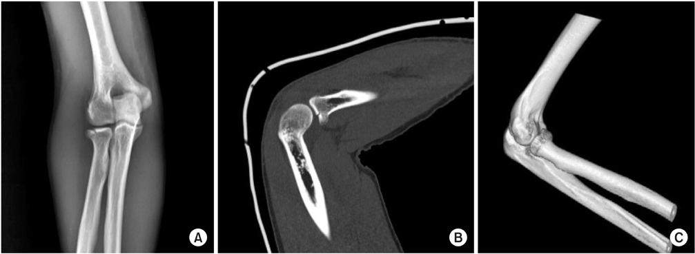
A 30-year-old male with Mason type 2 radial head fracture. Anteroposterior radiograph (A) and saggital computed tomography (CT) (B), and a 3-dimensional CT (C).
Fig. 2
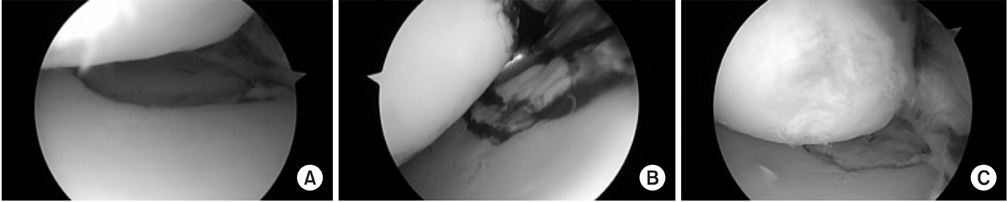
Right elbow arthroscopic photos. (A) Radial head fracture with articular step off. (B, C) The radial head was reduced with manipulation by the arthroscopic probe and micro curette.
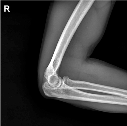
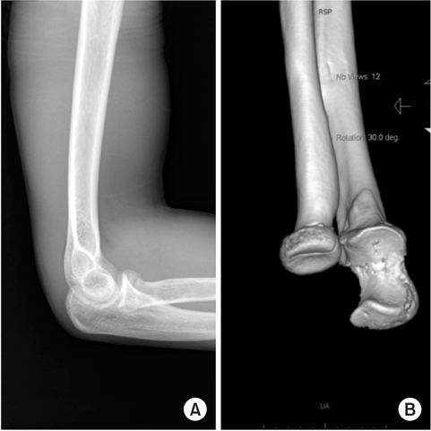
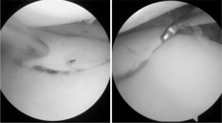
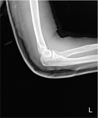
Figure & Data
REFERENCES
Citations
Citations to this article as recorded by 
- Bioabsorbable Screws Used in Hallux Valgus Treatment Using Proximal Chevron Osteotomy
Woo-Jin Shin, Young-Woo Chung, Ki-Yong An, Jae-Woong Seo
Journal of Korean Foot and Ankle Society.2018; 22(4): 181. CrossRef
 Cite
CiteArthroscopic Assisted Bioabsorbable Screw Fixation for Radial Head Fractures: A Report of Two Cases


Fig. 1
A 30-year-old male with Mason type 2 radial head fracture. Anteroposterior radiograph (A) and saggital computed tomography (CT) (B), and a 3-dimensional CT (C).
Fig. 2
Right elbow arthroscopic photos. (A) Radial head fracture with articular step off. (B, C) The radial head was reduced with manipulation by the arthroscopic probe and micro curette.
Fig. 3
Immediately postoperative right elbow lateral X-ray.
Fig. 4
A 42-year-old male with Mason type 2 radial head fracture. Lateral radiograph (A) and 3-dimensional computed tomography (B).
Fig. 5
The radial head reduced with manipulation by the arthroscopic probe and micro curette.
Fig. 6
Immediately postoperative left elbow lateral X-ray.
Fig. 1
Fig. 2
Fig. 3
Fig. 4
Fig. 5
Fig. 6
Arthroscopic Assisted Bioabsorbable Screw Fixation for Radial Head Fractures: A Report of Two Cases

