 E-submission
 TOTA
TOTA TOTS
TOTS
Articles
- Page Path
- HOME > J Musculoskelet Trauma > Volume 23(3); 2010 > Article
-
Original Article
- Open Reduction and Internal Fixation with AO Calcaneal Plate for Displaced Intra-articular Calcaneal Fracture
- Myung Jin Lee, M.D., Sung Keun Sohn, M.D., Kyu Yeol Lee, M.D., Sung Soo Kim, M.D., Min Soo Kang, M.D., Hyeon Jun Kim, M.D, Sang Kyu Sun, M.D.
-
Journal of the Korean Fracture Society 2010;23(3):303-309.
DOI: https://doi.org/10.12671/jkfs.2010.23.3.303
Published online: July 31, 2010
Department of Orthopedic Surgery, College of Medicine, Dong-A University, Busan, Korea.
*Department of Orthopedic Surgery, Dong-Eui Medical Center, Busan, Korea.
- Address reprint requests to: Hyeon Jun Kim, M.D. Department of Orthopedic Surgery, College of Medicine, Dong-A University, 1, Dongdaesin-dong 3-ga, Seo-gu, Busan 602-715, Korea. Tel: 82-51-240-5167, Fax: 82-51-254-6757, campbellkim@naver.com
• Received: November 19, 2009 • Revised: March 4, 2010 • Accepted: April 7, 2010
Copyright © 2010 The Korean Fracture Society
- 1,408 Views
- 3 Download
- 2 Crossref
Abstract
-
Purpose
- To evaluate the surgical outcomes of open reduction and internal fixation of AO calcaneal plate in displaced intra-articular fractures of the calcaneus.
-
Materials and Methods
- From January 2004 to July 2007, 25 patients with 27 displaced intra-articular calcaneal fractures were treated by open reduction and internal fixation using the AO calcaneal plate. Preoperative, postoperative evaluations and a follow-up after 1 year were done radiologically by the Böhler angle, Gissane angle, heel height and width among all patients. Their functional status was assessed by means of the Maryland foot score.
-
Results
- The mean Böhler angle, Gissane angle, heel height and width were restored comparing with preoperative data. However, in Sanders type IV, some losses of reduction occurred at 1 year follow-up (p<0.05). The mean Maryland foot scores were 85 points in type II, 82 points in type III and 63 points in type IV. Sanders types significantly affected the clinical results (p<0.05).
-
Conclusion
- The AO calcaneal plate fixation using extensile L-shpaed lateral approach shows satisfactory radiologic and clinical results in the treatment of displaced intra-articular calcaneal fractures.
- 1. Basile A. Operative versus nonoperative treatment of displaced intra-articular calcaneal fractures in elderly patients. J Foot Ankle Surg, 2010;49:25-32.Article
- 2. Boack DH, Wichelhaus A, Mittlmeier T, Hoffmann R, Haas NP. Therapy of dislocated calcaneus joint fracture with the AO calcaneus plate. Chirurg, 1998;69:1214-1223.ArticlePDF
- 3. Buckley R, Tough S, McCormack R, et al. Operative compared with nonoperative treatment of displaced intra-articular calcaneal fractures: a prospective, randomized, controlled multicenter trial. J Bone Joint Surg Am, 2002;84:1733-1744.
- 4. Cave EF. Fractures of the os calcis--the problem in general. Clin Orthop Relat Res, 1963;30:64-66.
- 5. Fernandez DL, Koella C. Combined percutaneous and "minimal" internal fixation for displaced articular fractures of the calcaneus. Clin Orthop Relat Res, 1993;290:108-116.Article
- 6. Frankel JP, Anderson CD. The use of a calcaneal reconstruction plate in intra-articular calcaneal fractures. J Foot Ankle Surg, 1996;35:318-330.Article
- 7. Huang PJ, Huang HT, Chen TB, et al. Open reduction and internal fixation of displaced intra-articular fractures of the calcaneus. J Trauma, 2002;52:946-950.Article
- 8. Huefner T, Thermann H, Geerling J, Pape HC, Pohlemann T. Primary subtalar arthrodesis of calcaneal fractures. Foot Ankle Int, 2001;22:9-14.ArticlePDF
- 9. Jung HG, Kim YJ, Jeon SH. Primary subtalar arthrodesis for the treatment of intra-articular calcaneal comminuted fractures. J Korean Fract Soc, 2006;19:418-423.Article
- 10. Kerr PS, Pape M, Jackson M, Atkins RM. Early experiences with the AO calcaneal fracture plate. Injury, 1996;27:39-41.Article
- 11. Kim IG, Kim JH, Kim CH, Kim JS. A critical analysis of long-term result and prognostic factors of fractures of the calcaneus. J Korean Soc Fract, 1998;11:354-361.Article
- 12. Kim ST, Youn TH, Park JB, Lee JY. Surgical outcomes of intra-articular fractures of calcaneus using AO calcaneal plate. J Korean Foot Ankle Soc, 2009;13:75-79.
- 13. Lee HJ, Kang SY, Kim JW. Surgical treatment of displaced intra-articular fracture of the calcaneus using a Y-plate. J Korean Soc Fract, 2002;15:433-438.Article
- 14. Longino D, Buckley RE. Bone graft in the operative treatment of displaced intraarticular calcaneal fractures: is it helpful? J Orthop Trauma, 2001;15:280-286.Article
- 15. Loucks C, Buckley R. Bohler's angle: correlation with outcome in displaced intra-articular calcaneal fractures. J Orthop Trauma, 1999;13:554-558.Article
- 16. Myerson M, Quill GF Jr. Late complications of fractures of the calcaneus. J Bone Joint Surg Am, 1993;75:331-341.Article
- 17. Parmar HV, Triffitt PD, Gregg PJ. Intra-articular fractures of the calcaneum treated operatively or conservatively. A prospective study. J Bone Joint Surg Br, 1993;75:932-937.ArticlePDF
- 18. Radnay CS, Clare MP, Sanders RW. Subtalar fusion after displaced intra-articular calcaneal fractures: does initial operative treatment matter? J Bone Joint Surg Am, 2009;91:541-546.Article
- 19. Robb CA, Deans V, Iqbal MJ, Cooper JP. Comparison of non-operative and surgical treatment of displaced calcaneal fractures. Foot, 2007;17:169-173.Article
- 20. Rodriguez SR, Garduno RB, Raygoza CO. Surgical treatment of calcaneal fractures with a special titanium AO plate. Acta Ortopedica Mexicana, 2004;18:34-38.
- 21. Ross SD, Sowerby MR. The operative treatment of fractures of the os calcis. Clin Orthop Relat Res, 1985;199:132-143.Article
- 22. Sanders R, Fortin P, Dipasquale T, Walling A. Operative treatment in 120 displaced intraarticular calcaneal fractures: Results using a prognostic computed tomography scan classification. Clin Orthop Relat Res, 1993;290:87-95.
- 23. Schepers T, van Lieshout EM, van Ginhoven TM, Heetveld MJ, Patka P. Current concepts in the treatment of intra-articular calcaneal fractures: results of a nationwide survey. Int Orthop, 2008;32:711-715.ArticlePDF
- 24. Song KS, Jeon SY, Chun JH. Radiologic evaluation of treatment outcome in intraarticular calcaneal fracture by open reduction without bone graft. J Korean Soc Fract, 2002;15:226-233.Article
- 25. Stephenson JR. Surgical treatment of displaced intra-articular fractures of the calcaneus. A combined lateral and medial approach. Clin Orthop Relat Res, 1993;290:68-75.
- 26. Thordarson DB, Krieger LE. Operative vs. non operative treatment of intra-articular fractures of the calcaneus: a prospective randomized trial. Foot Ankle Int, 1996;17:2-9.ArticlePDF
- 27. Yang KH, Chung JB, Yoon HK, Park SY, Yoon HS. Treatment of displaced intra-articular calcaneal fractures using a F-plate. J Korean Fract Soc, 2007;20:1-5.Article
- 28. Zwipp H, Tscherne H, Thermann H, Weber T. Osteosynthesis of displaced intraarticular fractures of the calcaneus. Results in 123 cases. Clin Orthop Relat Res, 1993;290:76-86.
REFERENCES
Figure 1
A 21-year-old female patient sustained left intra-articular calcaneal fracture by slip down.
(A) Preoperative X-ray shows tongue type intra-articular calcaneal fracture.
(B) Preoperative axial and sagittal CT scans show Sanders type IIb calcaneal fracture.
(C) Postoperative X-ray shows restoration of Böhler angle and heel width.
(D) At follow-up of 13months, X-ray shows no subtalar joint arthritis.

Figure 2
A 45-year-old male patient sustained right open comminuted intra-articular calcaneus fracture by fall down from 6 meter height.
(A) Preoperative X-ray shows joint depression type intra-articular calcaneal fracture with negative Böhler angle.
(B) Preoperative semicoronal and 3D CT scan shows Sanders type IV calcaneal fracture.
(C) Postoperative X-ray shows restoration of Böhler angle and stable fixation.
(D) Postoperative X-ray shows varus change of heel and malunion of calcaneus and losses of corrections.

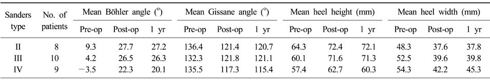
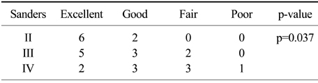
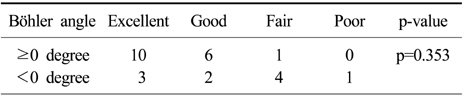
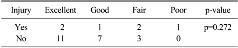
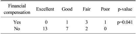
Figure & Data
REFERENCES
Citations
Citations to this article as recorded by 
- Surgical Treatment of Calcaneal Fractures of Sanders Type II and III by A Minimally Invasive Technique with 6.5 mm Cancellous Screw
Yong Seung Oh, Kyung Ho Lee, Jung Ho Kim, Myoung Jin Lee
Journal of Korean Foot and Ankle Society.2019; 23(3): 116. CrossRef - Usefulness of Treatment with 6.5 mm Cancellous Screw and Steinmann Pin Fixation for Calcaneal Joint Depression Fracture
Gi-Soo Lee, Chan Kang, Deuk-Soo Hwang, Chang-Kyun Noh, Gi-Young Lee
Journal of Korean Foot and Ankle Society.2015; 19(1): 11. CrossRef
 Cite
CiteOpen Reduction and Internal Fixation with AO Calcaneal Plate for Displaced Intra-articular Calcaneal Fracture
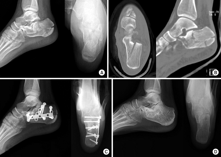
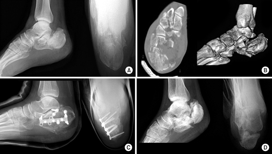
Figure 1
A 21-year-old female patient sustained left intra-articular calcaneal fracture by slip down.
(A) Preoperative X-ray shows tongue type intra-articular calcaneal fracture.
(B) Preoperative axial and sagittal CT scans show Sanders type IIb calcaneal fracture.
(C) Postoperative X-ray shows restoration of Böhler angle and heel width.
(D) At follow-up of 13months, X-ray shows no subtalar joint arthritis.
Figure 2
A 45-year-old male patient sustained right open comminuted intra-articular calcaneus fracture by fall down from 6 meter height.
(A) Preoperative X-ray shows joint depression type intra-articular calcaneal fracture with negative Böhler angle.
(B) Preoperative semicoronal and 3D CT scan shows Sanders type IV calcaneal fracture.
(C) Postoperative X-ray shows restoration of Böhler angle and stable fixation.
(D) Postoperative X-ray shows varus change of heel and malunion of calcaneus and losses of corrections.
Figure 1
Figure 2
Open Reduction and Internal Fixation with AO Calcaneal Plate for Displaced Intra-articular Calcaneal Fracture
Summary of radiologic parameters
The relationship between Sanders type and Maryland foot score (Linear by linear association)
The relationship between preoperative Böhler angle and Maryland foot score (Fisher's exact test)
The relationship between associated injury and Maryland foot score (Fisher's exact test)
The relationship between financial compensation and Maryland foot score (Fisher's exact test)
Table 1
Summary of radiologic parameters
Table 2
The relationship between Sanders type and Maryland foot score (Linear by linear association)
Table 3
The relationship between preoperative Böhler angle and Maryland foot score (Fisher's exact test)
Table 4
The relationship between associated injury and Maryland foot score (Fisher's exact test)
Table 5
The relationship between financial compensation and Maryland foot score (Fisher's exact test)

