 E-submission
 TOTA
TOTA TOTS
TOTS
Articles
- Page Path
- HOME > J Musculoskelet Trauma > Volume 24(3); 2011 > Article
-
Surgical Technique
- Avulsion Fracture of Calcaneal Tubercle Treated with Cannulated Cancellous Screws and Wire: Surgical Technique
- Chang Ho Yi, M.D., Jin Rok Oh, M.D.
-
Journal of the Korean Fracture Society 2011;24(3):262-266.
DOI: https://doi.org/10.12671/jkfs.2011.24.3.262
Published online: July 15, 2011
Department of Orthopedic Surgery, Wonju College of Medicine, Yonsei University, Wonju, Korea.
- Address reprint requests to: Jin Rok Oh, M.D. Department of Orthopedic Surgery, Yonsei University Wonju College of Medicine, 162, Ilsan-dong, Wonju 220-701, Korea. Tel: 82-33-741-1355, Fax: 82-33-746-7326, jroh@yonsei.ac.kr
• Received: August 16, 2010 • Revised: March 15, 2011 • Accepted: May 3, 2011
Copyright © 2011 The Korean Fracture Society
- 845 Views
- 10 Download
Abstract
- The incidence rate of calcaneal fracture consists about 2% of all fractures, and, of the fracture, calcaneal tubercle avulsion fracture is known to be rare. To treat non-displaced calcaneal tubercle avulsion fracture, conservative treatment such as cast fixation is applied. However, most cases accompany displacement of the avulsion fragment, and, usually, surgery is necessary to treat the displaced fracture. Although surgical fixation simply by cancellous screw or tension wire is widely used, fixation failure is potential complication in this method. Thus, this study wants to introduce a prospective and useful method that further strengthens the calcaneal fixation by using both cannulated screw and tension band wiring.
- 1. Biehl WC 3rd, Morgan JM, Wagner FW Jr, Gabriel R. Neuropathic calcaneal tuberosity avulsion fractures. Clin Orthop Relat Res, 1993;296:8-13.Article
- 2. Bierwag K. Avulsion fracture of the calcaneus. Report of an unusual case and discussion of the pathogenesis. Int Surg, 1970;54:424-427.
- 3. Dieterle J. A case of so-called "open-beak" fracture of the os calcis. J Bone Joint Surg Am, 1940;2:740.
- 4. Hedlund LJ, Maki DD, Griffiths HJ. Calcaneal fractures in diabetic patients. J Diabetes Complications, 1998;12:81-87.Article
- 5. Hess M, Booth B, Laughlin RT. Calcaneal avulsion fractures: complications from delayed treatment. Am J Emerg Med, 2008;26:254.e1-254.e4.Article
- 6. Lowy M. Avulsion fractures of the calcaneus. J Bone Joint Surg Br, 1969;51:494-497.ArticlePDF
- 7. Lui TH. Fixation of tendo Achilles avulsion fracture. Foot Ankle Surg, 2009;15:58-61.Article
- 8. Martini F, Kremling E, Sell S. Bilateral atraumatic avulsion fracture of the calcaneal tubercle in osteomalacia during fluoride therapy--a case report. Acta Orthop Scand, 1999;70:91-92.Article
- 9. Protheroe K. Avulsion fractures of the calcaneus. J Bone Joint Surg Br, 1969;51:118-122.
- 10. Rothberg A. Avulsion fracture of the os calcis. J Bone Joint Surg Am, 1939;2:218-220.
- 11. Rowe CR, Sakellarides H, Freeman P, Sorbie C. Fractures of os calcis: a long-term follow-up study of one hundred forty six patient. JAMA, 1963;184:920-923.Article
- 12. Slätis P, Santavirta S, Sandelin J. Surgical treatment of chronic dislocation of the peroneal tendons. Br J Sports Med, 1988;22:16-18.Article
- 13. Squires B, Allen PE, Livingstone J, Atkins RM. Fractures of the tuberosity of the calcaneus. J Bone Joint Surg Br, 2001;83:55-61.ArticlePDF
REFERENCES
Fig. 1
(A) The picture taken in the operating room of a 72-years old female shows the avulsion fracture of calcaneal tubercle.
(B) Open reduction and internal fixation was done by using 2 cannulated screws and wires.

Fig. 2The initial simple radiograph taken in the emergency room of a 72-years old female shows the avulsion fracture of calcaneal tubercle.

Fig. 3Open reduction and Internal fixation was done with 2 cannulated screws and wires 1 day after the injury.

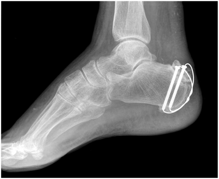
Fig. 5Initial radiograph of 68-eyars old female showed fracture of lateral malleolus (A) and the avulsion fracture of calcaneal tubercle (B).
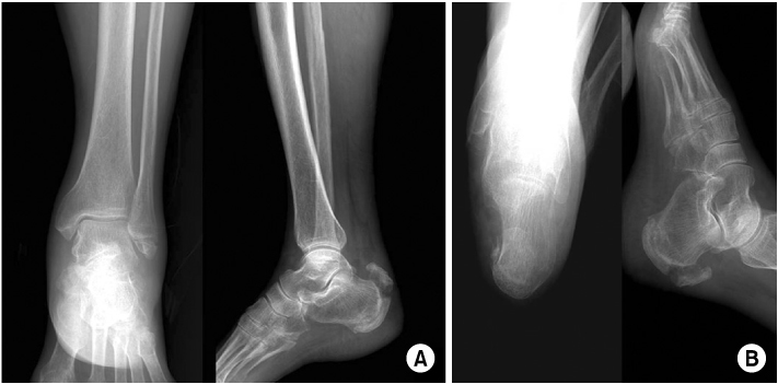

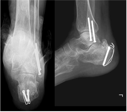
Figure & Data
REFERENCES
Citations
Citations to this article as recorded by 
 Cite
CiteAvulsion Fracture of Calcaneal Tubercle Treated with Cannulated Cancellous Screws and Wire: Surgical Technique
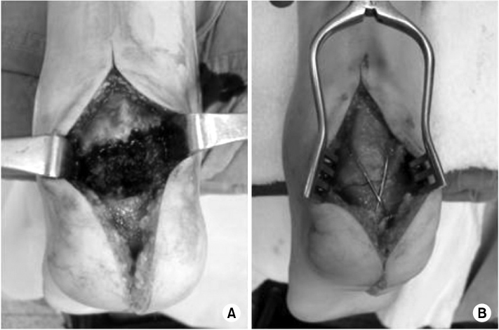
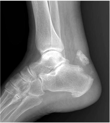
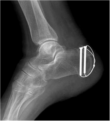


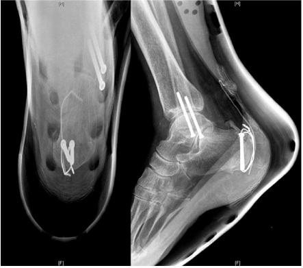

Fig. 1
(A) The picture taken in the operating room of a 72-years old female shows the avulsion fracture of calcaneal tubercle.
(B) Open reduction and internal fixation was done by using 2 cannulated screws and wires.
Fig. 2
The initial simple radiograph taken in the emergency room of a 72-years old female shows the avulsion fracture of calcaneal tubercle.
Fig. 3
Open reduction and Internal fixation was done with 2 cannulated screws and wires 1 day after the injury.
Fig. 4
At postoperative 10 weeks, findings of bony union were seen in plain radiograph.
Fig. 5
Initial radiograph of 68-eyars old female showed fracture of lateral malleolus (A) and the avulsion fracture of calcaneal tubercle (B).
Fig. 6
Immediate postoperative radiograph after closed reduction and internal fixation of lateral malleolus and open reduction and internal fixation of calcaneal tubercle.
Fig. 7
At postoperative 13 weeks, follow up radiograph showed bony union of calcaneus.
Fig. 1
Fig. 2
Fig. 3
Fig. 4
Fig. 5
Fig. 6
Fig. 7
Avulsion Fracture of Calcaneal Tubercle Treated with Cannulated Cancellous Screws and Wire: Surgical Technique

