 E-submission
 TOTA
TOTA TOTS
TOTS
Articles
- Page Path
- HOME > J Musculoskelet Trauma > Volume 24(4); 2011 > Article
-
Original Article
- Treatment of Distal Femoral Fractures Using Polyaxial Locking Plate
- Sang-Eun Park, M.D., Hyun-Taek Kang, M.D., Young-Yul Kim, M.D., Jae-Jung Jeong, M.D., Jung-U Lee, M.D., Weon-Yoo Kim, M.D.
-
Journal of the Korean Fracture Society 2011;24(4):321-327.
DOI: https://doi.org/10.12671/jkfs.2011.24.4.321
Published online: October 30, 2011
Department of Orthopedic Surgery, Daejeon St. Mary's Hospital, The Catholic University of Korea College of Medicine, Daejeon, Korea.
- Address reprint requests to: Weon-Yoo Kim, M.D. Department of Orthopedic Surgery, Daejeon St. Mary's Hospital, The Catholic University of Korea College of Medicine, 520-2, Daehung-dong, Joong-gu, Daejeon 301-723, Korea. Tel: 82-42-220-9530, Fax: 82-42-221-0429, weonkim@hotmail.com
• Received: January 4, 2011 • Revised: February 27, 2011 • Accepted: August 28, 2011
Copyright © 2011 The Korean Fracture Society
- 1,855 Views
- 11 Download
- 2 Crossref
Abstract
-
Purpose
- To report the clinical outcome of polyaxial locking plate (Noncontact bridging (NCB) plate (Zimmer, Warsaw, Indiana)) for the treatment of distal femur fracture with minimal invasive percutaneous periosteal osteosynthsis (MIPPO) technique.
-
Materials and Methods
- Between February 2008 to April 2010, twenty six patients (11 men, 15 women), twenty eight cases diagnosed as distal femoral fractures are enrolled in this retrospective study. The mean age of the patients was 63 years (34 to 85) and the mean follow-up was 20.3 months (12 to 32). According to the AO/ASIF classification, 15 fractures were type A, 1 type B and 9 type C. And there were 3 periprsthetic fractures around knee. The analysis of the clinical and radiologic outcome were performed by Sanders functional evaluation scale and radiologic follow up after operation, respectively.
-
Results
- Among 28 cases, 25 cases united without additional operation. According to Sanders functional evaluation scale, there were 11 excellent, 9 good, 4 fair, 2 poor. As complications, there were 1 knee stiffness, 1 delayed union, 1 implant failure with refracture, 1 implant loosening. Three patients except one knee stiffness, underwent a second LISS plating using NCB plate and and bone grafting, resulting in a satisfactory final outcome.
-
Conclusion
- Internal fixation using polyaxial locking plate with MIPO technique may be one of the most effective methods for the treatment of distal femoral fractures.
- 1. Apostolou CD, Papavasiliou AV, Aslam N, Handley RC, Willett KM. Preliminary results and technical aspects following stabilisation of fractures around the knee with liss. Injury, 2005;36:529-536.Article
- 2. Bong MR, Egol KA, Koval KJ, et al. Comparison of the LISS and a retrograde-inserted supracondylar intramedullary nail for fixation of a periprosthetic distal femur fracture proximal to a total knee arthroplasty. J Arthroplasty, 2002;17:876-881.Article
- 3. Button G, Wolinsky P, Hak D. Failure of less invasive stabilization system plates in the distal femur: a report of four cases. J Orthop Trauma, 2004;18:565-570.
- 4. Connolly JF, Dehne E, Lafollette B. Closed reduction and early cast-brace ambulation in the treatment of femoral fractures. II. Results in one hundred and forty-three fractures. J Bone Joint Surg Am, 1973;55:1581-1599.
- 5. Figgie MP, Goldberg VM, Figgie HE 3rd, Sobel M. The results of treatment of supracondylar fracture above total knee arthroplasty. J Arthroplasty, 1990;5:267-276.Article
- 6. Firoozbakhsh K, Behzadi K, DeCoster TA, Moneim MS, Naraghi FF. Mechanics of retrograde nail versus plate fixation for supracondylar femur fractures. J Orthop Trauma, 1995;9:152-157.Article
- 7. Haidukewych G, Sems SA, Huebner D, Horwitz D, Levy B. Results of polyaxial locked-plate fixation of periarticular fractures of the knee. Surgical technique. J Bone Joint Surg Am, 2008;90:117-134.
- 8. Helfet DL, Lorich DG. Retrograde intramedullary nailing of supracondylar femoral fractures. Clin Orthop Relat Res, 1998;350:80-84.Article
- 9. Kanabar P, Kumar V, Owen PJ, Rushton N. Less invasive stabilisation system plating for distal femoral fractures. J Orthop Surg (Hong Kong), 2007;15:299-302.ArticlePDF
- 10. Kim JJ. Treatment of distal femur fracture. J Korean Fract Soc, 2005;18:213-220.Article
- 11. Kobbe P, Klemm R, Reilmann H, Hockertz TJ. Less invasive stabilisation system (LISS) for the treatment of periprosthetic femoral fractures: a 3-year follow-up. Injury, 2008;39:472-479.Article
- 12. Koval KJ, Hoehl JJ, Kummer FJ, Simon JA. Distal femoral fixation: a biomechanical comparison of the standard condylar buttress plate, a locked buttress plate, and the 95-degree blade plate. J Orthop Trauma, 1997;11:521-524.Article
- 13. Marti A, Fankhauser C, Frenk A, Cordey J, Gasser B. Biomechanical evaluation of the less invasive stabilization system for the internal fixation of distal femur fractures. J Orthop Trauma, 2001;15:482-487.Article
- 14. Mize R. Treatment options for fractures of the distal femur. Instr Course Lect, 1994;43:109-117.
- 15. Mize RD, Bucholz RW, Grogan DP. Surgical treatment of displaced, comminuted fractures of the distal end of the femur. J Bone Joint Surg Am, 1982;64:871-879.Article
- 16. Ochsner PE, Pfister A. Use of the fork plate for internal fixation of periprosthetic fractures and osteotomies in connection with total knee replacement. Orthopedics, 1999;22:517-521.
- 17. Oh JK, Oh CW, Park SH, Roh KJ, Jeong CW. Conformity of the LCP-DF (Locking Compression Plate-Distal Femur) in Korean adult femur: a cadaver study. J Korean Fract Soc, 2005;18:399-404.Article
- 18. Otto RJ, Moed BR, Bledsoe JG. Biomechanical comparison of polyaxial-type locking plates and a fixed-angle locking plate for internal fixation of distal femur fractures. J Orthop Trauma, 2009;23:645-652.Article
- 19. Sanders R, Swiontkowski M, Rosen H, Helfet D. Double-plating of comminuted, unstable fractures of the distal part of the femur. J Bone Joint Surg Am, 1991;73:341-346.Article
- 20. Schandelmaier P, Partenheimer A, Koenemann B, Grün OA, Krettek C. Distal femoral fractures and LISS stabilization. Injury, 2001;32:SC55-SC63.Article
- 21. Schatzker J, Lambert DC. Supracondylar fractures of the femur. Clin Orthop Relat Res, 1979;138:77-83.Article
- 22. Schütz M, Müller M, Regazzoni P, et al. Use of the less invasive stabilization system (LISS) in patients with distal femoral (AO33) fractures: a prospective multicenter study. Arch Orthop Trauma Surg, 2005;125:102-108.ArticlePDF
- 23. Smith TO, Hedges C, MacNair R, Schankat K, Wimhurst JA. The clinical and radiological outcomes of the LISS plate for distal femoral fractures: a systematic review. Injury, 2009;40:1049-1063.Article
- 24. Stoffel K, Lorenz KU, Kuster MS. Biomechanical considerations in plate osteosynthesis: the effect of plate-to-bone compression with and without angular screw stability. J Orthop Trauma, 2007;21:362-368.Article
- 25. Syed AA, Agarwal M, Giannoudis PV, Matthews SJ, Smith RM. Distal femoral fractures: long-term outcome following stabilisation with the LISS. Injury, 2004;35:599-607.Article
- 26. Wilkens KJ, Curtiss S, Lee MA. Polyaxial locking plate fixation in distal femur fractures: a biomechanical comparison. J Orthop Trauma, 2008;22:624-628.Article
- 27. Wong MK, Leung F, Chow SP. Treatment of distal femoral fractures in the elderly using a less-invasive plating technique. Int Orthop, 2005;29:117-120.ArticlePDF
REFERENCES
Fig. 1
(A) The initial radiographs and 3D CT of a 60-yearold female who sustained a Type C2 supracondylar fracture of the distal femur showing a intra-articular supracondylar fracture.
(B) Postoperative 16 weeks radiographs shows solid union.

Fig. 2
A 44-year-old male suffering from multiple fractures, including left distal femoral open comminuted fracture (AO classification C3) was treated with open reduction and internal fixation using LCP. After 2 months, osteosynthesis with autologous iliac bone grafting was performed for scanty callus and remained bony defect which is augmented by medial NCB plate. Bony union was obtained during follow-up.
(A) Initial AP radiograph.
(B) Preoperative three-demensional CT scan.
(C) First postoperative (Internal fixation using LCP) AP radiograph.
(D) Second postoperative (Internal fixation and augmentation using NCB plate) AP radiograph.
(E) Last follow-up clinical photograph.

Fig. 3
(A) The initial radiographs of a 60-year-old female who sustained a Type A1 supracondylar fracture of the Lt. distal femur.
(B) The postoperative radiographs show the supracondylar fracture was reduced and fixed with 15 hole NCB plate.
(C) Postoperative 8 weeks, she suffered refracture with metallic breakage due to minor trauma during ambulation.
(D) Reoperation was done using same length NCB plate and wiring and showed callus formation.


Figure & Data
REFERENCES
Citations
Citations to this article as recorded by 
- Usefulness of Reduction and Internal Fixation Using a 2.4 mm Hand Plating System in Type AO 33-A3 Distal Femur Fracture: Technical Note
Bong-Ju Lee, Ja-Yeong Yoon, Seungha Woo
Journal of the Korean Fracture Society.2023; 36(1): 25. CrossRef - Incidence of nonunion after surgery of distal femoral fractures using contemporary fixation device: a meta‐analysis
Byung-Ho Yoon, In Keun Park, Youngwoo Kim, Hyoung-Keun Oh, Suk Kyu Choo, Yerl-Bo Sung
Archives of Orthopaedic and Trauma Surgery.2021; 141(2): 225. CrossRef
 Cite
CiteTreatment of Distal Femoral Fractures Using Polyaxial Locking Plate
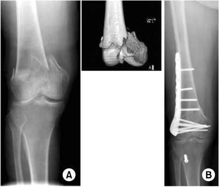
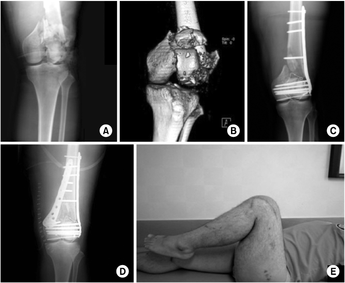
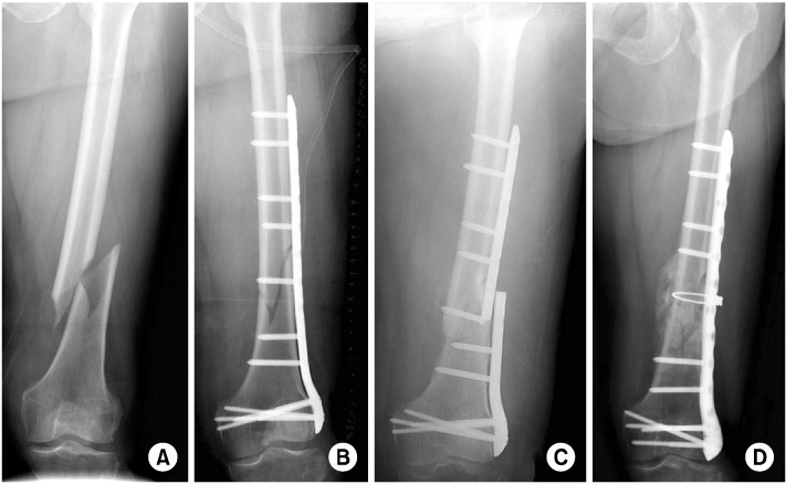
Fig. 1
(A) The initial radiographs and 3D CT of a 60-yearold female who sustained a Type C2 supracondylar fracture of the distal femur showing a intra-articular supracondylar fracture.
(B) Postoperative 16 weeks radiographs shows solid union.
Fig. 2
A 44-year-old male suffering from multiple fractures, including left distal femoral open comminuted fracture (AO classification C3) was treated with open reduction and internal fixation using LCP. After 2 months, osteosynthesis with autologous iliac bone grafting was performed for scanty callus and remained bony defect which is augmented by medial NCB plate. Bony union was obtained during follow-up.
(A) Initial AP radiograph.
(B) Preoperative three-demensional CT scan.
(C) First postoperative (Internal fixation using LCP) AP radiograph.
(D) Second postoperative (Internal fixation and augmentation using NCB plate) AP radiograph.
(E) Last follow-up clinical photograph.
Fig. 3
(A) The initial radiographs of a 60-year-old female who sustained a Type A1 supracondylar fracture of the Lt. distal femur.
(B) The postoperative radiographs show the supracondylar fracture was reduced and fixed with 15 hole NCB plate.
(C) Postoperative 8 weeks, she suffered refracture with metallic breakage due to minor trauma during ambulation.
(D) Reoperation was done using same length NCB plate and wiring and showed callus formation.
Fig. 1
Fig. 2
Fig. 3
Treatment of Distal Femoral Fractures Using Polyaxial Locking Plate
Summary of patient dermographics
TA: traffic accident.
Functional results by Sanders et al
Table 1
Summary of patient dermographics
TA: traffic accident.
Table 2
Functional results by Sanders et al

