 E-submission
 TOTA
TOTA TOTS
TOTS
Search
- Page Path
- HOME > Search
Original Articles
- Open reduction and internal fixation for distal humerus fractures in older adults: a retrospective comparative study by osteoporosis status
- Tong Joo Lee, Hee Beom Shin, Yongseok Lee
- J Musculoskelet Trauma 2026;39(3):227-235. Published online July 15, 2026
- DOI: https://doi.org/10.12671/jmt.2026.00101
-
 Abstract
 PDF
PDF - Background
Distal humerus fractures in older patients, particularly those with osteoporosis, pose substantial treatment challenges because of increased fracture complexity and compromised bone stock. Open reduction and internal fixation (ORIF) is the preferred treatment but may be complicated by fixation failure. This study investigated the outcomes of ORIF in older osteoporotic and nonosteoporotic patients with complete articular distal humerus fractures.
Methods
This retrospective study included 19 patients with Arbeitsgemeinschaft für Osteosynthesefragen/ Orthopaedic Trauma Association (AO/OTA) 13C distal humerus fractures who underwent surgical treatment between 2012 and 2024. The mean patient age was 79.1 years. Patients were divided into osteoporotic (n=11) and nonosteoporotic (n=8) groups based on the lowest recorded dual-energy X-ray absorptiometry T-score at the femoral neck or lumbar spine. Osteoporosis was defined as a T-score of −2.5 or lower, and the nonosteoporotic group included patients with osteopenia. All fractures were treated with ORIF using bicolumnar plating. All included patients completed 12 months of clinical follow-up for visual analog scale (VAS) and Mayo Elbow Performance Score (MEPS) assessments. Radiographic follow-up was recorded separately and continued until union was confirmed; imaging follow-up extended to 12 months or longer in 14 patients and was limited to 6 months in five asymptomatic patients after confirmed union.
Results
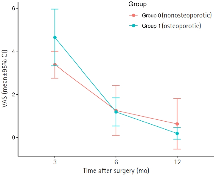
Both groups showed significant within-group improvement in pain, as assessed using the VAS, and function, as assessed using the MEPS, over time. Between-group comparisons at each follow-up time point showed no statistically significant differences in VAS or MEPS. No radiographic nonunion was observed during the available imaging follow- up, and complications were limited to one case of screw pullout/loosening and one case of postoperative stiffness.
Conclusions
ORIF provides reliable outcomes for older patients with complex distal humerus fractures, regardless of osteoporosis status, when stable reconstruction is achievable. These findings suggest that ORIF remains a viable treatment option, with satisfactory functional recovery and low complication rates in this population. Level of evidence: III.
- 164 View
- 7 Download

- Computational simulation of coracoclavicular screw insertion through the superior distal clavicular plate for clinical applications in Korean cadavers
- Hyung-Lae Cho, Ji Han Choi, Se-Lin Jeong, Gu-Hee Jung
- J Musculoskelet Trauma 2025;38(3):143-151. Published online July 22, 2025
- DOI: https://doi.org/10.12671/jmt.2025.00122
-
Abstract
PDF
- Background
The study was conducted to determine the practical area for inserting the coracoclavicular (CC) screw through the plate by analyzing three-dimensional (3D) shoulder models featuring virtually implanted, actual-size plates and screws.
Methods
Ninety cadaveric shoulders (41 males and 49 females) underwent continuous 1.0-mm slice computed tomography scans. The data were imported into image-processing software to generate a 3D shoulder model, including the scapula and clavicle. The overlapping area between the clavicle and the horizontal portion of the coracoid process (horizontal portion_CP) was analyzed in the cranial view. A curved pelvic recon plate was virtually placed on the upper surface of the distal clavicle, and an actual-size (3.5 mm) CC screw was inserted through the plate.
Results
The distal clavicle directly overlapped with the horizontal portion_CP in the vertical direction. The overlapping area was sufficient to place the 3.5 mm and 4.5 mm-sized screws. In all shoulder models, the CC screw could be inserted through the plate into the vertical direction, with an average length of 35.5 mm (range, 26.2–62.5 mm; standard deviation, 1.2 mm). In 87 models, the CC screw was inserted through the third hole from the lateral end of the plate. Two models were inserted through the second hole, and one model through the fourth hole.
Conclusions
The upper surface of the clavicle has sufficient overlapping area to place CC screws through the plate in the vertical direction in the corresponding hole. Supplemental CC screw fixation through the plate can be performed without additional or special equipment. Level of evidence: IV
- 1,254 View
- 26 Download
Review Article
- How to obtain the desired results from distal tibial nailing based on anatomy, biomechanics, and reduction techniques
- Jungtae Ahn, Se-Lin Jeong, Gu-Hee Jung
- J Musculoskelet Trauma 2025;38(2):74-85. Published online March 31, 2025
- DOI: https://doi.org/10.12671/jmt.2025.00024

-
Abstract
PDF
- Distal tibial metaphyseal fractures are commonly caused by high-energy injuries in young men and osteoporosis in older women. These fractures should be clearly distinguished from high-energy pilon fractures. Although the optimal surgical intervention methods for distal tibial metaphyseal fractures remain uncertain and challenging, surgical treatments for nonarticular distal tibia fractures can be broadly divided into two types: plate fixation and intramedullary nail (IMN) fixation. Once functional reduction is achieved using an appropriate technique, distal tibial nailing might be slightly superior to plate fixation in reducing postoperative complications. Thus, the surgical strategy should focus on functional realignment and proceed in the following sequence: (1) restoring the original tibial length, regardless of whether fibular fixation is to be done; (2) making the optimal entry point through an anteroposterior (AP) projection based on the overlapping point between the fibular tip and lateral plateau margin; (3) placing Kirschner wires (Ø2.4 mm) as blocking pins (in the AP orientation for coronal control and in the mediolateral [ML] orientation for sagittal control) as close to the upper locking hole as possible without causing further comminution on the concave aspect of the short fragment; and (4) making the the distal fixation construct with at least two ML and one AP interlocking screw or two ML interlocking screws and blocking screws. After the IMN is adequately locked, blocking pins (Ø2.4 mm) need to be replaced by a 3.5 mm screw.
-
Citations
Citations to this article as recorded by
- Rigid intramedullary nailing with suprapatellar approach for tibial shaft fractures in adolescents with open physes
Jong Wha Lee, Jae Ho Cho, Tae Hun Kim, Hyung Keun Song, Won-Tae Cho, Seungyeob Sakong, Hyun Il Choi, Sumin Lim
Injury.2026; 57(4): 113130. CrossRef - Impact of Foot Width on Patient-Reported Outcomes Assessed by 3-Dimensional Foot Morphometry in Hallux Valgus
Jungtae Ahn, Dae-Cheol Nam, Gu-Hee Jung
Clinics in Orthopedic Surgery.2025; 17(6): 1062. CrossRef
- Rigid intramedullary nailing with suprapatellar approach for tibial shaft fractures in adolescents with open physes
- 5,599 View
- 119 Download
- 2 Crossref
Original Articles
- The clinical outcome of treating elderly distal radius fractures by long volar locking plate with the elimination of irreducible metaphyseal comminuted volar cortical fragments: a retrospective case series
- Soo Min Cha
- J Musculoskelet Trauma 2025;38(1):13-22. Published online January 24, 2025
- DOI: https://doi.org/10.12671/jmt.2025.00003

-
Abstract
PDF
- Background
In severe comminuted metaphyseal distal radius fracture (DRF) of elderly patients, after maintaining only radiological parameters of the radius using long volar locking plates (VLPs), we inevitably eliminated a few volar cortical fragments of metaphysis. Here, we report the final radiological and clinical outcomes of our method. Methods: For the patients who were treated between 2014 and 2018, the demographic factors, the preoperative radiologic factors, area of the eliminated volar cortical fragment, and final radiologic parameter, were evaluated. Clinical outcomes and ranges of active motion were evaluated. Results: In total, 31 patients were included. The mean patient age was 77.3 years and the mean eliminated cortical area was 3.30 cm2. At the final follow-up, the mean volar tilt, radial inclination, articular step-off, and ulnar variance were 10.35°, 20.00°, 0.58 mm, and 0.71 mm, respectively. There were no definitive correlations between bone mineral density, fragment area, the largest cortical fragment diameter ratio and differences in final and immediate postoperative measurements of these radiological parameters, respectively. Visual analog scale and disabilities of the arm, shoulder, and hand (DASH) scores were satisfactory, and the mean arcs of flexion-extension and pronation-supination were 124.35° and 133.23°. Clinical outcomes were not significantly different according to the AO system category. Conclusions: For maintenance of radiological parameters of the radius, long VLPs are useful in older patients with DRFs who exhibit volar metaphyseal comminution, despite concurrent ulnar fractures. Inevitable elimination of irreducible free comminuted cortical fragments when filling the defect does not affect final radiological and clinical outcomes. Level of evidence: Level IV, case series. -
Citations
Citations to this article as recorded by- Open distal radius fracture with ulnar neurovascular injury treated with dorsal spanning plate fixation and vein interposition grafting: a case report
Kyeong-Eon Kim, Ho-Yong Lee, Shin-Woo Lee, Ji-Sup Kim
Journal of Musculoskeletal Trauma.2026; 39(3): 262. CrossRef
- Open distal radius fracture with ulnar neurovascular injury treated with dorsal spanning plate fixation and vein interposition grafting: a case report
- 3,783 View
- 52 Download
- 1 Crossref
- Does the Operator’s Experience Affect the Occurrence of Complications after Distal Radius Fracture Volar Locking Plate Fixation? A Comparative Study of the First Four Years and Thereafter
- Kee-Bum Hong, Chi-Hoon Oh, Chae Kwang Lim, Sungwoo Lee, Soo-Hong Han, Jun-Ku Lee
- J Musculoskelet Trauma 2024;37(4):175-183. Published online October 25, 2024
- DOI: https://doi.org/10.12671/jmt.2024.37.4.175
- Correction in: J Musculoskelet Trauma 2025;38(1):40
-
Abstract
PDF
- Purpose
The management of distal radius fractures (DRFs) has evolved with the introduction of volar locking plate (VLP) fixation, offering stable fixation and better outcomes. Nevertheless, the impact of the surgeon’s experience on the complication rates in VLP fixation remains to be determined, particularly for less-experienced surgeons. This study compared the complication rates during the initial four years and subsequent two years of a hand surgeon’s practice of VLP fixation for DRFs.
Materials and Methods
The data between March 2016 and December 2022 were analyzed retrospectively under the Institutional Review Board approval. A single surgeon performed all VLP fixation surgeries after finishing regular hand surgery training, with the first four years representing the less experienced phase (Group 1) and the following two years indicating the experienced phase (Group 2). The patients’ characteristics, operation-related factors, and postoperative complications, including tendon injuries, nerve-related complications, fixation and instrument-related issues, osteosynthesis-related problems, and infections, were compared. In addition, the authors compared the data with a large multicenter study conducted by experienced hand surgeons.
Results
Three hundred and nineteen patients (321 wrists) were included. The mean age was 63.3 years, and 26.3% were male and 73.7% were female. The operation time was 53.7±14.5 minutes and 74.4±26.5 minutes in groups 1 and 2, respectively, which was statistically significantly shorter (p<0.001). The complication rates between the two groups were similar, except for the higher implant removal rates in Group 1. A comparison with a previous multicenter study revealed higher reduction losses and carpal tunnel syndrome in this study, but the overall complication rate was low.
Conclusion
In DRF management, when the operating surgeon has completed an accredited training course, VLP fixation is a good treatment method that can be performed effectively even by less experienced surgeons with low complication rates. -
Citations
Citations to this article as recorded by- Epidemiological changes and surgical trends of distal radius fractures in adults over 50 years during the COVID-19 pandemic in Korea: a nationwide repeated cross-sectional study
Han-Kook Yoon, So Ra Yoon, Kee-Bum Hong, Youngsu Jung, SeongJu Choi, Jun-Ku Lee
Journal of Musculoskeletal Trauma.2026; 39(1): 12. CrossRef - Author correction: “Does the operator's experience affect the occurrence of complications after distal radius fracture volar locking plate fixation? A comparative study of the first four years and thereafter”
Kee-Bum Hong, Chi-Hoon Oh, Chae Kwang Lim, Sungwoo Lee, Soo-Hong Han, Jun-Ku Lee
Journal of Musculoskeletal Trauma.2025; 38(1): 40. CrossRef - Characteristics of patients with distal radius fracture requiring arthroscopic foveal repair after bone union
Min Jung Park, Cheungsoo Ha, Hyun Tak Kang, Yong Hyun Yoon, Jun-Ku Lee, Soo-Hong Han
Arthroscopy and Orthopedic Sports Medicine.2025; 12(2): 70. CrossRef
- Epidemiological changes and surgical trends of distal radius fractures in adults over 50 years during the COVID-19 pandemic in Korea: a nationwide repeated cross-sectional study
- 4,559 View
- 57 Download
- 3 Crossref
- Comparison of Results between Minimally Invasive Plate Fixation and Antegrade Intramedullary Nailing of Recon-Type in Low-Energy Injury Distal Femoral Shaft Fractures
- Hong Moon Sohn, Gwangchul Lee, Ba Rom Kim, Jung Soo Oh
- J Korean Fract Soc 2024;37(2):87-94. Published online April 30, 2024
- DOI: https://doi.org/10.12671/jkfs.2024.37.2.87
-
Abstract
PDF
- Purpose
This study compared the outcomes of minimally invasive plate osteosynthesis and antegrade intramedullary nailing for low-energy fracture of the distal femoral shaft.
Materials and Methods
A study was conducted on 30 patients who underwent surgery for low-energy fractures of the distal femoral shaft between January 2016 and April 2022. The study compared 15patients who underwent minimally invasive plate osteosynthesis (Group P) with 15 patients who underwent recon-type antegrade intramedullary nailing (Group N). We evaluated intraoperative blood loss, operative time, C-arm exposure time, bone density, final union status, anatomical reduction, and clinical evaluation. The complications were also examined, and statistical analysis was conducted to compare the two groups.
Results
The blood loss, surgery time, and C-arm time were similar in the two groups. The radiographic assessments and clinical evaluations were also similar in the two groups. The clinical results showed no difference between the groups. Group N had one case of nonunion and one case of delayed union, while Group P had one case of nonunion and one case of peri-prosthetic fracture.
Conclusion
Antegrade intramedullary nailing of the recon-type demonstrated comparable results to minimally invasive plate osteosynthesis. Hence, antegrade intramedullary nailing of the recon-type, which enhances stability by fixing the entire femur and providing additional fixation in the distal portion, is deemed appropriate for treating distal femoral shaft fractures.
- 1,159 View
- 18 Download
- Biomechanical Investigation to Establish Stable Fixation Strategies for Distal Tibial Fractures in Various Situations: Finite Element Analysis Studies
- Sung Hun Yang, Jun Young Lee, Gu-Hee Jung, Hyoung Tae Kim, Ba Woo Ko
- J Korean Fract Soc 2024;37(2):71-81. Published online April 30, 2024
- DOI: https://doi.org/10.12671/jkfs.2024.37.2.71
-
Abstract
PDF
- Purpose
This study examined the structural and mechanical stability as well as the clinical significance of various fixation constructs for distal tibial fractures using finite element analysis.
Materials and Methods
Fracture models with 20 mm and 120 mm defects were produced, and implants of an intramedullary nail and anatomical plate model were applied. An axial load of 800 N with 60% distribution in the medial compartment and 40% in the lateral compartment was applied and analyzed using Ansys ® software.
Results
In the intramedullary nail model, the maximum von Mises stress occurred at the primary lag screw hole and adjacent medial cortex, while in the plate model, it occurred at the locking holes around the fracture. The maximum shear stress on the bone and metal implant in the fracture model with a 20 mm defect was highest in the plate assembly model, and in the fracture model with a 120 mm defect, it was highest in the two-lag screw assembly model.
Conclusion
Based on an analysis of the maximum shear stress distribution, securing the fixation strength of the primary lag screw hole is crucial, and the assembly model of the intramedullary nail with two lag screws and a blocking screw applied was the model that best withstood the optimal load. Securing the locking hole directly above the fracture is believed to provide the maximum fixation strength because the maximum pressure in the plate model is concentrated in the proximal locking hole and the surrounding cortex. -
Citations
Citations to this article as recorded by- Loading-Condition Hierarchy of Anisotropy and Heterogeneity in Tibial Biomechanics: A Finite Element Study
Azadeh Ghouchani, Saba Mirzakhani, Amir M. Rashnavadi
Results in Engineering.2026; : 112129. CrossRef - How to obtain the desired results from distal tibial nailing based on anatomy, biomechanics, and reduction techniques
Jungtae Ahn, Se-Lin Jeong, Gu-Hee Jung
Journal of Musculoskeletal Trauma.2025; 38(2): 74. CrossRef
- Loading-Condition Hierarchy of Anisotropy and Heterogeneity in Tibial Biomechanics: A Finite Element Study
- 1,527 View
- 26 Download
- 2 Crossref
- Effect of Additional Medial Locking Plate Fixation and Autogenous Bone Graft for Distal Femur Nonunion after Lateral Locking Plate Fixation
- Ho Min Lee, Jong Pil Kim, In Hwa Baek, Han Sol Moon, Sun Kyo Nam
- J Korean Fract Soc 2024;37(1):30-38. Published online January 31, 2024
- DOI: https://doi.org/10.12671/jkfs.2024.37.1.30
-
Abstract
PDF
- Purpose
This study examined the outcomes of additional medial locking plate fixation and autogenous bone grafting in the treatment of nonunions that occurred after initial fixation for distal femoral fractures using lateral locking plates.
Materials and Methods
The study involved eleven patients who initially underwent minimally invasive lateral locking plate fixation for distal femoral fractures between January 2008 and December 2020. The initial procedure was followed by additional medial locking plate fixation and autogenous bone grafting for clinically and radiographically confirmed nonunions, while leaving the stable lateral locking plate in situ. A clinical evaluation of the bone union time, knee joint range of motion, visual analog scale (VAS) pain scores, presence of postoperative complications, and functional evaluations using the lower extremity functional scale (LEFS) were performed.
Results
In all cases, bone union was achieved in an average of 6.1 months after the secondary surgery. The range of knee joint motion, weight-bearing ability, and VAS and LEFS scores improved at the final follow-up compared to the preoperative conditions. All patients could walk without walking assistive devices and did not experience pain at the fracture site. On the other hand, three patients complained of pain in the lateral knee joint caused by irritation by the lateral locking plate; hence, lateral hardware removal was performed. One patient complained of mild paresthesia at the anteromedial incision site. Severe complications, such as deep infection or metal failure, were not observed.
Conclusion
For nonunion with stable lateral locking plates after minimally invasive lateral locking plate fixation of distal femur fractures, additional medial locking plate fixation and autogenous bone grafting, while leaving the lateral locking plate intact, can achieve successful bone union.
- 803 View
- 9 Download
- Bone Union Time of Simple Distal Femur Fractures in the Elderly according to Fracture Gap after Treated with Minimally Invasive Plate Osteosynthesis
- Young Ho Cho, Sangwoo Kim, Jaewook Koo
- J Korean Fract Soc 2023;36(4):133-138. Published online October 31, 2023
- DOI: https://doi.org/10.12671/jkfs.2023.36.4.133
-
Abstract
PDF
- Purpose
This study examined the difference in bone union time according to the fracture gap after minimally invasive plate osteosynthesis (MIPO) for simple distal femoral fractures in elderly patients.
Materials and Methods
From January 2010 to December 2019, patients aged 60 years or older who underwent surgical treatment for distal femoral fractures due to a low-energy injury were investigated retrospectively. Forty patients were enrolled in the study. The patients were divided into two groups according to the fracture gap after reduction: no more than 2 mm (Group A) and more than 2 mm (Group B) in the anteroposterior and lateral plane. The demographic, operation time, presence or absence of cerclage wiring, plate screw density, plate span ratio, plate length, bone union period, non-union, and complications were evaluated.
Results
No statistical differences in operation time, cerclage wiring, plate screw density, plate span ratio, and plate length were observed between the two groups, and the bone union was achieved in all patients without complication. The bone union period was 17.24±1.48 weeks in Group A and 24.53± 5.20 weeks in Group B, which was statistically significant (p<0.001).
Conclusion
The bone union time in treating geriatric simple distal femur fractures using the MIPO tech-nique was significantly shorter in the 2 mm or less fracture gap than in the greater than 2 mm group.
- 1,209 View
- 7 Download
- Demographic and Radiographic Parameters as Predictors of Reduction Loss after Conservative Treatment of Distal Radius Fractures in Adults
- Kyu Jin Kim, Dae Won Shin, Seong Kee Shin
- J Korean Fract Soc 2023;36(2):45-51. Published online April 30, 2023
- DOI: https://doi.org/10.12671/jkfs.2023.36.2.45
-
Abstract
PDF
- Purpose
This study examined the demographic and radiological risk factors for later reduction loss of distal radius fractures treated conservatively. Materials and Methods This study enrolled patients treated for distal radius fractures between January 2017 and December 2019. Seventy-eight patients were included in the analysis and divided into two groups. The patients who showed minimal reduction loss within an acceptable radiologic angle after initial manual reduction were classified as Group A. The patients who showed reduction loss out of an acceptable radiologic angle and finally malunited or converted to surgical treatments were classified as Group B. The patient’s age and bone marrow density were used as demographic data. The initial X-ray images were evaluated to determine the fracture type. Various radiological parameters were measured. Results The 78-patient study cohort consisted of nine men and 69 women with a mean age of 67 years. Forty-eight cases were sorted into Group A, and 30 cases into Group B. On logistic regression analysis, the age of 80 or older was a risk factor for later fracture displacement among the demographic factors (p=0.037, odds ratio=4.937). Among the radiographic factors, the presence of distal ulnar fracture and dorsal cortical comminution were disclosed as risk factors of later displacement (p=0.049, 0.003, odds ratio=3.429, 7.196). Conclusion When conservative management for distal radius fracture is decided in patients more than 80 years of age or accompanied by a distal ulnar fracture or with dorsal cortical comminution, the possibility of later displacement of the distal radius should be considered.
- 873 View
- 5 Download
Technical Note
- Usefulness of Reduction and Internal Fixation Using a 2.4 mm Hand Plating System in Type AO 33-A3 Distal Femur Fracture - Technical Note -
- Bong-Ju Lee, Ja-Yeong Yoon, Seungha Woo
- J Korean Fract Soc 2023;36(1):25-28. Published online January 31, 2023
- DOI: https://doi.org/10.12671/jkfs.2023.36.1.25
-
Abstract
PDF
- Open reduction in an AO 33-A3 class distal femur transverse and comminuted fracture is often difficult due to frequent reduction loss during surgery, leading to longer operative time and increased blood loss intra-operation. In this study, the authors report a case in which the use of an offset grid plate (OsteoMed, USA) using 2.4 mm HPS (hand plating system) eased the process of fracture reduction and achieved a stable internal fixation, ultimately leading to successful osteosynthesis. The authors experienced no need for temporary fixation devices such as K-wires or screws, which are otherwise required to stabilize the reduction. The fracture reduction was stable throughout the primary fixation of the fracture using a locking plate and screws. The authors report that the advantage of the HPS plate is fitting into the cortical contour and providing stable maintenance of fracture reduction intra-operation, which would be beneficial in certain distal femoral fracture patterns.
- 899 View
- 10 Download
Original Article
- Distal Femur Fractures Treated with Distal Femoral Locking Plate Fixation: A Retrospective Study of One Year Mortality and Risk Factors
- Kwang-Hwan Jung, Yoon-Seok Youm, Seung-Hyun Jung, Jae-Min Oh, Ki Bong Park
- J Korean Fract Soc 2023;36(1):10-16. Published online January 31, 2023
- DOI: https://doi.org/10.12671/jkfs.2023.36.1.10
-
Abstract
PDF
- Purpose
This study examined the one-year mortality after locking plate fixation for distal femur fractures and the risk factors related to death.
Materials and Methods
From July 2011 to June 2020, 128 patients who underwent locking plate fixation for distal femur fractures were analyzed retrospectively. Epidemiologic information of the patients, characteristics related to fracture and surgery, and death were investigated. The risk factors related to death were investigated using Cox analysis, and a subgroup analysis was also performed based on the age of 65 years.
Results
The one-year mortality rate after locking plate fixation for distal femur fractures was 3.9%, and the mortality rates in patients younger than 65 years and older than 65 years were 0% and 6.7%, respectively. There were no significant risk factors related to death in the total population. On the other hand, in patients aged 65 years or older, however, high-energy fracture and high comorbidity index increased the risk of death after surgery by 6.9-fold and 1.9-fold, respectively.
Conclusion
The one-year mortality rate for the total patients was 3.9%, but the mortality rate for patients over 65 years of age increased to 6.7%. High-energy fractures and high comorbidity index were risk factors related to death after surgery for distal femur fractures in patients aged 65 years or older.
- 921 View
- 6 Download
Review Article
- Pediatric Fractures around the Wrist
- Gihun Kim, Kun-Bo Park
- J Korean Fract Soc 2021;34(2):80-86. Published online April 30, 2021
- DOI: https://doi.org/10.12671/jkfs.2021.34.2.80
-
Abstract
PDF
- Fractures around the wrist are the third most common fracture among all pediatric fractures. Furthermore, distal radius fractures, a type of wrist fracture, are the most common fractures in children. Understanding pediatric fractures around the wrist is very important considering their prevalence. There is a specific belief that pediatric fractures can heal easily because of remodeling, but not all fractures can heal without proper treatment. Complications such as growth problems, nonunion can occur if the fracture is not treated properly. This paper reviewed recent articles about distal radius fractures, Galeazzi-equivalent fractures, and carpal bone fractures, including scaphoid fractures in children and adolescents. Successful treatment can be achieved without complications when an accurate diagnosis and proper non-surgical or surgical treatment are performed based on this article.
- 2,264 View
- 49 Download
Original Articles
- Primary Open Reduction and Plate Fixation in Open Comminuted Intra-Articular Distal Radius Fracture
- Jun-Ku Lee, Soonchul Lee, Weon Min Cho, Minkyu Kil, Soo-Hong Han
- J Korean Fract Soc 2021;34(1):16-22. Published online January 31, 2021
- DOI: https://doi.org/10.12671/jkfs.2021.34.1.16
-
Abstract
PDF
- Purpose
There are no standard surgical treatments for open distal radius fractures (DRFs), and the fracture fixator is chosen by the surgeon’s own experience. This study compared the outcomes of open reduction and volar locking plating (OR VLP) between closed and open AO-OTA type C3 DRFs. Materials and Methods: Patient data were retrospectively collected between January 2010 and December 2018. Only patients aged >18 years with AO-OTA C3 DRFs were included. After further exclusion, the patients with DRFs were divided into two groups: 13 patients with open DRFs in Group 1 and 203 patients with closed DRFs in Group 2. Data on the patient characteristics and treatment-related factors were further investigated. For the radiological evaluation, the radial height, volar height, and volar titling were measured based on the final plain radiography, and the union time was measured. The wrist range of motion (ROM), pain visual analogue scale score, and modified Mayo wrist score for function were measured at the final outpatient follow-up. Finally, the complications associated with OR VLP fixa-tion were investigated. Results: In the demographic comparison, the patients with open fractures were older (mean age, 62years) than those with closed fractures (mean age, 57 years), without a statistically significant differ-ence. The patients with open DRFs had longer antibiotic therapy and hospital stay durations. Although they presented a higher radial inclination, with statistical significance, the clinical implication was low with a mean difference of 3°. No significant differences were observed for the remaining radiological parameters, wrist ROM, and functional scores. An open DRF did not increase the complication rates,including deep infection. Conclusion: Depending on the expertise of the operating surgeon, the primary OR VLP fixation in open intra-articular comminuted DRF did not increase the incidence of deep infections and yielded similar outcomes to a closed intra-articular comminuted DRF.
- 1,825 View
- 12 Download
- Results of Intramedullary Nailing for Distal Metaphyseal Intra-Articular Fractures of Tibia
- Jun Young Lee, Yongjin Cho, Hyung Seok Park, Se Woong Jang
- J Korean Fract Soc 2020;33(4):196-203. Published online October 31, 2020
- DOI: https://doi.org/10.12671/jkfs.2020.33.4.196
-
Abstract
PDF
- Purpose
This study examined the results of internal fixation using an intramedullary nail in the treatment of distal metaphyseal fractures involving the articular surface.
Materials and Methods
From November 2009 to November 2018, distal tibia fractures involving the articular surface were treated with intramedullary nailing only for fractures corresponding to AO type 43 B and 43 C1, twenty-four cases were studied retrospectively. The tibial alignment was measured preoperatively and postoperatively, and the bone union time and nonunion were assessed. In addition, the clinical evaluation of ankle joint function was assessed using the Olerud and Molander ankle score (OMAS).
Results
Complete bone union was obtained in all cases, and the mean union time was 17.7±1.87 weeks (range, 15-20 weeks). The average preoperative coronal alignment was 6.4°±1.0° (range, 5.2°-8.4°), and sagittal alignment was 2.7°±0.6° (range, 1.9°-3.8°). The average postoperative coronal alignment was 2.5°±0.13° (range, 2.2°-2.6°) and sagittal alignment was 0.4°±0.25° (range, 0.09°-0.95°). There was no nonunion. The OMAS had an average of 85±7.9 points (range, 70-95 points).
Conclusion
In the treatment of distal metaphyseal fractures involving the articular surface, internal fixation using an intramedullary nail reduces complications and achieves satisfactory reduction and union. This method is considered an excellent treatment to obtain good clinical results.
- 927 View
- 6 Download
- Comparison of a Novel Box-Frame External Fixator and Conventional Delta-Frame External Fixator in the Staged Treatment of Distal Tibia Fractures
- Yong-Cheol Yoon, MinKyu Shin, Chang-Wug Oh, Jong-Keon Oh
- J Korean Fract Soc 2020;33(3):125-133. Published online July 31, 2020
- DOI: https://doi.org/10.12671/jkfs.2020.33.3.125
-
Abstract
PDF
- Purpose
Distal tibia fractures with severe soft-tissue edema or intra-articular fractures are treated by staged operations using external fixators. Definitive surgery that maintains ligamentotaxis has been difficult using existing fixators. This study introduced a novel ‘box-frame’ external fixator and evaluated its clinical usefulness.
Materials and Methods
This study included 45 patients (32 males, 13 females) diagnosed with distal tibia fractures who underwent staged operations between March 2012 and March 2016, with a follow-up of at least one year. The patients were divided into two groups. In one group, fixation was performed with a box-frame external fixator (Group A). In the other group, fixation was performed with a delta-frame external fixator (Group B). The following outcomes were evaluated: the time until definitive surgery, operative time of the definitive surgery, radiation exposure time, bone union, time to achieve bone union, postsurgical complications, American Orthopaedic Foot & Ankle Society anklehindfoot score, and ankle range of motion.
Results
Compared to the delta-frame, the box-frame showed a statistically significant reduction in the mean radiation-exposure time and operative time during the definitive surgery by 58 seconds and 25 minutes, respectively. The differences in the time until definitive surgery, bone union, time to achieve bone union, postsurgical complications, and functional scores were not significant.
Conclusion
The box-frame external fixator can be a useful treatment method in the staged surgery of distal tibia fractures. -
Citations
Citations to this article as recorded by- Temporary Circular External Fixation for Spanning the Traumatized Ankle Joint
Nando Ferreira, Niel Bruwer, Adriaan Jansen van Rensburg, Ernest Muserere, Shao-Ting Jerry Tsang
JBJS Essential Surgical Techniques.2024;[Epub] CrossRef - Temporary circular external fixation for spanning the traumatised ankle joint: A cohort comparison study
William D. Harrison, Franklin Fortuin, Matthieu Durand-Hill, Etienne Joubert, Nando Ferreira
Injury.2022; 53(10): 3525. CrossRef
- Temporary Circular External Fixation for Spanning the Traumatized Ankle Joint
- 4,619 View
- 34 Download
- 2 Crossref
- Results of Single Small Incision Minimally Invasive Plate Osteosynthesis in the Treatment of the Distal Radius Fractures
- Young Sung Kim, Jong Pil Kim, Phil Hyun Chung, Ho Min Lee, Bo Sung Go
- J Korean Fract Soc 2020;33(2):72-80. Published online April 30, 2020
- DOI: https://doi.org/10.12671/jkfs.2020.33.2.72
-
Abstract
PDF
- Purpose
This study compared minimally invasive plate osteosynthesis (MIPO) using a single small skin incision and conventional open volar locking plate fixation (OP) for distal radius fracture to identify outcome difference.
Materials and Methods
Forty-three patients who underwent MIPO using a single small skin incision or OP for distal radius fractures were evaluated retrospectively. Of the patients, 21 were treated with MIPO using a single small skin incision and 22 with the OP method through the conventional volar approach. The postoperative radiographic results and clinical outcomes at the final follow-up in each group were compared.
Results
All patients achieved bone union in the MIPO and OP groups. No significant differences in the bone union time, alignment, range of motion, QuickDASH, or pain score were observed. On the other hand, the size of the incision was significant: 23 mm in the MIPO group and 55 mm in the OP group (p<0.001).
Conclusion
MIPO technique using a single small incision showed similar satisfactory radiographic and functional outcomes compared to conventional OP for distal radius fractures. The MIPO technique using a single small incision offered advantages, including cosmetic benefits and minimal soft tissue damage, is recommended, particularly in young women and high functional demand patients.
- 1,216 View
- 13 Download
Case Report
- Tension Band Wiring Technique for Distal Radius Fracture with a Volar Articular Marginal Fragment: Technical Note
- Neunghan Jeon, Jong Keon Oh, Jae Woo Cho, Youngwoo Kim
- J Korean Fract Soc 2020;33(1):38-42. Published online January 31, 2020
- DOI: https://doi.org/10.12671/jkfs.2020.33.1.38
-
Abstract
PDF
- Most distal radius fractures are currently being treated with anterior plating using anatomical precontoured locking compression plates via the anterior approach. However, it is difficult to fix the volar articular marginal fragment because these anatomical plates should be placed proximally to the watershed line. There were just a few methods of fixation for this fragment on medical literature. Herein, we introduced a tension band wiring technique for fixation of a volar articular marginal fragment in the distal radius.
-
Citations
Citations to this article as recorded by- Hook plate versus periarticular-type volar locking plate for distal radius fractures involving the volar lunate facet in Korea: a retrospective cohort study
Hyun-Jae Park, Joo-Hak Kim
Journal of Musculoskeletal Trauma.2025; 38(4): 221. CrossRef
- Hook plate versus periarticular-type volar locking plate for distal radius fractures involving the volar lunate facet in Korea: a retrospective cohort study
- 1,491 View
- 10 Download
- 1 Crossref
Original Articles
- Use of Miniplate for Severe Comminuted Metadiaphyseal Fractures of the Distal Radius
- Jong Ryoon Baek, Yong Cheol Yoon, Seung Hyun Baek
- J Korean Fract Soc 2019;32(4):204-210. Published online October 31, 2019
- DOI: https://doi.org/10.12671/jkfs.2019.32.4.204
-
Abstract
PDF
- PURPOSE
This study investigated the clinical and radiological outcomes of patients undergoing provisional fixation in conjunction with locking plate fixation. Miniplates were used as the reduction plates for the surgical treatment of severe comminuted metadiaphyseal fractures with an intra-articular fracture of the distal radius.
MATERIALS AND METHODS
The radial length, radial inclination, volar tilt, and radial intra-articular step-off were measured preoperatively, postoperatively, and at one year after surgery in 12 patients (eight males, four females, mean age 55.4 years old). The patients underwent volar locking plate fixation with miniplate as a reduction plate for severe comminuted metadiaphyseal fractures with an intra-articular fracture of the distal radius. Clinical evaluations were conducted using the modified Mayo wrist score (MMWS).
RESULTS
Bone union was achieved in all cases. The mean MMWS was 81.8 points, including two excellent, three good, and seven fair cases. Radiological improvements were observed in the average radial length (preoperative, 6.4 mm; postoperative, 11.8 mm), average radial inclination (10.2° to 22.4°), average volar tilt (−4.5° to 10.6°), and average radial intra-articular step-off (4.8–0.8 mm) (all, p<0.05). Radiographic measurements obtained immediately after surgery and at the final follow-up revealed insignificant decreases in radial length (0.6 mm), radial inclination (0.4°), and volar tilt (0.9°) (all, p>0.05).
CONCLUSION
Miniplate fixation can be an effective treatment option as a reduction plate for the treatment of distal radial fractures, which is challenging to reduce and maintain due to severely comminuted metadiaphysis fractures with the intra-articular fracture.
- 1,274 View
- 5 Download
- Outcomes following Treatment of Geriatric Distal Femur Fractures with Analyzing Risk Factors for the Nonunion
- Soo young Jeong, Jae Ho Lee, Ki Chul Park
- J Korean Fract Soc 2019;32(4):188-195. Published online October 31, 2019
- DOI: https://doi.org/10.12671/jkfs.2019.32.4.188
- Correction in: J Musculoskelet Trauma 2020;33(1):62
-
Abstract
PDF
- PURPOSE
Many international journals have published studies on the results of distal femoral fractures in elderly people, but only a few studies have been conducted on the Korean population. The aim of this study was to determine the factors that are associated with the outcomes and prognosis of fixation of distal femur fractures using the minimally invasive plate osteosynthesis (MIPO) technique in elderly patients (age≥60) and to determine the risk factors related witht he occurrence of nonunion.
MATERIALS AND METHODS
This study is a retrospective study. From January 2008 to June 2018, distal femur fracture (AO/OTA 33) patients who underwent surgical treatment (MIPO) were analyzed. A total of 52 patients were included in the study after removing 121 patients that met with the exclusion criteria. Medical records, including surgical records, were reviewed to evaluate the patients' underlying disease, bone mineral density, the number of days delayed from surgery, complications and mortality. In addition, follow-up radiographs were used to determine bone union, delayed union and nonunion.
RESULTS
The average time to achieve bone union was 19.95 weeks, the rate of nonunion was 20.0% (10/50) and the overall mortality was 3.8% (2/52). There were no significant differences in the clinical and radiological results of those patients with or without periprosthetic fracture. On the univariate analysis, which compared the union group vs. the nonunion group, no factors were identified as significant risk factors for nonunion. On the multiple logistic regression analysis, medical history of cancer was identified as a significant risk factor for nonunion (p=0.045).
CONCLUSION
The rate of nonunion is high in the Korean population of elderly people suffering from distal femur fracture, but the mortality rate appears to be low. A medical history of cancer is a significant risk factor for nonunion. Further prospective studies are required to determine other associated factors. -
Citations
Citations to this article as recorded by- Comparison of Clinical Outcomes for Femoral Neck System and Cannulated Compression Screws in the Treatment of Femoral Neck Fracture
Jae Kwang Hwang, KiWon Lee, Dong-Kyo Seo, Joo-Yul Bae, Myeong-Geun Song, Hansuk Choi
Journal of the Korean Fracture Society.2023; 36(3): 77. CrossRef
- Comparison of Clinical Outcomes for Femoral Neck System and Cannulated Compression Screws in the Treatment of Femoral Neck Fracture
- 2,325 View
- 11 Download
- 1 Crossref
- Paratricipital Approach for AO/OTA Type C2 Intra-Articular Fracture of Distal Humerus
- Chul Hyung Lee, Doo Hun Sun, Deukhee Jung, Chung Han An
- J Korean Fract Soc 2019;32(3):128-134. Published online July 31, 2019
- DOI: https://doi.org/10.12671/jkfs.2019.32.3.128
-
Abstract
PDF
- PURPOSE
The aim of this study was to determine the outcomes of fixation of AO/OTA type C2 fractures among intra-articular fractures of the distal humerus using the paratricipital approach (side to side retraction of the triceps).
MATERIALS AND METHODS
From June 2008 to January 2018, 12 patients underwent an open reduction and internal fixation with the paratricipital approach and were followed-up for more than 10 months after surgery. According to the AO/OTA classification, type C2 fractures were chosen among the intraarticular distal humerus fractures. An extended posterior incision was used over the olecranon in the prone position, preserving the insertion site of the triceps brachii muscle. The fracture site was exposed by retracting the muscle side-to side through a dissection of the medial and lateral intermuscular septum of the triceps brachii muscle. The therapeutic results were assessed by the anatomical reduction of the articular surface and integrity of the metaphyseal contour in postoperative simple radiographs, complications, such as neuropathy or non-union, and the Mayo elbow performance score (MEPS) were checked to estimate the functional outcome.
RESULTS
In the postoperative simple radiographs, no case showed more than 1 mm step-off and the disrupted contour of the distal humerus was recovered to normal alignment in most cases. The range of elbow joint motion in the last follow-up was 133.8° on average with a mean flexion contracture of 5.0°. The clinical results depending on the MEPS were excellent, except for two cases, which were good. Neuropathy of the ulnar nerve was observed in one patient, which was resolved after metal removal.
CONCLUSION
The paratricipital approach is useful technique in AO/OTA type C2 intra-articular distal humerus fractures that provides sufficient exposure of the surgical field, without injury to the triceps brachii muscle and postoperative complications associated with the trans-olecranon approach. -
Citations
Citations to this article as recorded by- Short-Term Results After Intra-Articular Fractures of the Distal Humerus Treated by a Paratriceps Approach
Petar Petkov
Scripta Scientifica Medica.2025; 57(1): 48. CrossRef
- Short-Term Results After Intra-Articular Fractures of the Distal Humerus Treated by a Paratriceps Approach
- 1,916 View
- 16 Download
- 1 Crossref
Review Article
- Locked Plating in Elderly Patients with Distal Femur Fracture: How to Avoid Complications?
- Chul Young Jang, Je Hyun Yoo
- J Korean Fract Soc 2019;32(2):112-119. Published online April 30, 2019
- DOI: https://doi.org/10.12671/jkfs.2019.32.2.112
-
Abstract
PDF
- Distal femur fractures in elderly patients with osteoporosis are complicated because poor bone quality makes screw purchase and fixation less secure, presenting many clinical challenges to the orthopedic surgeon. Minimally invasive locked plating using an angularly stable locking compression plate has become an integral tool for achieving secure fixation in osteoporotic distal femur fractures with improved biomechanical performance. On the other hand, complications, such as implant failure and periplate fracture, have still occurred. This paper describes the principles of internal fixation in minimally invasive lateral locked plating in elderly patients with osteoporotic distal femur fractures as well as how to avoid complications.
- 1,846 View
- 38 Download
Case Report
- Pseudoaneurysm of the Anterior Tibial Artery after Reduction with Pointed Bone Reduction Forceps on a Spiral Fracture of the Distal Tibia: A Case Report
- Hyunseung Yoo, Youngho Cho, Seongmun Hwang
- J Korean Fract Soc 2019;32(1):43-46. Published online January 31, 2019
- DOI: https://doi.org/10.12671/jkfs.2019.32.1.43
-
Abstract
PDF
- This paper reports a pseudoaneurysm of the anterior tibial artery after reduction with pointed bone reduction forceps on a spiral fracture of the distal tibia. Most reported injuries occurred at the proximal part of anterior tibial artery during drilling of the proximal tibia. To the best of the authors' knowledge, injury of the distal part of anterior tibial artery has never been reported. This paper describes a 54-year-old woman with a pseudoaneurysm of the anterior tibial artery clinically detected 11 weeks after the index surgery. This report highlights the need for surgeons to be aware of and careful about this complication during and after surgical intervention.
- 1,072 View
- 1 Download
Original Articles
- Results after Less Invasive Locking Plating in Intra-Articular Fractures of the Distal Femur
- Sung Hyun Kim, Sung Hyun Yoon, Hee Gon Park, Jae Uk Jung
- J Korean Fract Soc 2019;32(1):14-20. Published online January 31, 2019
- DOI: https://doi.org/10.12671/jkfs.2019.32.1.14
-
Abstract
PDF
- PURPOSE
The purpose of this study was to determine the clinical outcomes after a less invasive locking plating technique in intra-articular fractures of the distal femur.
MATERIALS AND METHODS
This was a retrospective 19 case series of patients with distal femoral intraarticular fractures treated with a less invasive locking plating technique in a single center (Dankook University Hospital) from June 2010 to April 2016. Nineteen patients (11 males and 8 females) with a mean age of 55.9 years were enrolled. The functional outcomes were evaluated using the visual analogue scale (VAS), range of knee joint motion (flexion & extension), and Knee Society score. The radiology outcomes were evaluated with parameters measured in a plain radiograph (deviation angle of alignment axis on coronal and sagittal plane, mechanical lateral distal femur angle).
RESULTS
The mean follow-up period was 26.4 months (range, 12–72 months) and the mean duration to union was 15.94 weeks (range, 11–28 weeks). The mean VAS was 1.36 (range, 0–8) and the range of motion of the knee joint was extension 4.73° (range, 0°–30°) and flexion 107.36° (range, 60°–135°). The mean Knee Society score was 85.47 (range, 47–100). The mean deviation angle of the coronal alignment axis was 4.07° (range, 1.3°–8.8°), the mean deviation angle of the sagittal alignment axis was 3.23° (range, 0.7°–7.0°), and the mechanical lateral femoral angle was 87.75° (range, 82.8°–95.5°). Six patients had traumatic osteoarthritis at the final follow-up.
CONCLUSION
The purpose of this study was to evaluate the clinical and radiologic outcomes of intraarticular fractures of the distal femur in patients who underwent an anatomical reduction through an open reduction, and converted to an extra-articular fracture with rigid internal fixation. The results were relatively satisfactory.
- 978 View
- 3 Download
- Comparative Analysis of Minimally Invasive Plate Osteosynthesis and Intramedullary Nailing in the Treatment of the Distal Tibia Fractures
- Ho Min Lee, Young Sung Kim, Jong Pil Kim, Phil Hyun Chung, Suk Kang, Kaung Suk Jo
- J Korean Fract Soc 2018;31(3):94-101. Published online July 31, 2018
- DOI: https://doi.org/10.12671/jkfs.2018.31.3.94
-
Abstract
PDF
- PURPOSE
This study compared the radiological and clinical results of minimally invasive plate osteosynthesis (MIPO) and intramedullary nailing (IMN) of distal tibial fractures, which were classified as the simple intra-articular group and extra-articular group.
MATERIALS AND METHODS
Fifty patients with distal tibial fractures, who could be followed-up more than 12 months, were evaluated. Group A consisted of 19 patients treated with MIPO and group B consisted of 31 patients treated with IMN. The results of each group were analyzed by radiological and clinical assessments.
RESULTS
The mean operation times in groups A and B were 72.4 minutes and 65.7 minutes, respectively. The mean bone union times in groups A and B were 16.4 weeks and 15.7 weeks, respectively. The bone union rate in groups A and B were 100% and 93%, respectively. The ranges of ankle motion were similar in the two groups at the last follow-up. The mean American Orthopaedic Foot and Ankle Society score was similar: 90.1 in group A and 90.5 in group B. The radiological and clinical results were similar in the intra and extra-articular groups. In groups A and B, two cases of posterior angulation and five cases of valgus deformity of more than 5° were encountered.
CONCLUSION
Both MIPO and IMN achieved satisfactory results in extra-articular AO type A and simple articular extension type C1 and C2 distal tibia fractures. -
Citations
Citations to this article as recorded by- Intramedullary Nailing versus Minimally Invasive Plate Osteosynthesis for Distal Tibia Shaft Fractures: Retrospective Comparison of Functional and Cosmetic Outcomes
Kahyun Kim, In Hee Kim, Geon Jung Kim, SungJoon Lim, Ji Young Yoon, Jong Won Kim, Yong Min Kim
Journal of Korean Foot and Ankle Society.2023; 27(3): 93. CrossRef
- Intramedullary Nailing versus Minimally Invasive Plate Osteosynthesis for Distal Tibia Shaft Fractures: Retrospective Comparison of Functional and Cosmetic Outcomes
- 918 View
- 2 Download
- 1 Crossref
- A Comparison of the Results between Internal Fixation and External Fixation in AO C Type Distal Radius Fractures
- Yoon min Lee, Hwa Sung Lee, Seok Whan Song, Jae Hoon Choi, Jong Tae Park
- J Korean Fract Soc 2018;31(3):87-93. Published online July 31, 2018
- DOI: https://doi.org/10.12671/jkfs.2018.31.3.87
-
Abstract
PDF
- PURPOSE
The purpose of this study was to evaluate the radiological and clinical results of plate fixation and external fixation with additional devices for treating distal radius fracture in AO type C subtypes, and propose a treatment method according to the subtypes.
MATERIALS AND METHODS
Two hundred and one AO type C distal radius fracture patients were retrospectively reviewed. Eighty-five patients in group 1 were treated with volar or dorsal plate, and 116 patients in group 2, were treated with external fixation with additional fixation devices. Clinical (range of mtion, Green and O'Brien's score) and radiological outcomes were evaluated.
RESULTS
At the 12-month follow-up, group 1 showed flexion of 64.4°, extension of 68.3°, ulnar deviation of 30.6°, radial deviation of 20.8°, supination of 76.1°, and pronation of 79.4° in average; group 2 showed flexion of 60.5°, extension of 66.9°, ulnar deviation of 25.5°, radial deviation of 18.6°, supination of 73.5°, and pronation of 75.0° in average. The mean Green and O'Brien score was 92.2 in group 1 and 88.6 in group 2. The radial height of group 1 and group 2 was 11.6/11.4 mm; radial inclination was 23.2°/22.5°; volar tilt was 11.6°/8.7°; and the ulnar displacement was 1.27/0.93 mm.
CONCLUSION
Judicious surgical techniques during device application and tips for postoperative management during external fixation can produce similar clinical results compared with internal fixation patients. -
Citations
Citations to this article as recorded by- Intra-articular fracture distal end radius external fixation versus locking volar radius plate: A comparative study
S.P.S Gill, Manish Raj, Santosh Singh, Ajay Rajpoot, Ankit Mittal, Nitin Yadav
Journal of Orthopedics, Traumatology and Rehabilitation.2019; 11(1): 31. CrossRef
- Intra-articular fracture distal end radius external fixation versus locking volar radius plate: A comparative study
- 939 View
- 1 Download
- 1 Crossref
Review Article
- Nonsurgical Treatment of a Distal Radius Fracture: When & How?
- Young Ho Shin, Jun O Yoon, Jae Kwang Kim
- J Korean Fract Soc 2018;31(2):71-78. Published online April 30, 2018
- DOI: https://doi.org/10.12671/jkfs.2018.31.2.71
-
Abstract
PDF
- Distal radius fractures are a common upper extremity fracture and a considerable number of patients have a stable fracture. In the treatment of distal radius fractures, there is considerable disagreement regarding the need for a strict anatomical restoration with operation in elderly patients. Therefore, nonsurgical treatment is a still important treatment option in distal radius fractures. The radiological parameters of before or after manual reduction are important for deciding whether to perform operation or not. The radiological parameters include dorsal angulation of the articular surface, radial shortening, extent of dorsal comminution, intra-articular displacement, concomitant ulnar metaphyseal fracture, shear fracture, and fracture-dislocation of the distal radio-ulnar joint. In addition, clinical situations of patients, including age, activity level, underline disease, and recovery level, which the patients wish should be considered, comprehensively. For the duration of a splint or cast, three to four weeks are recommended in impacted or minimally displaced fractures and five to six weeks in displaced fractures. After reduction of the displaced fractures, patients should undergo a radiologicical examination every week to check the redisplacement or deformity of the fracture site until two or three weeks post trauma. Arm elevation is important for controlling fracture site swelling and finger exercises, including metacarpophalangeal joint motion, are needed to prevent hand stiffness. Active range of motion exercise of the wrist should be initiated immediately after removing the splint or cast.
-
Citations
Citations to this article as recorded by- The Clinical Effect of Complex Korean Medical Admission Treatment in Patients with Fractures of Distal Radius by Traffic Accident: 2 Cases Series Report
Gyu-cheol Choi, Ji-won Lee, Ji-Eun Bae, Dong-jin Kim, Jeong-su Hong, Da-hyun Kyung
Journal of Korean Medicine Rehabilitation.2021; 31(1): 187. CrossRef - The Clinical Effect of Rehabilitation Protocol for Distal Radius Fracture in Korean Medicine: A Report of 3 Cases
Won-Bae Ha, Ji-Hye Geum, Nak-Yong Koh, Jung-Han Lee
Journal of Korean Medicine Rehabilitation.2018; 28(3): 97. CrossRef
- The Clinical Effect of Complex Korean Medical Admission Treatment in Patients with Fractures of Distal Radius by Traffic Accident: 2 Cases Series Report
- 1,053 View
- 7 Download
- 2 Crossref
Original Articles
- Factors Affecting Posterior Angulation in Retrograde Intramedullary Nailing for Distal Femoral Fractures
- Hohyoung Lee, Ji Ho Jeong, Min Su Kim, Bum Soo Kim
- J Korean Fract Soc 2018;31(2):50-56. Published online April 30, 2018
- DOI: https://doi.org/10.12671/jkfs.2018.31.2.50
-
Abstract
PDF
- PURPOSE
To analyze the factors that cause a posterior angulatory deformity in the retrograde intramedullary nailing of distal femoral fractures.
MATERIALS AND METHODS
Fifty-five patients with distal femur fractures who were treated with retrograde intramedullary nailing were enrolled in this study. They were followed-up for at least one year postoperatively. The posterior angulatory deformity was evaluated according to the fracture location, pattern, and insertion point and the insertion point was compared with the ideal point derived from the radiographs of 100 normal adults. The correlation between the posterior angulation and the entry point of the nail was analyzed.
RESULTS
The posterior angulation was similar in terms of the fracture location; a meaningful difference was noted among the fracture patterns (p=0.047). The posterior angulation was significantly greater when the entry point was located more posteriorly, accepting a malreduced state (p=0.012).
CONCLUSION
Posterior angulation was smaller in the transverse fracture and the posterior location of the entry point from the apex of the Blumensaat's line increased the posterior angulation.
- 693 View
- 8 Download
- How Difficult Is It to Surgically Treat AO-C Type Distal Humerus Fractures for Inexperienced Orthopedic Surgeons?
- Seong Ho Yoo, Suk Woong Kang, Moo Ho Song, Young Jun Kim, Hyuck Bae
- J Korean Fract Soc 2018;31(2):45-49. Published online April 30, 2018
- DOI: https://doi.org/10.12671/jkfs.2018.31.2.45
-
Abstract
PDF
- PURPOSE
Twenty early surgical management cases of distal humerus type-C fractures were analyzed.
MATERIALS AND METHODS
This study analyzed 20 early patients, who received surgical management of distal humerus type-C fractures, and could be followed-ups for more than one year between March of 2013 and May of 2015. The operative time, bone union time, and elbow range of motion were analyzed. The Mayo's functional score was used to evaluate their postoperative function. The primary and secondary complications of each patient immediately after each of their surgery were also reviewed.
RESULTS
All patient groups achieved bone union within an average period of 16.4 weeks. Based on the Mayo functional score, 6, 10, and 4 patients scored excellent, good, and fair, respectively. The average range of motion was a flexion contracture of 14.5° with a follow-up improvement averaging 120.7°. Six patients received nine revision operations due to major and minor complications. Two patients received revision fixation from an inadequate fixating power, and another patient received an ulnar nerve transposition. Other complications included olecranon osteotomy site displacement, superficial operational site infection, and pin loosening.
CONCLUSION
Distal humerus fractures of the AO-C type can cause a range of complications and has a very high rate of revision due to its difficult nature of surgical manageability. Therefore, it is imperative for a surgeon to expect various complications beforehand and a careful approach to their postoperative rehabilitation is essential.
- 576 View
- 1 Download
- Anatomical Reduction with Brick-Work Technique in Comminuted Intraarticular Distal Radius Fractures
- Hyoung Min Kim, Hyung Lae Cho, Jong Woo Chae, Myung Ji Shin
- J Korean Fract Soc 2018;31(1):1-8. Published online January 31, 2018
- DOI: https://doi.org/10.12671/jkfs.2018.31.1.1
-
Abstract
PDF
- PURPOSE
This study examined the clinical outcomes of comminuted intraarticular distal radius fractures treated by an anatomical reduction using a brick-work technique.
MATERIALS AND METHODS
Seventeen patients with AO/OTA type 23-C3 distal radius fractures were enrolled in this study. An anatomical reduction of the articular surface was achieved using a brick-work technique through the dorsal approach and dorsal plates were used for fixation. The postoperative functional results were assessed with the range of motion of the wrist and the modified Mayo wrist score (MMWS). In addition, the radial length, radial inclination, volar tilt, and Lidstrom score were evaluated from the radiology results. The mean postoperative follow-up period was 13.6 months.
RESULTS
All patients showed bony union and the mean range of motion of the injured wrists was 94% (92% to 95%) of the uninjured side. The mean MMWS was 85.3, and the functional results were excellent in 12 patients, good in 4, and fair in one at the final follow-up. Based on the final radiographic measurements, the radial length, volar tilt, and radial inclination were 11.4 mm (10.0 to 13.5 mm), 6.6° (−1.8° to 9.2°), and 21.3° (20.1° to 25.7°), respectively. The radiologic results according to the Lidstrom score were excellent in 14 patients and good in three.
CONCLUSION
An anatomical reduction with the brick-work technique is relatively easy, results in a reproducible clinical outcome, and could be a safe and effective treatment option for severe comminuted intraarticular distal radius fractures that are not amenable to volar plate fixation.
- 648 View
- 1 Download
- Posterior Dual Plating for Distal Shaft Fractures of the Humerus
- Chul Hyun Cho, Kwang Yeung Jeong, Beom Soo Kim
- J Korean Fract Soc 2017;30(3):117-123. Published online July 31, 2017
- DOI: https://doi.org/10.12671/jkfs.2017.30.3.117
-
Abstract
PDF
- PURPOSE
To evaluate the results and efficacy of posterior dual plating for distal shaft fractures of the humerus.
MATERIALS AND METHODS
We retrospectively analyzed 12 patients, who underwent open reduction and internal fixation using posterior dual plating for distal shaft fractures of the humerus, between July 2007 and July 2015, with at least 6 months of follow-up. After locating the radial nerve without dissection via posterior triceps splitting, the fracture was stabilized using a short 3.5 mm locking compression plate. Then additional fixation, using a long 3.5 mm locking compression plate, was performed. The clinical outcomes were assessed in accordance with the Mayo Elbow Performance Index (MEPI) scoring system, and the radiological outcomes were assessed using serial plain radiographs.
RESULTS
Eleven patients (91.7%) had bony union, and the mean union period was 13.9 weeks. In one patient, delayed union was treated by autogenous iliac bone graft at 8 months after surgery, which resulted in bony union. The mean MEPI score was 95.8, and the clinical outcomes were excellent in 9 patients and good in 3 patients. Postoperative complications included 1 elbow stiffness by heterotopic ossification and 1 temporary radial nerve palsy. One patient with temporary radial nerve palsy was completely recovered within the first 4 days after surgery.
CONCLUSION
Posterior dual plating for distal shaft fractures of the humerus revealed satisfactory clinical and radiological outcomes. It can be a useful alternative to provide stable fixation without the need for a dissection of the radial nerve. -
Citations
Citations to this article as recorded by- Quantitative Analysis of the Radial Nerve Pathway Relative to the Humerus Shaft Length: A Cadaveric Study Aimed at Minimizing Nerve Injury in Fracture Treatment
Chang-Jin Yon, Beom-Soo Kim, Eun-Seok Son, Chul-Hyun Cho
Clinics in Orthopedic Surgery.2026; 18(3): 405. CrossRef
- Quantitative Analysis of the Radial Nerve Pathway Relative to the Humerus Shaft Length: A Cadaveric Study Aimed at Minimizing Nerve Injury in Fracture Treatment
- 1,387 View
- 32 Download
- 1 Crossref
- The Result of Using an Additional Mini-Locking Plate for Tibial Pilon Fractures
- Suenghwan Jo, Jun Young Lee, Boseon Kim, Kang Hyeon Ryu
- J Korean Fract Soc 2017;30(2):75-82. Published online April 30, 2017
- DOI: https://doi.org/10.12671/jkfs.2017.30.2.75
-
Abstract
PDF
- PURPOSE
We evaluated the usefulness of an additional, 2.7 mm mini-locking plate for tibial pilon fractures.
MATERIALS AND METHODS
We studied 21 patients (14 males and 7 females), who were treated with a 2.7 mm mini-locking plate via the anterolateral approach for tibial pilon fractures between September 2012 and April 2014. The mean age was 43.85 years, and the mean follow-up period was 16.6 months. The radiologic outcomes were graded by the Burwell and Charnley modified system and clinical outcomes were evaluated by the American Orthopaedic Foot and Ankle Society (AOFAS) ankle-hind foot score and visual analogue scale (VAS) score.
RESULTS
The mean union period was 14.3 weeks. At the final follow-up, radiologic results showed 16 excellent results, 4 fair results, and 1 poor result. The average VAS was 3.4 points; the average AOFAS score was 81.8 points. During the follow-up period, there were three cases of posttraumatic osteoarthritis and one case of superficial skin infection.
CONCLUSION
Additional anterolateral, 2.7 mm mini-locking plate may be a good treatment method to manage tibial pilon fractures.
- 1,004 View
- 11 Download
- Ultrasonographic Assessment of the Pronator Quadratus Muscle after Surgical Treatment for Distal Radius Fractures
- Dong Hyuk Choi, Hyun Kyun Chung, Ji Won Lee, Cheol Hwan Kim, Yong Soo Choi
- J Korean Fract Soc 2017;30(2):69-74. Published online April 30, 2017
- DOI: https://doi.org/10.12671/jkfs.2017.30.2.69
-
Abstract
PDF
- PURPOSE
This study was to assess the morphological changes of the pronator quadratus (PQ) muscle using an ultrasonography in the volar locking plate fixation group and in the percutaneous K-wire fixation group for distal radius fracture, and to evaluate the impact on clinical outcomes.
MATERIALS AND METHODS
Fifty-four patients who received surgical treatment for distal radius fracture were enrolled in this study. They were divided into two groups according to treatment modality: Group 1 included 34 patients who underwent internal fixation with volar locking plate and Group 2 included 20 patients with percutaneous K-wire fixation. Thickness of the PQ muscle was measured using an ultrasonography at the final follow-up. We evaluated the outcomes using the Mayo wrist score, wrist range of motion, and grip strength at the final follow-up.
RESULTS
Compared with the uninjured side, thickness of the PQ muscle showed 31.9% of mean atrophy in Group 1 and 11.4% in Group 2. The atrophy of PQ muscle was severe in Group 1 (p=0.01). However, there was no significant difference in the mean Mayo wrist score between the two groups (83.1±10.9 in Group 1 and 80.2±8.9 in Group 2, p=0.28), except a mild limitation of pronation in Group 1.
CONCLUSION
The healed PQ muscle from fracture itself after distal radius fracture revealed a morphological atrophy. Moreover, the volar locking plate resulted in greater atrophy of the PQ muscle, but there was no specific impact on clinical outcomes. -
Citations
Citations to this article as recorded by- Quantitative analysis of radial torsion angle according to location with CT scan
Eic Ju Lim, Seungyeob Sakong, Jeong Seok Choi, Wonseok Choi, Jong-Keon Oh, Jae-Woo Cho
Injury.2025; 56(10): 112634. CrossRef
- Quantitative analysis of radial torsion angle according to location with CT scan
- 1,193 View
- 9 Download
- 1 Crossref
- Radiologic Analysis of Distal Radius Fracture Accompanying Spontaneous Extensor Pollicis Longus Rupture
- Jun Ku Lee, In Tae Hong, Young Woo Kwon, Gyu Chol Jang, Soo Hong Han
- J Korean Fract Soc 2017;30(2):63-68. Published online April 30, 2017
- DOI: https://doi.org/10.12671/jkfs.2017.30.2.63
-
Abstract
PDF
- PURPOSE
The spontaneous extensor pollicis longus (EPL) tendon rupture is a well-documented complication of non-displaced or minimally displaced distal radius fracture. Authors analyzed the radiographs of patients treated for closed EPL rupture after distal radius fracture.
MATERIALS AND METHODS
Twenty-eight patients (21 females, 7 males; average age of 58 years) with tendon transfer for spontaneous rupture of EPL after distal radius fracture were included. Wrist radiographs were taken at the first visit with EPL rupture. On the lateral view, posterior cortical displacement, distance from highest point in Lister's tubercle to fracture line, and height of the Lister's tubercle were measured. The distance from the lunate facet to the fracture line was measured on anteroposterior view. Radiologic change at the time of EPL rupture around the Lister's tubercle was evaluated by comparing it with the contra lateral wrist radiograph. Radial beak fracture pattern was also identified.
RESULTS
The interval between the injury and the spontaneous EPL rupture varied from 2 to 20 weeks, with an average of 6.7 weeks. There were 25 cases of non-displacement, 3 cases of mean 2.0 mm cortical displacement. The average distance from the lunate facet to the fracture line was 9.1 mm (3-12.1 mm), from the highest point in Lister's tubercle to the fracture line was 3.0 mm toward proximal radius (1.7-4.9 mm). The average height of the Lister's tubercle was 3.4 mm in the injured wrist and 3.1 mm in the opposite wrist. Radial beak fracture pattern was shown at 11 cases.
CONCLUSION
All cases presented no or minimal displaced fracture, and the fracture line was in the vicinity of the Lister's tubercle. Those kinds of fractures can highlight the possibility of spontaneous EPL rupture, depites its rarity.
- 938 View
- 2 Download
- The Mid-Term Result after Osteosynthesis of Intra-Articular Fractures of Distal Femur
- Sam Guk Park, Jeong Jae Moon, Oog Jin Shon
- J Korean Fract Soc 2016;29(4):242-249. Published online October 31, 2016
- DOI: https://doi.org/10.12671/jkfs.2016.29.4.242
-
Abstract
PDF
- PURPOSE
This study was to evaluate the radiological and clinical mid-term results and the presence of post-traumatic osteoarthritis after osteosynthesis in patients under the age of 50 years undergoing osteosynthesis for distal femur intra-articular fractures (AO/OTA 33-B & C) from high-energy trauma.
MATERIALS AND METHODS
Between January 2008 and January 2013, a total of twenty-one patients with more than three years of follow-up were enrolled. Recovery of the alignment of the lower extremity, union period, and the presence of post-traumatic osteoarthritis were confirmed by follow-up radiographs. Clinically, the range of motion, pain on fracture lesion, and Knee Society score (KSS) were evaluated.
RESULTS
The average duration of union was 18.2 weeks (10-28 weeks), and the alignment of the lower extremity was within normal range in all patients. Seven patients showed post-traumatic osteoarthritis at the final follow-up after more than three years. The presence of post-traumatic osteoarthritis was associated with the classification of fractures, coronal plane fracture, and age. The average range of motion, knee score among KSS, and function score at the last follow-up were 128.7°, 86.1, and 85.1, all showing a greater improvement when compared with the one-year follow-up scores.
CONCLUSION
The mid-term result was radiologically and clinically satisfactory. Furthermore, only 33.3% of patients showed a slight progress of post-traumatic osteoarthritis, which critically effects the prognosis. -
Citations
Citations to this article as recorded by- Incidence of nonunion after surgery of distal femoral fractures using contemporary fixation device: a meta‐analysis
Byung-Ho Yoon, In Keun Park, Youngwoo Kim, Hyoung-Keun Oh, Suk Kyu Choo, Yerl-Bo Sung
Archives of Orthopaedic and Trauma Surgery.2021; 141(2): 225. CrossRef
- Incidence of nonunion after surgery of distal femoral fractures using contemporary fixation device: a meta‐analysis
- 1,155 View
- 3 Download
- 1 Crossref
Case Report
- Medial Plating of Distal Femoral Fracture with Locking Compression Plate-Proximal Lateral Tibia: Cases' Report
- Se Ang Jang, Young Soo Byun, In Ho Han, Dongju Shin
- J Korean Fract Soc 2016;29(3):206-212. Published online July 31, 2016
- DOI: https://doi.org/10.12671/jkfs.2016.29.3.206
-
Abstract
PDF
- Generally, lateral plating is used for a comminuted fracture of the distal femur. However, in some cases, it has been shown that using a medial plate is necessary to achieve better outcome. Nevertheless, there are no available anatomical plates that fit either the distal medial femoral condyle or fracture fixation, except for the relatively short plate developed for distal femoral osteotomy. We found that locking compression plate-proximal lateral tibia (LCP-PLT) fits anatomically well for the contour of the ipsilateral medial femoral condyle. Moreover, LCP-PLT has less risk of breaking the thread holes since it rarely needs to be bent. We report a plastic bone model study and two cases of distal femoral fractures fixed with medial plating using LCP-PLT.
-
Citations
Citations to this article as recorded by- A novel anatomical locked medial femoral condyle plate: a biomechanical study
M. A. Ozer, S. Keser, D. Barıs, O. Yazoglu
European Journal of Orthopaedic Surgery & Traumatology.2024; 34(5): 2767. CrossRef - Medial plating of distal femur: which pre-contoured angular stable plate fits best?
Shaam Achudan, Rex Premchand Antony Xavier, Sze Ern Tan
European Journal of Orthopaedic Surgery & Traumatology.2024; 34(6): 3297. CrossRef - Medial augmentation of distal femur fractures using the contralateral distal femur locking plate: A technical note
Jaime Andrés Leal
OTA International.2024;[Epub] CrossRef - The missing piece of the trauma armoury-medial femoral condyle plate
Piyush Upadhyay, Farhan Syed, Darryl N Ramoutar, Jayne Ward
Injury.2022; 53(3): 1237. CrossRef - Surgical Tips and Tricks for Distal Femur Plating
Christopher Lee, Dane Brodke, Ajay Gurbani
Journal of the American Academy of Orthopaedic Surgeons.2021; 29(18): 770. CrossRef - Medial minimally invasive helical plate osteosynthesis of the distal femur – a new technique
G.M. Hohenberger, A.M. Schwarz, P. Grechenig, B. Clement, Mario Staresinic, Bore Bakota
Injury.2021; 52: S27. CrossRef - Feature-Based Design of Personalized Anatomical Plates for the Treatment of Femoral Fractures
Xiaozhong Chen, Zhijian Mao, Xi Jiang
IEEE Access.2021; 9: 43824. CrossRef
- A novel anatomical locked medial femoral condyle plate: a biomechanical study
- 2,776 View
- 131 Download
- 7 Crossref
Original Articles
- Usefulness of the Additional K-Wire Fixation and Suture for Reinforce the Treatment of Distal Clavicle Fracture Using Modified Tension Band Wiring
- Seung Bum Chae, Chang Hyuk Choi, Dong Young Kim
- J Korean Fract Soc 2016;29(2):107-113. Published online April 30, 2016
- DOI: https://doi.org/10.12671/jkfs.2016.29.2.107
-
Abstract
PDF
- PURPOSE
We attempted to evaluate the clinical results of modified tension band wiring (MTBW) with additional K-wire fixation and suture for distal clavicle fracture.
MATERIALS AND METHODS
Fifty-nine patients with a distal clavicle fracture from May 2009 to December 2013 treated with MTBW were enrolled in this study. Their fracture types were type 2, 12; and type 3, 33; type 4, 8; and type 5, 6 according to Craig classification group II; average age was 47.2 years with a mean follow-up period of 27.9 months. The operations were performed within a mean of 3.1 days a fter t rauma. The c linical results were evaluated u sing University of California at Los Angeles scores (UCLA), American Shoulder and Elbow Surgeons scores (ASES) and Korean Shoulder Society scores (KSS) at 1 year after surgery.
RESULTS
Radiographic bone union was achieved at a mean of 3.7 months after the operation. In the last observation, their range of motion was forward flexion 159.0°, external rotation 59.8°, and internal rotation 4.3 points, and there were 2 cases of nonunion. Each average functional score was UCLA 31.3 points, KSS 91.6 points, and ASES 93.0 points.
CONCLUSION
For the surgical treatment of distal clavicle fractures, MTBW with additional K-wire fixation and suture is a useful technique allowing early range of motion exercises, minimizing soft tissue damage, and preserving the acromio-clavicular joint. -
Citations
Citations to this article as recorded by- Treatment Results for Unstable Distal Clavicle Fractures Using Hybrid Fixations with Finger Trap Wire and Plate
Jeong-Seok Yu, Bong-Seok Yang, Byeong-Mun Park, O-Sang Kwon
Journal of the Korean Orthopaedic Association.2022; 57(2): 135. CrossRef - Comparison of Locking Compression Plate Superior Anterior Clavicle Plate with Suture Augmentation and Hook Plate for Treatment of Distal Clavicle Fractures
Jun-Cheol Choi, Woo-Suk Song, Woo-Sung Kim, Jeong-Muk Kim, Chan-Woong Byun
Archives of Hand and Microsurgery.2017; 22(4): 247. CrossRef
- Treatment Results for Unstable Distal Clavicle Fractures Using Hybrid Fixations with Finger Trap Wire and Plate
- 1,423 View
- 7 Download
- 2 Crossref
- Operative Treatment of Pediatric Distal Forearm Bothbone Fracture
- Sang Uk Lee, Changhoon Jeong, Il Jung Park, Jaeyoung Lee, Seman Oh, Kyung Hoon Lee, Sanghyun Jeon
- J Korean Fract Soc 2015;28(4):237-244. Published online October 31, 2015
- DOI: https://doi.org/10.12671/jkfs.2015.28.4.237
-
Abstract
PDF
- PURPOSE
Pediatric patients with distal forearm bothbone fractures of surgical indication were treated with the Kapandji reduction technique for radius and flexible intramedullary nail for ulna at our institution. The purpose of this study is to evaluate clinical and radiological results.
MATERIALS AND METHODS
From February 2012 to June 2014, we retrospectively evaluated 16 out of 18 cases with distal forearm bothbone fractures treated with the Kapandji reduction technique for radius and flexible intramedullary nail for ulna with at least 1-year follow-up. The average age at operation was 9.1 years (7-13 years).
RESULTS
Adequate reduction for both radius and ulna was achieved for all cases, and none of the cases showed re-displacement until the last follow-up. Mean 6.6 weeks lapsed until bony union was observed for the radius. For the ulna, the mean was 6.5 weeks. All patients gained full wrist range of motion at the last visit.
CONCLUSION
For pediatric distal forearm bothbone fractures, intrafocal Kapandji reduction and internal fixation with Kirschner wire for radius and reduction and internal fixation with a flexible intramedullary nail for ulna is the technique for handy reduction. Use of this technique can prevent re-displacement during the union process and achieve excellent clinical and radiologic results.
- 771 View
- 1 Download
- Outcomes of Severe Comminuted Distal Radius Fractures with Pronator Preserving Approach
- Seung Hyun Cho, Hong Gi Park, Deuk Soo Jun, Jae Ang Sim, Young Hak Roh, Yong Cheol Yoon, Jong Ryoon Baek
- J Korean Fract Soc 2015;28(3):178-185. Published online July 31, 2015
- DOI: https://doi.org/10.12671/jkfs.2015.28.3.178
-
Abstract
PDF
- PURPOSE
We investigate the outcomes of treatment of patients with severe comminuted distal radius fractures with volar plate fixation using a pronator-preserving approach.
MATERIALS AND METHODS
Fourteen patients with severe comminution of the distal radius fractures for whom anatomical reduction of the fractures was deemed difficult to achieve with traditional approaches were enrolled. The gender ratio was 8 males to 6 females, and the average age of the patients was 64.9 years. According to the AO/OTA classification of fractures, 2 patients had 23-A3 fractures, 7 patients had 23-C2, and 5 patients had 23-C3. Radial length, radial inclination, and volar tilt were measured for radiologic evaluation. Modified Mayo wrist score (MMWS) was used for clinical outcome.
RESULTS
Bony union was achieved in all 14 patients without signs of complications. The average time-to-union was 4.3 months (3-6 months). The radiological findings at the final follow-up were as follows: the average radial inclination was 20.5degrees; the average volar tilt, 7.57degrees; and the average radial length, 11.8 mm. At the final follow-up, the results of the MMWS were 'Fair' in 1 patient, 'Good' in 4, and 'Excellent' in 9.
CONCLUSION
We propose that a pronator-preserving approach is an effective treatment for severe comminuted distal radius fracture. -
Citations
Citations to this article as recorded by- Use of Miniplate for Severe Comminuted Metadiaphyseal Fractures of the Distal Radius
Jong-Ryoon Baek, Yong-Cheol Yoon, Seung Hyun Baek
Journal of the Korean Fracture Society.2019; 32(4): 204. CrossRef
- Use of Miniplate for Severe Comminuted Metadiaphyseal Fractures of the Distal Radius
- 1,458 View
- 2 Download
- 1 Crossref
- Treatment of the Communited Distal Radius Fracture Using Volar Locking Plate Fixation with Allogenic Cancellous Bone Graft in the Elderly
- Je Kang Hong, Chang Hyun Shin
- J Korean Fract Soc 2015;28(1):8-16. Published online January 31, 2015
- DOI: https://doi.org/10.12671/jkfs.2015.28.1.8
-
Abstract
PDF
- PURPOSE
We studied results of the communited distal radius fracture treated with allogenic cancellous bone graft and volar locking plate in the elderly.
MATERIALS AND METHODS
We studied 29 cases of communited distal radius fracture treated with allogenic cancellous bone graft and volar locking plate from April 2009 to April 2013. Fracture was classified according to AO/OTA classification. Postoperative clinical evaluation was performed with measurement of wrist range of motion (ROM) at last follow-up, modified Mayo wrist scoring system (MMWS), and visual analogue pain scale (VAS). Radiologic evaluation was performed with measurement of radial length on immediate postoperation and last follow-up, radial inclination, volar tilt and ulnar variance checked at the last follow-up using Sarmiento criteria.
RESULTS
Using the MMWS, 13 cases were classified as 'good', 10 'fair', and 5 'normal'. The average wrist ROM was 88.5% for flexion, 92.2% for extension, 90.5% for adduction, and 94.0% for abduction. The average VAS was 1.7. On the last follow-up, average radius length, radial inclination and volar tilt did not show statistically significant improvement (p>0.05) compared to immediate post operation measurements, and according to Sarmiento criteria, 5 cases were classified as 'good', 14 'fair', and 7 'normal'.
CONCLUSION
Treatment of severe communited distal radius fracture accompanied by bone defect with volar locking plate and allogenic cancellous bone graft is a satisfying and effective treatment method in the elderly.
- 1,408 View
- 5 Download
- Treatment of Fractures of the Distal Radius Using Variable-Angle Volar Locking Plate
- Jae Cheon Sim, Sung Sik Ha, Ki Do Hong, Tae Ho Kim, Min Chul Sung
- J Korean Fract Soc 2015;28(1):46-52. Published online January 31, 2015
- DOI: https://doi.org/10.12671/jkfs.2015.28.1.46
-
Abstract
PDF
- PURPOSE
The purpose of this study is to evaluate outcome of variable-angle volar locking plate for treatment of distal radius fractures.
MATERIALS AND METHODS
We retrospectively analyzed the results in 45 cases treated by variable-angle volar locking plate. We evaluated the clinical results according to the Mayo wrist performance scoring system and radiographic results.
RESULTS
All cases had bony union. The mean Mayo wrist performance scoring system was 84.8. Between preoperative and immediate postoperative radiographic measurement, the mean radial length improved from 8.4 to 11.8 mm, radial inclination from 14.2degrees to 22.4degrees, volar tilt from -4.5degrees to 9.6degrees, and intraarticular step-off from 1.8 to 0.3 mm (p<0.05). Between immediate postoperative and latest follow-up radiographic measurements, the mean loss of radial length measured 0.8 mm, radial inclination 0.4degrees, and volar tilt 0.9degrees (p>0.05). All cases showed bone union with no evidence of malunion, nonunion, or metal failure.
CONCLUSION
Treatment of distal radius fractures using variable angle volar locking plate showed satisfactory outcomes. It is a good option to obtain stable fixation without significant loss of reduction.
- 920 View
- 6 Download
- The Surgical Outcome of Unstable Distal Clavicle Fractures Treated with 2.4 mm Volar Distal Radius Locking Plate
- Suk Kyu Choo, Ji Ho Nam, Youngwoo Kim, Hyoung Keun Oh
- J Korean Fract Soc 2015;28(1):38-45. Published online January 31, 2015
- DOI: https://doi.org/10.12671/jkfs.2015.28.1.38
-
Abstract
PDF
- PURPOSE
This study evaluated the surgical outcomes of unstable distal clavicular fractures treated with a 2.4 mm volar distal radius locking plate.
MATERIALS AND METHODS
From August 2009 to August 2012, 16 patients with distal clavicle fractures underwent surgical treatment. Mean age was 36 years (18-62 years) and mean follow-up period was 12.9 months (6-32 months). Two cases were Neer type I, six cases IIa, three cases IIb, three cases III, and two cases V. For the radiologic assessment, union time and metal failure were evaluated, and coracoidiologic assessment, union time and metal failure were evaluatethe acromioclavicular joint. The clinical results were evaluated by range of motion, postoperative complication, and University of California at Los Angeles (UCLA) score.
RESULTS
Mean time to fracture union was 7.4 weeks (6-14 weeks) in all cases. No statistical difference in coracoid-clavicle distance was observed between immediate post-operation group and contra-lateral group (p=0.6), but an increase of 2.1 mm was observed in the last follow up group compared with the contra-lateral group (p<0.01). The UCLA scoring system showed excellent results in 15 cases and good results in one case. Acromial-clavicle instability occurred in one case so that metal removal and distal clavicle resection were performed.
CONCLUSION
A 2.4 mm volar distal radius locking plate can provide rigid fixation through several screw fixation in the short distal fragment and lead to satisfactory clinical outcomes in unstable distal clavicular fractures. -
Citations
Citations to this article as recorded by- Estudo retrospectivo da placa anterior superior como tratamento para fraturas instáveis da clavícula distal (tipo 2 de Neer)
Syed Ibrahim, Jimmy Joseph Meleppuram
Revista Brasileira de Ortopedia.2018; 53(3): 306. CrossRef - Retrospective study of superior anterior plate as a treatment for unstable (Neer type 2) distal clavicle fractures
Syed Ibrahim, Jimmy Joseph Meleppuram
Revista Brasileira de Ortopedia (English Edition).2018; 53(3): 306. CrossRef
- Estudo retrospectivo da placa anterior superior como tratamento para fraturas instáveis da clavícula distal (tipo 2 de Neer)
- 1,096 View
- 1 Download
- 2 Crossref
- Concomitant Carpal Injuries in Distal Radius Fractures: Retrospective Analysis by Plain Radiographs and Computed Tomography
- Chul Hyun Cho, Eun Seok Son
- J Korean Fract Soc 2015;28(1):1-7. Published online January 31, 2015
- DOI: https://doi.org/10.12671/jkfs.2015.28.1.1
-
Abstract
PDF
- PURPOSE
The purpose of this study is to evaluate the incidence and characteristics of concomitant carpal bone fractures and ligament injuries and to analyze risk factors for carpal injuries in patients with distal radius fractures.
MATERIALS AND METHODS
A total of 362 patients with 379 distal radius fractures were reviewed retrospectively. Associated carpal bone fractures and ligament injuries were evaluated by plain radiographs and computed tomography at the time of initial trauma. Correlation between associated carpal injuries and various parameters was also analyzed.
RESULTS
Of 379 distal radius fractures, 39 cases (10.3%) had one or more carpal bone fracture and 40 cases (10.6%) had carpal ligament injuries. Overall, carpal injuries occurred in 59 cases (15.6%) distal radius fractures. Associated carpal ligament injuries showed correlation with young age and associated carpal bone fractures showed correlation with AO type B distal radius fractures. Carpal injuries including fracture and ligament injury showed correlation with male, high energy trauma, or associated injuries beyond wrist.
CONCLUSION
The incidence of concomitant carpal injuries in patients with distal radius fractures is relatively high. Concomitant carpal injuries were more common in young age, male, high energy trauma, AO type B distal radius fractures, or associated injuries beyond wrist. -
Citations
Citations to this article as recorded by- A Classification of Extra-articular Distal Radius Fractures With Associated Injuries
Galal Hegazy, Mahmoud Seddik, Rashed Emam El-Sadek, Tarek Mansour, Elsayed Shaheen, Ibrahem El-Sebaey, Mohammed Alnahas, Mohamed Elhalawany, Abdulhamid Elzoghby, Mohamed Abdelaziz, Ahmed Darweash
The Journal of Hand Surgery.2026; 51(7): 762.e1. CrossRef - Korean Medicine Treatments for the Angular Deformity of Wrist Fracture with Disuse Osteopenia: A Case Report
Myung Jin Oh
Korean Journal of Acupuncture.2018; 35(4): 234. CrossRef - Comparison of Distal Radius Fractures with or without Scaphoid Fractures
Jin Rok Oh, Dong Woo Lee, Jun Pyo Lee
Journal of the Korean Society for Surgery of the Hand.2016; 21(1): 23. CrossRef
- A Classification of Extra-articular Distal Radius Fractures With Associated Injuries
- 1,583 View
- 23 Download
- 3 Crossref
- The Fate of Pronator Quadratus Muscle after Volar Locking Plating of Unstable Distal Radius Fractures
- Chae Hyun Lim, Heun Guyn Jung, Ju Yeong Heo, Young Jae Jang, Yong Soo Choi
- J Korean Fract Soc 2014;27(3):191-197. Published online July 31, 2014
- DOI: https://doi.org/10.12671/jkfs.2014.27.3.191
-
Abstract
PDF
- PURPOSE
The purpose of this study is to evaluate the pronator quadrates muscle in patients who underwent internal fixation with a volar locking plate for unstable distal radius fractures.
MATERIALS AND METHODS
Forty patients who underwent internal fixation with a volar locking plate for unstable distal radius fracture were enrolled. We evaluated the clinical results according to the Mayo wrist score, the wrist range of motion, and the grip strength at the last follow-up. Using ultrasonography, muscle thickness of the pronator quadrates was compared between injured and uninjured arm.
RESULTS
Bone union was achieved in all cases. The mean Mayo wrist score was 82.79 points. The grip strength of the injured arm was decreased to 89.1% of the uninjured side. The decrease of pronation range of the injured wrist motions was significant (82.3degrees, p=0.004). There was significant atrophy of the pronator quadrates muscle on the injured side (injured side: 3.19 mm, uninjured side: 4.72 mm, p=0.001); and the decrement of muscle thickness in pronator quadrates showed an association with the Mayo wrist score (r=-0.35, p=0.042).
CONCLUSION
These results suggest that continuity of the muscle is maintained after use of the volar locking plating for unstable distal radius fractures with repair of pronator quadrates; however, there is atrophy of pronator quadrates muscle and limitation of pronation in the injured wrist.
- 843 View
- 1 Download
- Intramedullary Nailing of Distal Tibial Fractures with Percutaneous Reduction by Pointed Reduction Forceps
- Jae Kwang Hwang, Chung Hwan Kim, Young Joon Choi, Gi Won Lee, Hyun Il Lee, Tae Kyung Kim
- J Korean Fract Soc 2014;27(2):144-150. Published online April 30, 2014
- DOI: https://doi.org/10.12671/jkfs.2014.27.2.144
-
Abstract
PDF
- PURPOSE
The purpose of this study is to analyze the radiographic and clinical results of intramedullary nailing after percutaneous reduction using pointed reduction forceps for spiral or oblique fractures of the distal tibia. The benefit of percutaneous reduction using pointed reduction forceps in anatomical reduction and maintenance was assessed.
MATERIALS AND METHODS
From January 2005 to December 2009, 47 cases of distal one-third tibial fracture were managed by intramedullary nailing using pointed reduction forceps. Thirty-eight cases were spiral fracture and nine cases were oblique fracture. In all cases, the percutaneous reduction was achieved using pointed reduction forceps under fluoroscopy control. While maintaining the reduction with the pointed reduction forceps, the intramedullary nail was inserted. The pointed reduction forceps were removed after insertion of proximal and distal inter-locking screws. Alignment was evaluated with anterior-posterior and lateral radiographs taken immediately post-operation and at the time of union.
RESULTS
At immediate post-operation, the mean displacement of valgus and anterior angulation was 0.57degrees and 0.24degrees, respectively. That of valgus and anterior angulation at bone union was 0.37degrees and 0.16degrees, respectively. The average duration of bone union was 16.1 weeks.
CONCLUSION
Intramedullary nailing with percutaneous reduction using pointed reduction forceps for distal tibial fractures was an easy and effective method for achievement of accurate alignment intra-operatively. Accurate alignment was successfully maintained until bone union.
- 939 View
- 3 Download
Case Report
- Transscaphoidal Dorsal Perilunar Dislocation Associated with Dislocation of Distal Radioulnar Joint: A Case Report
- Chul Hyung Kang, Chul Hyun Cho, Dong Wan Kim
- J Korean Fract Soc 2014;27(1):77-81. Published online January 31, 2014
- DOI: https://doi.org/10.12671/jkfs.2014.27.1.77
-
Abstract
PDF
- Dorsal perilunar dislocations are rare traumatic entities. Associated fractures such as carpal bones and radial styloid processes can occur. However, the dorsal perilunar dislocation associated with dislocation of distal radioulnar joint is extremely rare. The authors herein report the case of a 34-year-old man who was presented with transscaphoidal perilunar dislocation which is associated with dislocation of distal radioulnar joint.
- 646 View
- 0 Download
Original Articles
- The Clinical Results of Opening Wedge Osteotomy in the Volarly Malunited Distal Radius
- Seoung Joon Lee, Jin Ho Choi
- J Korean Fract Soc 2014;27(1):29-35. Published online January 31, 2014
- DOI: https://doi.org/10.12671/jkfs.2014.27.1.29
-
Abstract
PDF
- PURPOSE
To report the clinical results of opening wedge osteotomy graft in the volarly malunited distal radius.
MATERIALS AND METHODS
Ten patients with volarly malunited distal radius fractures treated by opening wedge osteotomy were included in this study. Grip power, range of motion of the wrist, radiographic parameter and Mayo wrist scores were retrospectively evaluated.
RESULTS
At the final follow-up, the rotation of the forearm, the range of motion of wrist, and the grip power were improved. The average radial inclination improved to 22.2degrees, the average volar tilting improved to 5.6degrees, and the average ulnar variance improved to 0.8 mm. The average Mayo wrist score was improved to 85.6.
CONCLUSION
Opening wedge osteotomy for volarly malunited distal radius was considered as one of the good treatments to restore anatomy of the distal radius and distal radioulnar joint and also to improve the function of the wrist joint.
- 928 View
- 6 Download
- Clinical Assessment after the Volar Locking Plate Removal of Distal Radius Fracture
- Hee Chul Gwak, Joo Yong Kim, Gyu Min Kong, Jung Won Kim, Jae Yong Kwak, Dong Gyun Kim
- J Korean Fract Soc 2014;27(1):23-28. Published online January 31, 2014
- DOI: https://doi.org/10.12671/jkfs.2014.27.1.23
-
Abstract
PDF
- PURPOSE
The purpose of this study is to evaluate the clinical outcomes after removing the volar locking plate for distal radius fracture.
MATERIALS AND METHODS
We reviewed retrospectively the medical records of 34 patients, 36 cases after removing the plates among 150 patients, with 162 cases that underwent open reduction and internal fixation using the volar locking plate between January 2006 and May 2011. We performed preoperative and postoperative clinical assessments using the quick-disabilities of the arm, shoulder and hand (Q-DASH), the visual analog scale (VAS) score, and the range of motion on wrist, grip and pinch power.
RESULTS
The major reason for plate removal was the time to remove the plate according to the fracture union and the patient's demand without other specific complaints (28 cases). The mean preoperative VAS score was 1.78 and the mean postoperative VAS score 1.81 (p=0.64). The mean preoperative Q-DASH score was 30.02 and the mean postoperative Q-DASH score 38.46 (p<0.001). The mean preoperative grip and pinch power were 18.14 kg and 7.67 kg. The mean postoperative grip and pinch power were 15.27 kg and 6.94 kg (p=0.23).
CONCLUSION
The removal of the volar locking plate for distal radius fracture should be decided by considering the patient's clinical and socioeconomic conditions carefully.
- 1,285 View
- 0 Download
Case Report
- Rupture of the Extensor Pollicis Longus Tendon at the Proximal Screw of Volar Plate Fixation for Distal Radius Fracture: A Case Report
- Dong Ju Shin, Seung Oh Nam, Hun Sik Cho
- J Korean Fract Soc 2013;26(4):338-342. Published online October 31, 2013
- DOI: https://doi.org/10.12671/jkfs.2013.26.4.338
-
Abstract
PDF
- As volar plate fixation of distal radius fracture becomes more common, reports of ruptured extensor pollicis longus tendon by a protruding distal screw tip are also increasing steadily. Authors have experienced a rare case of ruptured extensor pollicis longus tendon at the prominent proximal screw of fixed volar plate for distal radius fracture, and we report it herein with a review of the literature.
- 893 View
- 1 Download
Original Article
- The Comparison of Minimally Invasive Percutaneous Plate Osteosynthesis versus Open Plate Fixation in the Treatment of in the Distal Femur Fracture
- Seong Jun Ahn, Suk Woong Kang, Bu Hwan Kim, Moo Ho Song, Seong Ho Yoo, Kwan Taek Oh
- J Korean Fract Soc 2013;26(4):314-320. Published online October 31, 2013
- DOI: https://doi.org/10.12671/jkfs.2013.26.4.314
-
Abstract
PDF
- PURPOSE
To evaluate the efficacy of surgical treatment through retrospective comparison of minimally invasive percutaneous plate osteosynthesis (MIPPO) vs open plate fixation in the treatment of the distal femur fractures.
MATERIALS AND METHODS
Thirty-one patients with distal femur fractures from January 2002 to December 2010 were divided into two groups depending on the surgical method. Minimum follow up was 12 months. Group A consisted of 17 patients treated with MIPPO, and group B was comprised of 14 patients treated with open plate fixation. Clinical outcomes including operation time, transfusion rate, rehabilitation, range of motion, and interval change of postoperative C-reactive protein (CRP) were evaluated to assess postoperative inflammatory reaction, postoperative complications and clinical results with the use of Sanders criteria.
RESULTS
The operative time was 86/135 min and transfusion volume was 0.8/1.9 unit respectively. The postoperative 3-day and 7-day CRP were 7.4/1.5 mg% in group A and 10.3/2.4 mg% in group B, showing more minimal tissue injury and early recovery in group A. There were no significant differences in clinical results by Sanders criteria in both groups.
CONCLUSION
Both MIPPO and open plate fixation for the treatment of distal femur fractures showed comparably good results. However, the MIPPO technique is superior to group B in view of minimal tissue injury and operation time and was proven to lessen the transfusion rate. -
Citations
Citations to this article as recorded by- Usefulness of Reduction and Internal Fixation Using a 2.4 mm Hand Plating System in Type AO 33-A3 Distal Femur Fracture: Technical Note
Bong-Ju Lee, Ja-Yeong Yoon, Seungha Woo
Journal of the Korean Fracture Society.2023; 36(1): 25. CrossRef
- Usefulness of Reduction and Internal Fixation Using a 2.4 mm Hand Plating System in Type AO 33-A3 Distal Femur Fracture: Technical Note
- 1,278 View
- 5 Download
- 1 Crossref
 First
First Prev
Prev

