 E-submission
 TOTA
TOTA TOTS
TOTS
Search
- Page Path
- HOME > Search
Original Articles
- Relationship of lateral malleolar fracture patterns to posterior malleolar fracture morphology in supination-external rotation ankle fractures in Korea: a retrospective cohort study
- Jong-Eun Kim, Chan-Jin Park, Jun-Young Lee, Keun-Bae Lee, Gun-Woo Lee
- J Musculoskelet Trauma 2025;38(4):212-220. Published online October 24, 2025
- DOI: https://doi.org/10.12671/jmt.2025.00234
-
 Abstract
 PDF
PDF - Background
Posterior malleolar fractures frequently accompany rotational ankle fractures. However, the morphological relationship between lateral and posterior malleolar fractures in supination-external rotation (SER) ankle fractures remains unclear. This study aimed to classify lateral malleolar fracture patterns in SER type 3 and 4 ankle fractures and investigated their associations with posterior malleolar fracture morphology.
Methods
We retrospectively reviewed 132 patients with SER type 3 or 4 ankle fractures and concurrent posterior malleolar fractures between January 2016 and December 2021. Lateral malleolar fractures were categorized as fibular fractures extending <4.5 cm proximal to the ankle joint (102 ankles) or fibular fractures extending ≥4.5 cm proximal to the ankle joint (30 ankles) based on posterior cortex height measured using three-dimensional computed tomography (3D-CT). Posterior malleolar fracture morphology was assessed using the Haraguchi and Bartonicek classifications. Quantitative parameters—including fracture height, angle, and articular involvement—were analyzed using 3D-CT imaging.
Results
Fibular fractures extending ≥4.5 cm proximal to the ankle joint were associated with a significantly higher frequency of Haraguchi type II and Bartonicek types 3 and 4 posterior malleolar fractures. This group also exhibited greater articular involvement (19.2% vs. 12.0%) and posterior cortical height (55.4 mm vs. 24.8 mm) compared to the <4.5 cm group (all P<0.001).
Conclusions
In SER type 3 and 4 ankle fractures, a fibular fracture extending ≥4.5 cm proximal to the ankle joint may be associated with posterior malleolar fractures exhibiting greater articular involvement and medial extension. Preoperative evaluation of the lateral malleolar fracture pattern may provide useful insights into posterior malleolar morphology and assist in surgical planning. However, these findings should be interpreted with caution due to inherent study limitations. Level of evidence: IV
- 1,484 View
- 33 Download

- Correlation of bone mineral density with ankle fractures in older adults in Korea: a retrospective cohort study
- Seung Hyun Lee, Chae Hun Lee, Seo Jin Park, Jun Young Lee
- J Musculoskelet Trauma 2025;38(4):186-192. Published online October 24, 2025
- DOI: https://doi.org/10.12671/jmt.2025.00150
-
Abstract
PDF
- Background
Bone mineral density (BMD) is well-documented in relation to fractures of the spine, hip, distal radius, and proximal humerus; however, its correlations with other fracture types are less established. This study aimed to analyze BMD and associated risk factors in older adults (≥65 years of age) with osteoporotic ankle fractures. These fractures involve low-energy trauma, resulting from falls from a standing height or lower, and occur from impacts which typically do not cause fractures in individuals with normal bone.
Methods
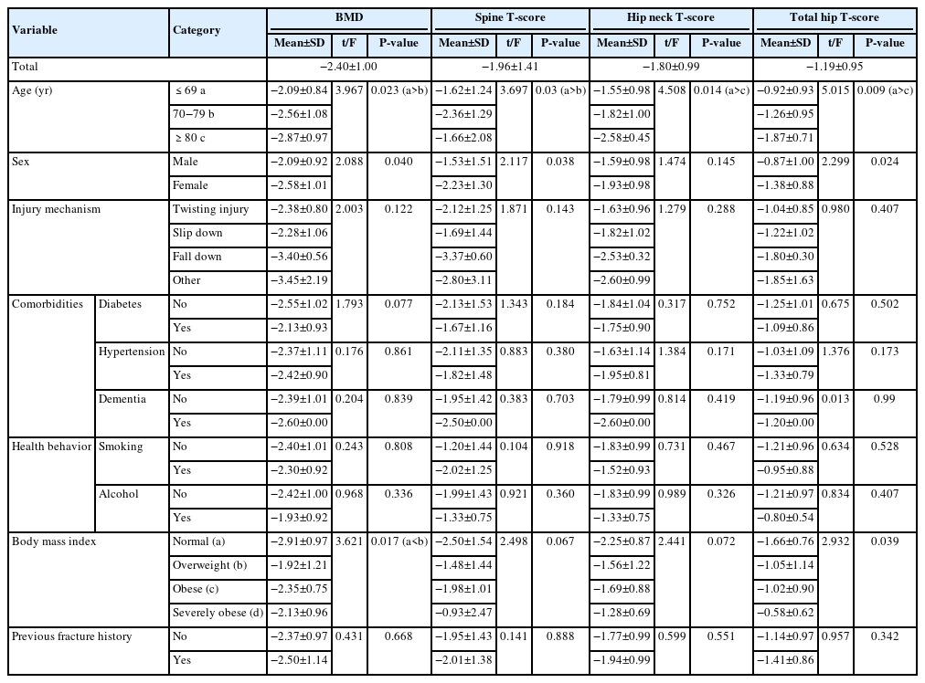
This retrospective study analyzed data from 1,411 patients diagnosed with ankle fractures admitted to Chosun University Hospital between February 2012 and April 2023. After applying inclusion criteria (age ≥65 years; low energy ankle fracture) and exclusion criteria (high energy trauma, open/multiple fractures, missing dual X-ray absorptiometry [DXA]), 73 of 1,411 patients were analyzed. Lumbar spine, femoral neck, and total hip T scores were obtained with a Horizon Wi DXA scanner, and associations with age, sex, mechanism of injury, comorbidities, smoking status, alcohol consumption, body mass index (BMI), and history of fractures were tested by ANOVA with Scheffe post hoc and Fisher exact tests.
Results
Lower BMD correlated significantly with older age, female sex, and lower BMI (P<0.05) in older adults with ankle fractures. No significant associations were observed for comorbidities (diabetes, hypertension, dementia), smoking, alcohol consumption, injury mechanism, or prior fractures.
Conclusion
These results indicate that older age, female, and lower BMI are linked to reduced BMD in ankle fracture patients over 65 years of age. Focused osteoporosis screening and management may therefore be most beneficial for older, low BMI women presenting with ankle fractures. Level of evidence: IV.
- 1,223 View
- 37 Download
- Risk factors for ankle fractures in older adults based on clinical components of the Fracture Risk Assessment (FRAX) tool and comorbidities in Korea: a retrospective case-control study
- Myeong Jun Song, Se Woong Jang, Jun Young Lee, Seojin Park
- J Musculoskelet Trauma 2025;38(4):193-202. Published online October 24, 2025
- DOI: https://doi.org/10.12671/jmt.2025.00143
-
Abstract
PDF
- Background
Ankle fractures are common in older adults; however, their relationship with osteoporotic fractures remains unclear. This study aimed to evaluate potential risk factors for ankle fractures in older adults by analyzing individual clinical components of the Fracture Risk Assessment (FRAX) tool and comorbidities.
Methods
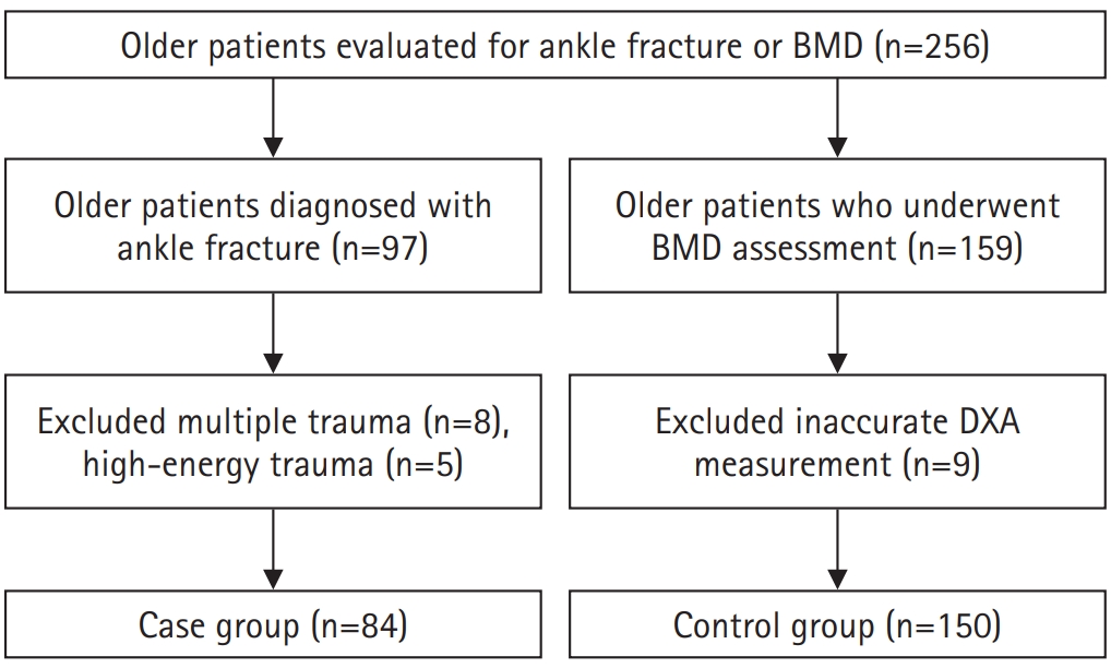
We conducted a retrospective case-control study including 84 patients aged ≥65 years with ankle fractures and 150 controls who underwent bone mineral density (BMD) testing without prior ankle fractures. The variables analyzed included age, sex, body mass index, smoking, alcohol consumption, prior fracture history, and comorbidities such as hypertension, diabetes mellitus, and dementia. BMD was measured at the spine, total hip, and femoral neck.
Results
Univariate analysis showed that alcohol consumption, diabetes mellitus, and total hip T-score categories were significantly associated with ankle fractures. In binary logistic regression, alcohol consumption remained significantly associated with higher ankle fracture risk (odds ratio [OR], 5.302; 95% confidence interval [CI], 1.778–15.811; P=0.003), and both osteopenia and osteoporosis at the total hip were also associated with increased risk (OR, 3.260, P=0.049; OR, 3.561, P=0.031, respectively). Diabetes mellitus did not reach statistical significance in the adjusted model (P=0.074). Model fit was adequate (Hosmer-Lemeshow P=0.377), and post hoc power analysis confirmed sufficient sample size.
Conclusions
These findings suggest that lower total hip BMD and alcohol-related factors may be associated with ankle fracture risk in older adults. The FRAX score itself was not calculated; instead, this study focused on analyzing selected clinical components. Limitations include the retrospective design, lack of fall and medication data, and cross-sectional BMD assessment. Level of evidence: III.
- 1,633 View
- 30 Download
Review Article
- Avulsion Fractures in the Ankle and Foot
- Gyeong Hoon Lim, Jae Won Kim, Sung Hyun Lee
- J Korean Fract Soc 2024;37(2):102-116. Published online April 30, 2024
- DOI: https://doi.org/10.12671/jkfs.2024.37.2.102
-
Abstract
PDF
- An avulsion fracture occurs when a muscle-tendon unit attached to a bone produces sufficient force to tear a fragment of the bone. If not treated properly, this injury can lead to deformity, nonunion, malunion, pain, and disability. Although avulsion fractures around the foot and ankle can occur anywhere there are tendon and ligament attachments, they are common in the anterior talofibular ligament, anterior-inferior tibiotalar ligament, calcaneal tuberosity, the base of the fifth metatarsal, and navicular bone. The optimal treatment for each fracture depends on the location and severity of the fracture. Conservative treatment involves limiting weight bearing for a period, splint immobilization, and using various orthoses. Surgical treatment is usually reserved for cases of severe displacement or when nonsurgical treatment has failed. The goals of surgery include reduction of the fracture fragment, prevention of nonunion or malunion and soft tissue injury, and early return to function. The decision for each treatment modality may depend on the patient demographics or preferences and the surgeon experience. This review summarizes previous and current views on the pathogenesis, diagnosis, and treatment of common avulsion fractures to guide the treatment and diagnosis.
- 3,363 View
- 64 Download
Original Article
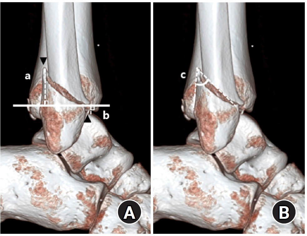
- Prediction of Syndesmotic Instability according to the Lateral Malleolus Fracture Pattern in Supination-External Rotation Type Ankle Fractures: Short Oblique versus Long Oblique Fracture
- Chan-Jin Park, Min-Su Lee, Keun-Bae Lee
- J Korean Fract Soc 2024;37(1):39-45. Published online January 31, 2024
- DOI: https://doi.org/10.12671/jkfs.2024.37.1.39
-
Abstract
PDF
- Purpose
This study examined whether preoperative radiological evaluations can predict syndesmotic instability according to the lateral malleolus fracture pattern in supination-external rotation-type ankle fractures.
Materials and Methods
This study enrolled 132 patients (132 ankles) with supination-external rotation stage 3 and 4 ankle fractures. Three-dimensional computed tomography was used for the morphological classification of the lateral malleolus fractures. A long oblique fracture was defined when the posterior cortical bone height of the fracture was 4.5 cm or more from the plafond of the distal tibial articular surface. A short oblique fracture was defined when the height was less than 4.5 cm. The demographic characteristics and syndesmotic instability of the two groups were evaluated.
Results
Short oblique fractures were confirmed in 102 cases, and long oblique fractures were confirmed in 30 cases. Long oblique fractures occurred at a statistically significantly higher incidence in younger ages and among males compared to short oblique fractures. Syndesmotic instability was more common in long oblique fractures.
Conclusion
In supination-external rotation-type ankle fractures, syndesmotic instability was observed in approximately 13%. Specifically, when the fracture pattern of the lateral malleolus is long oblique, the incidence of syndesmotic instability is approximately three times higher than in short oblique fractures. Therefore, meticulous evaluations of the lateral malleolus fracture pattern and establishing an appropriate treatment plan before surgery are crucial. -
Citations
Citations to this article as recorded by
- Relationship of lateral malleolar fracture patterns to posterior malleolar fracture morphology in supination-external rotation ankle fractures in Korea: a retrospective cohort stduy
Jong-Eun Kim, Chan-Jin Park, Jun-Young Lee, Keun-Bae Lee, Gun-Woo Lee
Journal of Musculoskeletal Trauma.2025; 38(4): 212. CrossRef
- Relationship of lateral malleolar fracture patterns to posterior malleolar fracture morphology in supination-external rotation ankle fractures in Korea: a retrospective cohort stduy
- 940 View
- 11 Download
- 1 Crossref
Case Report
- Irreducible Ankle Fracture Dislocation due to Dislocated Tibialis Posterior Tendon - A Case Report -
- Seungyup Shin, Bum-Soo Kim, Ji-Won Lee, Euisun Yoon
- J Korean Fract Soc 2023;36(2):52-56. Published online April 30, 2023
- DOI: https://doi.org/10.12671/jkfs.2023.36.2.52
-
Abstract
PDF
- An irreducible ankle dislocation is a rare injury. The cause is a dislocation of the distal fibula anteriorly or posteriorly or the insertion of soft tissue, such as the deltoid ligament or posteromedial tendon. The tibialis posterior tendon can be dislocated through distal tibiofibular diastasis and prevent reduction of the ankle joint. The authors experienced anterolateral ankle fracture dislocation with a diastasis of the distal tibiofibular joint, and reduction was impossible because of impingement of the tibialis posterior tendon dislocated anteriorly through the distal tibiofibular diastasis. This paper reports the treatment of this injury.
- 780 View
- 15 Download
Original Articles
- Analysis of Clinical and Functional Outcomes according to the Blood Sugar Control Status at the Time of Ankle Fractures Resulting from Rotational Injuries
- Jun Young Lee, Dong Seop Lim, Seung Hyun Lee, Seo Jin Park
- J Korean Fract Soc 2022;35(4):135-141. Published online October 31, 2022
- DOI: https://doi.org/10.12671/jkfs.2022.35.4.135
-
Abstract
PDF
- Purpose
Patients with diabetes are known to have poor clinical outcomes due to the high incidence of complications after ankle joint fracture surgery. This study reports the clinical and functional outcomes based on glycemic control status among patients with ankle joint fractures who underwent surgical treatment.
Materials and Methods
Among patients who underwent surgical treatment due to ankle joint fractures from January 2015 to October 2019, 253 patients with a minimum follow-up of 12 months were identified. We divided them into 3 groups: 195 patients with no diabetes (Group A), 26 patients with well-controlled diabetes (Group B), and 32 patients with uncontrolled diabetes (Group C). In addition, patients with lateral, medial malleolar, bimalleolar, and trimalleolar fractures were identified using radi-ography. The functional outcome measures used for evaluation were the Revised Foot Function Index (FFI), Short Musculoskeletal Function Assessment (SMFA), and the Foot and Ankle Outcome Score (FAOS).
Results
Bone union at 3 months after surgery was high in Group A, showing significant differences compared to the other groups. There was a significant difference between the groups in the incidence of arthropathy and one or more complications. However, the FFI, SMFA, and FAOS did not show significant differences between the groups.
Conclusion
The incidence of complications was high in patients with uncontrolled diabetes compared to the patients with well-controlled diabetes and those with no diabetes. However, functional outcomes showed no significant difference.
- 873 View
- 6 Download
- Comparison of the Size of the Posterior Malleolar Fragment in Trimalleolar Ankle Fractures Measured Using Lateral Plain Radiography and Three-Dimensional Computed Tomography
- Gun-Woo Lee, Dong-Min Jung, Woo Kyoung Kwak, Keun-Bae Lee
- J Korean Fract Soc 2022;35(3):91-96. Published online July 31, 2022
- DOI: https://doi.org/10.12671/jkfs.2022.35.3.91
-
Abstract
PDF
- Purpose
This study aimed to evaluate and compare the accuracy of the size of the posterior malleolar fragment measured using lateral plain radiography and three-dimensional computed tomography (3DCT) in patients with ankle trimalleolar fractures.
Materials and Methods
This study enrolled 80 patients (80 ankles) with ankle trimalleolar fractures and analyzed the size of the posterior malleolar fragments using plain radiography and 3D-CT. The articular involvement of the posterior malleolar fragments was measured as a percentage of the articular surface in the sagittal length of the tibial plafond using lateral plain radiography, and the articular surface area was directly measured using 3D-CT. In addition, we classified the patients into three groups based on the morphology of the posterior malleolar fracture, according to the Haraguchi classification method, and evaluated and compared the accuracy of the size of the posterior malleolar fragments.
Results
The mean articular involvement of the posterior malleolar fragments on plain radiography was 27.6% (range, 6.0%-53.1%), which was significantly higher than the mean of 21.9% (range, 4.7%-47.1%) measured using 3D-CT (p=0.004). In the analysis, according to the fracture morphology, the mean difference between the two methods was the largest for type I fractures at 9.1% (range, 1.8%-19.5%) and the smallest for type II fractures at 1.1% (range, –7.7% to 8.8%).
Conclusion
The articular involvement of posterior malleolar fragments measured using plain radiography showed low accuracy and significantly higher values than the actual articular involvement. Therefore, careful evaluation using 3D-CT is crucial for accurate analysis and optimal treatment in patients with ankle trimalleolar fractures. -
Citations
Citations to this article as recorded by- Relationship of lateral malleolar fracture patterns to posterior malleolar fracture morphology in supination-external rotation ankle fractures in Korea: a retrospective cohort stduy
Jong-Eun Kim, Chan-Jin Park, Jun-Young Lee, Keun-Bae Lee, Gun-Woo Lee
Journal of Musculoskeletal Trauma.2025; 38(4): 212. CrossRef
- Relationship of lateral malleolar fracture patterns to posterior malleolar fracture morphology in supination-external rotation ankle fractures in Korea: a retrospective cohort stduy
- 727 View
- 10 Download
- 1 Crossref
Review Articles
- Treatment of Ankle Fracture and Dislocation
- Chan Kang
- J Korean Fract Soc 2022;35(1):38-49. Published online January 31, 2022
- DOI: https://doi.org/10.12671/jkfs.2022.35.1.38
-
Abstract
PDF
- Ankle fractures are the most common type of foot and ankle fracture injury. Several types of fractures occur in the ankle structures (medial malleolus, lateral malleolus, posterior malleolus, and Chaput’s tubercle) with various mechanisms and extent of fracture force. Moreover, fractures can be accompanied by other injuries, such as distal tibiofibular syndesmotic injury, medial deltoid ligament rupture, and lateral ligament complex rupture. Ankle dislocation can be accompanied when an injury is caused by a greater fracture force. Non-surgical treatments or combined surgeries may be performed depending on the mechanism and fracture type. Generally, a stable fracture maintaining anatomical reduction is treated conservatively, but surgical treatment is performed when this is not the case. Furthermore, surgeries for stable fractures can be offered when the patients demand early weight bearing due to their occupation, age, and performance state. Restoring the ankle mortise in its anatomical shape before the injury and starting early rehabilitation for functional recovery simultaneously until a union is achieved is important. Traumatic arthritis can occur if the treatment focuses only on fractures and neglects ligament injuries, such as distal tibiofibular syndesmotic injury and medial deltoid ligament rupture. Shortening, angular deformation, and rotational deformation of the fibular promote the progression of traumatic ankle arthritis in the long term, which may further cause chronic ankle pain. An overlooked displaced posterior malleolus fracture also causes traumatic arthritis through anteroposterior instability of the ankle joint.
- 1,504 View
- 53 Download
- Ankle Fractures in Children: Classification and Treatment
- Ha-Yong Kim, Yong-Han Cha, Woo-Suk Kim, Won-Sik Choy
- J Korean Fract Soc 2021;34(2):87-95. Published online April 30, 2021
- DOI: https://doi.org/10.12671/jkfs.2021.34.2.87
-
Abstract
PDF
- Pediatric ankle fractures are defined as damage to the metaphysis, epiphyseal plate, and epiphysis of the distal tibia and fibula. Although the injury mechanism could be similar, the fracture patterns and treatment of pediatric ankle fractures are different from those of adults. In children, growth plate injuries are more common with a force that would cause sprains in adults because the ligaments are stronger than the growth plate cartilage in children. In the adolescent period, unique fractures, called “transitional fractures”, occur while the physis is closed. For a diagnosis, plain images of the anteroposterior, lateral, and mortise views are essential. Stress radiographs, ultrasound, and magnetic resonance imaging can be used for suspected ligament injuries. The treatment goal is to restore the articular congruity, normal bony alignment, and preserve the epiphyseal plate to ensure normal growth. Pediatric ankle fractures frequently lead to premature physeal arrest, angular deformities, malunion, and posttraumatic arthritis even after anatomic reduction. Treating surgeons should follow-up children for a sufficient time and explain to the caregiver the possible complications before treatment.
- 2,962 View
- 75 Download
Original Articles
- Comparing Outcomes of Screw Fixation and Non-Fixation for Small-Sized Posterior Malleolar Fragment in Ankle Trimalleolar Fractures
- Jee-Wook Ko, Gun-Woo Lee, Keun-Bae Lee
- J Korean Fract Soc 2021;34(1):8-15. Published online January 31, 2021
- DOI: https://doi.org/10.12671/jkfs.2021.34.1.8
-
Abstract
PDF
- Purpose
This study was undertaken to compare outcomes of screw fixation and non-fixation of a small-sized posterior malleolar fragment involving less than 25% articular surface in ankle trimalleolar fractures. Materials and Methods: A total of 32 consecutive ankles (32 patients), with posterior malleolar fragment involving 15%-25% of the joint surface, were enrolled in the study. Patients were divided into 2 groups according to whether the fragment was fixed or not (fixed: 20 ankles, non-fixed: 12 ankles). The minimum follow-up period was 12 months. Median size of the posterior malleolar fragment in the fixed and non-fixed groups were 24.6% (range, 22.3%-25.0%) and 22.1% (range, 17.4%-24.3%), respectively. Complications as well as clinical and radiographic outcomes were compared and analyzed between the two groups. Results: Clinical outcomes, including American Orthopaedic Foot & Ankle Society (p=0.501), visual analogue scale (p=0.578), and ankle range of motion (p=0.552), showed no difference between groups at the final follow-up. No differences were obtained in the radiographic outcomes, including joint stepoff (p=0.289) and fragment gap (p=0.289). Complications, including 1 case of delayed union and 1 case of wound infection, were reported in the fixed group. Conclusion: Clinical outcomes and radiographic outcomes of the non-fixation group were satisfactory and comparable to the fixation group. Our results indicate that anatomical reduction with small-sized posterior malleolar fragment in ankle trimalleolar fractures is sufficient for satisfactory outcomes, without the need for additional internal fixation.
- 1,020 View
- 13 Download
- Treatment of Isolated Lateral Malleolar Fractures Using Locking Compression Plate Fixation and Tension Band Wiring Fixation
- Woojin Shin, Seondo Kim, Jiyeon Park
- J Korean Fract Soc 2020;33(1):16-21. Published online January 31, 2020
- DOI: https://doi.org/10.12671/jkfs.2020.33.1.16
-
Abstract
PDF
- PURPOSE
The purpose of this study was to compare the clinical and radiological outcomes of locking compression plate (LCP)-screw fixation and tension band wiring (TBW) fixation in isolated lateral malleolar fractures.
MATERIALS AND METHODS
From May 2016 to August 2018, 52 patients with isolated lateral malleolar fracture were retrospectively reviewed. They were divided into 30 cases of the LCP fixation group (Group I) and 22 cases of the TBW fixation group (Group II). The clinical and radiological results of those groups were compared. Pearson chi-square tests and independent t-tests were used in the statistical analysis.
RESULTS
The mean length of the surgical incision was 8.3 cm in Group I and 4.9 cm in Group II. Radiological union was obtained at a mean of 8.4 weeks in both groups. The mean American Orthopaedic Foot and Ankle Society score was 90 (range, 85–97) and 92 (range, 85–100) in Groups I and II, respectively, at the last follow up.
CONCLUSION
Both the LCP-screw and TBW techniques revealed excellent results in isolated lateral malleolar fractures. The tension band technique may be a fine alternative method of fixation in the treatment of isolated lateral malleolar fracture.
- 1,447 View
- 13 Download
- Ankle Fracture Associated with Tibia Shaft Fractures
- Ji Wan Kim, Hong Joon Choi, Dong Hyun Lee, Young Chang Kim
- J Korean Fract Soc 2014;27(2):136-143. Published online April 30, 2014
- DOI: https://doi.org/10.12671/jkfs.2014.27.2.136
-
Abstract
PDF
- PURPOSE
The purpose of this study is to evaluate the incidence of ankle injury in ipsilateral tibial shaft fractures and to assess the risk factors for ankle injury associated with tibial shaft fractures.
MATERIALS AND METHODS
Sixty patients with tibial shaft fractures were enrolled in this retrospective study. The incidence and characteristics of ankle injury were evaluated, and fracture classification, fracture site, and fracture pattern of the tibial shaft fractures were analyzed for assessment of the risk factors for ankle injury combined with tibial shaft fractures.
RESULTS
Ankle injury occurred in 20 cases (33%). There were four cases of lateral malleolar fracture, four cases of posterior malleolar fracture, two cases of distal tibiofibular ligament avulsion fracture, and 10 cases of complex injury. Fourteen cases (70%) of 20 cases of ankle injury were diagnosed from x-ray films, and the other six cases were recognized in ankle computed tomography (CT). Ankle injury occurred in 45.1% of distal tibial shaft fractures and found in 41.4% of A type, but there was no statistical significance. Ankle injury was observed in 54% of cases of spiral pattern of tibial shaft fracture and the incidence was statistically higher than 19% of cases of non-spiral pattern tibial shaft fracture.
CONCLUSION
Ankle injury was observed in 33% of tibial shaft fractures; however, only 70% could be diagnosed by x-ray. Ankle injury occurred frequently in cases of spiral pattern of tibial shaft fracture, and evaluation of ankle injury with CT is recommended in these cases. -
Citations
Citations to this article as recorded by- Usefulness of Computed Tomography on Distal Tibia Intra-Articular Fracture Associated with Spiral Tibia Shaft Fracture
Seong-Eun Byun, Sang-June Lee, Uk Kim, Young Rak Choi, Soo-Hong Han, Byong-Guk Kim
Journal of the Korean Fracture Society.2016; 29(2): 114. CrossRef
- Usefulness of Computed Tomography on Distal Tibia Intra-Articular Fracture Associated with Spiral Tibia Shaft Fracture
- 1,067 View
- 7 Download
- 1 Crossref
- Radiological Assessment for Morphological Diversity of Distal Fibula
- Su Young Bae, Jin Hee Yoo
- J Korean Fract Soc 2014;27(1):1-9. Published online January 31, 2014
- DOI: https://doi.org/10.12671/jkfs.2014.27.1.1
-
Abstract
PDF
- PURPOSE
The purpose of this study is to determine whether the morphological consistency of distal fibula could be defined by measurement through radiological assessment as there was doubt regarding the adequacy of anatomical distal fibular plates.
MATERIALS AND METHODS
Plain radiographs and computed tomography (CT) images of 300 cases from 2009 to 2012 were reviewed. The distance from the lateral vertex to the tip of the distal fibula and to the lateral margin of the shaft was measured, respectively, in order to understand the shape of the lateral curve of the distal fibula on plain radiographs. The neutral ridge was defined as a point of the lateral ridge located in the center of the antero-posterior diameter and the distance from the tip of the distal fibula to the neutral ridge was measured for determining the shape of the ridge on CT images. The angle of the lateral and posterior surface of the fibular incisura at the level of the neutral ridge was also measured.
RESULTS
A statistically significant difference in the lateral vertex and margin of the fibular shaft on plain radiographs and distance from the tip of the distal fibula to the neutral ridge, angle of the fibular lateral surface on CT images was observed between male and female. The mean distance from the lateral vertex to the tip of distal fibula was 12.2+/-3.0 mm, to the lateral margin of the fibular shaft was 5.6+/-1.7 mm, distance from tip of the distal fibula to the neutral ridge was 54.9+/-6.4 mm, the fibular lateral surface angle was 52.2degrees+/-9.1degrees, and the fibular posterior surface angle was 32.5degrees+/-9.3degrees.
CONCLUSION
Based on the various radiologic parameters, it was concluded that there was a wide morphological diversity of shape of lateral curve and fibular ridge.
- 638 View
- 3 Download
Case Report
- Delayed Foreign-body Reaction of Ankle Fracture Treated with a Biodegradable Plate and Screws: A Case Report
- Chul Hyun Park, Dae Hyun Song, Jae Ho Cho
- J Korean Fract Soc 2012;25(2):142-145. Published online April 30, 2012
- DOI: https://doi.org/10.12671/jkfs.2012.25.2.142
-
Abstract
PDF
- Biodegradable implants made of co-polymers composed of L-lactide, D-lactide, and trimethylene carbonate were used in the present case. To our knowledge, only one reported tissue reaction has been associated with ankle fracture treated with third-generation implants internationally and none yet domestically. We report a delayed foreign-body reaction of ankle fracture treated with a third-generation biodegradable plate and screws. We suggest that ankle fracture patients treated with biodegradable implants should be advised of this possible complication and should be followed for at least 2 years.
- 865 View
- 5 Download
Original Article
- Treatment of the Trimalleolar Fracture Using Posterolateral Approach: Minimum 2-year Follow Up Results
- Gwang Chul Lee, Jun Young Lee, Sang Ho Ha, Jae Won You, Sang Hong Lee, Hong Moon Sohn, Ki Young Nam, Kwang Hyo Seo
- J Korean Fract Soc 2011;24(4):328-334. Published online October 31, 2011
- DOI: https://doi.org/10.12671/jkfs.2011.24.4.328
-
Abstract
PDF
- PURPOSE
To analyze the long term follow up results of treatment with posterolateral approach and to investigate its usefulness in the patients of trimalleolar fracture with posterior fragment which is above 25% of articular involvement.
MATERIALS AND METHODS
There were 34 cases of trimalleolar fracture in our hospital from May 2004 to April 2008. We investigated 20 patients who underwent operation with the posterolateral approach and over-2 years follow up cases. The mean follow up period was 34 (24~58) months. Preoperative posterior malleolar fragment involved above 25% of articular surface in all cases and displaced more than 2 mm in 11 cases. We analyzed the radiologic type of posterior malleolar fragments and evaluated the function and pain through AOFAS score and complications.
RESULTS
All cases showed primary union at mean 13.1 weeks. The complications are that partial ankylosis result of soft tissue contracture is seen in 2 cases (10%) and post-traumatic arthritis is seen in 1 cases (5%) and 17 cases (85%) of all patients are showed excellent AOFAS score.
CONCLUSION
The posterolateral approach is a valuable method because that it enables us to easily reduction and internal fixation of the posterior malleolus and lateral malleolus at one time and the results are satisfied for a long time follow up. -
Citations
Citations to this article as recorded by- Outcomes of Immediate Operative Treatment of Ankle Trimalleolar Open Fractures
Jun-Young Lee, Yong-Jin Cho, Sin-Wook Kang, Yung-Min Cho, Hyun-Bai Choi
Journal of Korean Foot and Ankle Society.2020; 24(1): 25. CrossRef
- Outcomes of Immediate Operative Treatment of Ankle Trimalleolar Open Fractures
- 888 View
- 3 Download
- 1 Crossref
Case Report
- Checkrein Deformity by Incarcerated Posterior Tibial Tendon and Displaced Flexor Hallucis Longus Tendon following Ankle Dislocation: A Case Report
- Su Young Bae, Hyung Jin Chung, Man Young Kim
- J Korean Fract Soc 2011;24(3):271-276. Published online July 31, 2011
- DOI: https://doi.org/10.12671/jkfs.2011.24.3.271
-
Abstract
PDF
- We report a case of 20 year-old man who had unusual equinus and checkrein deformity following dislocation of his right ankle joint. He had been treated with distal tibiofibular screw fixation and external fixation. After removal of external fixator, he had suffered from progressive deformity of foot and ankle. Widening of distal tibiofibular joint and medial clear space was found on radiograph and it was revealed that posterior tibial tendon had been dislocated and incarcerated into the distal tibiofibular joint on MRI. We corrected the deformity with excision of incarcerated posterior tibial tendon, adhesiolysis and lengthening of flexor hallucis longus tendon, reconstruction of deltoid ligament and flexor digitorum longus tendon transfer.
-
Citations
Citations to this article as recorded by- Management of Checkrein Deformity
Min Gyu Kyung, Yun Jae Cho, Dong Yeon Lee
Clinics in Orthopedic Surgery.2024; 16(1): 1. CrossRef - A Neglected Extensor Hallucis Longus Tendon Rupture Caused by Arthritic Adhesion
Sung Hun Won, Sung Hwan Kim, Young Koo Lee, Dong-Il Chun, Byung-Ryul Lee, Woo-Jong Kim
Medicina.2023; 59(6): 1069. CrossRef - The Checkrein Deformity of Extensor Hallucis Longus Tendon and Extensor Retinaculum Syndrome with Deep Peroneal Nerve Entrapment after Triplane Fracture: A Case Report
Hyungon Gwak, Jungtae Ahn, Jae Hoon Lee
Journal of Korean Foot and Ankle Society.2021; 25(3): 145. CrossRef - Checkrein Deformity Due to Flexor Digitorum Longus Adhesion after Comminuted Calcaneus Fracture: A Case Report
Jin Su Kim, Han Sang Lee, Ki Won Young, Keun Woo Lee, Hun Ki Cho, Sang Young Lee
Journal of Korean Foot and Ankle Society.2015; 19(1): 35. CrossRef
- Management of Checkrein Deformity
- 902 View
- 1 Download
- 4 Crossref
Original Articles
- Treatment of the Posterior Malleolar Fracture Using Posterior Approach
- Hyun Wook Chung, Dong Hwan Kim, Si Hoon Yoo, Jin Soo Suh
- J Korean Fract Soc 2010;23(1):50-56. Published online January 31, 2010
- DOI: https://doi.org/10.12671/jkfs.2010.23.1.50
-
Abstract
PDF
- PURPOSE
For fixation of the large posterior malleolar fracture fragment, indirect anterior fixation with cannulated screw has been widely used, but the anatomical reduction is not always obtained. The purpose of this article is to evaluate the clinical result of posterior malleolar fractures treated with anatomical reduction and internal fixation using posterior approach.
MATERIALS AND METHODS
We have analyzed the 15 patients with posterior malleolar fractures, treated with posterior approach from August 2005 to August 2008. The mean follow up period was 17.6 months, We have reviewed the perioperative joint integrity, method of operation, postoperative care, bony union and complication. A clinical outcome was evaluated by AOFAS (American orthopedic foot and ankle society) scaling system and Olerud & Molander scoring system.
RESULTS
Among 15 cases, posterolateral approach and posteromedial approach were chosen in 9 cases and 6 cases respectively. The radiologic unions were achieved at 12.4 (12~18) weeks. Mean AOFAS score was 90.3 (72~98), and Olerud & Molander score was "excellent" in 5 cases, "good" in 7 cases, "fair" in 1 case and "poor" in 2 cases. Postoperative complications in 2 cases revealed a posttraumatic arthritis and a scar band contracture respectively.
CONCLUSION
In posterior malleolar fracture of ankle joint, the integrity of joint has closely affected clinical outcomes. We suggest that a posterior approach for posterior malleolar fracture with especially incarcerated fragments and comminuted fractures, can be a useful method for anatomical reduction and stable fixation, and satisfactory clinical results. -
Citations
Citations to this article as recorded by- Single lateral approach for open reduction and internal fixation of posterior malleolar fragment in Weber B rotational ankle fracture
Jaehyung Lee, Hwan Ryu, Jae Yong Park
Medicine.2023; 102(3): e32725. CrossRef - Posterior Malleolus Fractures in Trimalleolar Ankle Fractures: Malleolus versus Transyndesmal Fixation
Bilgehan Tosun, Ozgur Selek, Umit Gok, Halil Ceylan
Indian Journal of Orthopaedics.2018; 52(3): 309. CrossRef - Single Oblique Posterolateral Approach for Open Reduction and Internal Fixation of Posterior Malleolar Fractures With an Associated Lateral Malleolar Fracture
Jun Young Choi, Ji Hoon Kim, Hyeong Tak Ko, Jin Soo Suh
The Journal of Foot and Ankle Surgery.2015; 54(4): 559. CrossRef
- Single lateral approach for open reduction and internal fixation of posterior malleolar fragment in Weber B rotational ankle fracture
- 972 View
- 3 Download
- 3 Crossref
- Radiologic Analysis and Treatment of Posterior Malleolar Fractures of the Ankle
- Jae Sung Lee, Soo Yong Kang, Han Jun Lee, Young Bong Ko
- J Korean Fract Soc 2009;22(2):98-103. Published online April 30, 2009
- DOI: https://doi.org/10.12671/jkfs.2009.22.2.98
-
Abstract
PDF
- PURPOSE
The purpose of this study was to classify posterior malleolar fractures according to the position of fragments and to analyze radiologic features of each type.
MATERIALS AND METHODS
We analyzed forty-six patients of ankle fractures involving a posterior malleolus who were treated between January 2004 and December 2007. The posterior malleolar fractures were categorized into three types (posterolateral, posteromedial, shell) based on the major fracture line. In each type, we analyzed amount of displacement, involvement of articular surface, existence of subluxation and osteochondral impacted fragments.
RESULTS
The forty-six patients were categorized into three types: Posterolateral (PL) type (33 cases, 72%), Posteromedial (PM) type (8 cases, 17%), shell type (5 cases, 11%). Of the 8 cases with PM type, 7 cases showed displacement more than Grade II, 4 cases showed subluxation of ankle joint, and 3 cases showed osteochondral impacted fragment. Average involvement of articular surface of PM type is 35% (15~65%).
CONCLUSION
Posterior malleolar fractures with medial extension tended to have adverse effect on ankle stability and Preoperative CT scan is essential for evaluation of fracture type and determination of appropriate surgical approach. -
Citations
Citations to this article as recorded by- Treatment of Isolated Posterior Malleolus Fracture in the Ankle
Ji Hoon Kim, Seong Mu Cha, Dae Yeon Jo, Jin Soo Suh
Journal of the Korean Orthopaedic Association.2014; 49(1): 29. CrossRef
- Treatment of Isolated Posterior Malleolus Fracture in the Ankle
- 909 View
- 2 Download
- 1 Crossref
- Surgical Fixation with Biodegradable Plate for the Treatment of Ankle Fractures
- Jae Young Cho, Jin Whan Kim, Sang Eun Kim, Kyung Chil Jung, Seung Hyun Choi
- J Korean Fract Soc 2008;21(1):31-36. Published online January 31, 2008
- DOI: https://doi.org/10.12671/jkfs.2008.21.1.31
-
Abstract
PDF
- PURPOSE
The purpose of this article is to show the efficacy of a biodegradable plate for treating lateral malleolar fractures in the ankle joint.
MATERIALS AND METHODS
The 20 patients who underwent an open reduction and internal fixation for lateral malleolar fractures in the ankle joint from February, 2006 to February, 2007 in our hospital were enrolled into the study. The average age of the patients was 49.7 years and the average follow-up period was 5.6 months. The cases were analyzed by radiological bone union time and clinical results according to the criteria of Meyer et al.
RESULTS
Average radiological bone union time was 10.5 weeks. The clinical result was excellent in 19 cases (95%), good in 1 case (5%). There was one case of minimal displacement less than 1 mm, associated with anterior distal tibio-fibular ligament avulsion fracture.
CONCLUSION
For proper patients, a biodegradable plate is an effecttive alternative implant for stabilizing lateral malleolar fractures in the ankle joint, because there is no requirement for subsequent removal and slow resorption in vivo. -
Citations
Citations to this article as recorded by- Delayed Foreign-body Reaction of Ankle Fracture Treated with a Biodegradable Plate and Screws - A Case Report -
Chul-Hyun Park, Dae-Hyun Song, Jae Ho Cho
Journal of the Korean Fracture Society.2012; 25(2): 142. CrossRef
- Delayed Foreign-body Reaction of Ankle Fracture Treated with a Biodegradable Plate and Screws - A Case Report -
- 1,018 View
- 2 Download
- 1 Crossref
- Nonoperative Treatment of Isolated Lateral Malleolar Fracture
- Woo Chun Lee, Jong Ho Ahn
- J Korean Fract Soc 2005;18(3):291-293. Published online July 31, 2005
- DOI: https://doi.org/10.12671/jkfs.2005.18.3.291
-
Abstract
PDF
- PURPOSE
To evaluate the results of conservative treatment for isolated lateral malleolus fracture without medial ankle injury.
MATERIALS AND METHODS
From March 1999 to February 2003, 25 ankles in 25 patients were treated for isolated lateral malleolus fracture and followed for more than one year. Mean age was 46.9 years (range, 20~71 years). Cases without any swelling or tenderness on the deltoid area, or cases with minimal pain, swelling or tenderness on the deltoid area and medial clear space 1 mm or less on stress radiograph were included for the study. Immediate weight bearing was allowed with below-knee cast immobilization in all cases.
RESULTS
All were supinatin-external rotation stage II injury and mean duration of cast immobilization was 6.3+/-1.6 weeks after injury. There was no case which showed widening of medial clear space during routine radiographic follow-up. There was no change in the degree of displacement in spite of immediate weight bearing with short leg cast on.
CONCLUSION
Because the lateral malleolus fracture without medial injury can be managed nonoperatively, we need to differentiate this type of fracture to avoid unnecessary surgery, and for early return to normal daily activity. -
Citations
Citations to this article as recorded by- Posterior Plating in Distal Fibular Fracture
Choong-Hyeok Choi, Young-A Cho, Jae-Hoon Kim, Il-Hoon Sung
Journal of the Korean Fracture Society.2007; 20(2): 161. CrossRef
- Posterior Plating in Distal Fibular Fracture
- 1,183 View
- 5 Download
- 1 Crossref
- Comparison between X-ray and Three Dimensional Computed Tomography in Trimalleolar Ankle Fractures
- Sang Jun Song, Hyung Ku Yoon, Dong Eun Shin, Soo Hong Han, Jae Hwa Kim, Hyung Kun Park, Yong Sub Han
- J Korean Fract Soc 2005;18(2):160-164. Published online April 30, 2005
- DOI: https://doi.org/10.12671/jkfs.2005.18.2.160
-
Abstract
PDF
- PURPOSE
To evaluate the accuracy of X-ray evaluation in classification, displacement and size of posterior malleolar fragment, comparing with three dimensional computed tomography (3D CT) in trimallelar ankle fractures.
MATERIALS AND METHODS
20 cases of trimalleolar ankle fractures evaluated with preoperative 3D CT, and followed up periods were at least 2 years. All cases were classified according to the Danis-Weber and Lauge-Hansen classification. Displacement and size of posterior malleolar fragment were measured using PACS. The reliability between simple X-ray and 3D CT was evaluated in the Danis-Weber and Lauge-Hansen classification (kappa analysis). The correlation between simple X-ray and 3D CT was evaluated in displacement and size of posterior malleolar fragment (correlation analysis).
RESULTS
Degree of agreement of Danis-Weber classification in simple X-ray and 3D CT was 0.700 kappa value, and that of Lauge-Hansen was 0.605 kappa value. Measurement of simple X-ray and 3D CT about displaced status of posterior malleolar fragment showed statistically significant positive linear correlation (p= 0.000), but correlation of measurement of size in simple X-ray and CT was not statistically significant (p=0.102).
CONCLUSION
CT or operative field will be more accurate than simple X-ray to select the method of treatment and operation, especially when the displacement and size of posterior malleolar fragment are important to decide. -
Citations
Citations to this article as recorded by- Comparison of the Size of the Posterior Malleolar Fragment in Trimalleolar Ankle Fractures Measured Using Lateral Plain Radiography and Three-Dimensional Computed Tomography
Gun-Woo Lee, Dong-Min Jung, Woo Kyoung Kwak, Keun-Bae Lee
Journal of the Korean Fracture Society.2022; 35(3): 91. CrossRef
- Comparison of the Size of the Posterior Malleolar Fragment in Trimalleolar Ankle Fractures Measured Using Lateral Plain Radiography and Three-Dimensional Computed Tomography
- 882 View
- 3 Download
- 1 Crossref
- Modified Tension Band Wiring using Cortical Screw for Medial Malleolar Fractures
- Ho Rim Choi, Hyun Woo Doh, Byoung Heum Kim, Kyou Hyeun Kim, Jong Seok Park, Joon Min Song
- J Korean Fract Soc 2004;17(4):319-322. Published online October 31, 2004
- DOI: https://doi.org/10.12671/jkfs.2004.17.4.319
-
Abstract
PDF
- PURPOSE
To evaluate the clinical results of modified tension band wire technique using cortical screw for treatment of displaced medial malleolar fractures of the ankle.
MATERIALS AND METHODS
From January 2001 to January 2003, 24 patients were treated by modified tension band wiring using cortical screw for medial malleolar fracture. The follow-up period was 12~35 months (average 18 months). There were 13 males and 11 females, and the mean age was 46 years. Fractures were classified by Lauge-Hansen's classification. The results were analyzed by Meyer and Kumler's criteria.
RESULTS
There were 13 cases (54%) of excellent, 9 cases (38%) of good, and one case of fair because of limitation of motion of the ankle joint and one case of poor which showed post-traumatic arthritis of the ankle.
CONCLUSION
Modified tension band wire technique using cortical screw can be an effective operative method for the treatment of displaced medial malleolar fractures of the ankle.
- 499 View
- 1 Download
- Surgical Treatment of Internal Malleolar Fracture of the Ankle: Rush Rod Versus Plate Osteosynthesis
- Hak Jun Kim, Kwon Ick Ha, Jae Ik Shim, Taik Seon Kim, Jeong Ro Yoon, Young Bae Kim, Woo Seung Lee, Jae Young Chang
- J Korean Soc Fract 2003;16(4):519-525. Published online October 31, 2003
- DOI: https://doi.org/10.12671/jksf.2003.16.4.519
-
Abstract
PDF
- PURPOSE
We evaluated the results between the methods of open reduction and internal fixation using plate and screws and the methods of closed reduction and fixation with rush pin in lateral malleolar fractures.
MATERIALS AND METHODS
We analysed the 33 fractures of lateral malleolus which had been treated by open reduction and internal fixation using plate and screws or closed reduction and fixation with rush pin from January 1995 to January 2002 and had been observed over 1 year. The 33 patients were observed for the comparison of radiologic and clinical results in according to the measure of McLennan and Ungersma.
RESULTS
Among the 33 cases, 15 cases were treated by open reduction and internal fixation with plate, and 18 cases were treated by closed reduction and Rush rods fixation. In according to the measure of McLennan and Ungersma, good radiologic result was 60% (9 cases) and excellent clinical result was 27% (4 cases) in plate fixation, and good radiologic result was 61% (11 in 18 cases) and excellent clinical result was 39% (7 in 18 cases) in Rush rods fixation.
CONCLUSION
In ankle fractures of elderly patients who have soft tissue problems and osteoporotic bony quality, radiologic and clinical results of internal fixation of distal fibula were relatively same between fixation with plate and screws and Rush rods. Therefore, closed reduction and internal fixation with Rush rods is one of the good treatment modalities of distal fibular fracture.
- 590 View
- 2 Download
- The role of posterior malleolar fragments in ankle pain after trimalleolar fractures
- Su Young Bae, Dong Hoon Sihn
- J Korean Soc Fract 2003;16(1):59-66. Published online January 31, 2003
- DOI: https://doi.org/10.12671/jksf.2003.16.1.59
-
Abstract
PDF
- PURPOSE
There are some criticisms of indication for internal fixation of the posterior malleolar fragments in trimalleolar fractures. We tried to find out clinical and radiologic factors which affect on a clinical outcome of trimalleolar fractures.
MATERIALS AND METHODS
Thirty three patients who were treated for trimalleolar fractures and given anatomical reduction of lateral and medial malleolus were included. We divided patients into two groups, a group without the pain and the other group with the pain. Preoperative and postoperative lateral plain radiographic films were used to estimate fragment size, post-reduction gap and step off. By reviewing the medical records, other factors such as the time of ankle motion, weight loading and whether posterior malleolus was fixed. or not were stucdied. A clinical outcome was evaluated by AOFAS(American Orthopaedic Foot and Ankle Society) scaling system. We performed statistical analysis using Logistic regression analysis and Chi-square test on each factors.
RESULTS
There was no definite difference between two groups on the functional outcome. There was one case showing limited ankle motion. Seven patients were involved in the group with the pain and 23 in the group without the pain. The remnant fracture gap and step off of joint surface statistically showed the meaningful corellation with the pain but a fragment size and a surgical fixation, time of motion and weight loading did not show any significances.
CONCLUSION
We doubt the significance of the size of posterior malleolar fragment. We concluded that anatomical reduction of posterior malleolus is the most significant factor of a clinical outcome regardless of the size or internal fixation, especially the pain after trimalleolar injuries. -
Citations
Citations to this article as recorded by- Treatment of Isolated Posterior Malleolus Fracture in the Ankle
Ji Hoon Kim, Seong Mu Cha, Dae Yeon Jo, Jin Soo Suh
Journal of the Korean Orthopaedic Association.2014; 49(1): 29. CrossRef - Treatment of the Posterior Malleolar Fracture Using Posterior Approach
Hyun Wook Chung, Dong Hwan Kim, Si Hoon Yoo, Jin Soo Suh
Journal of the Korean Fracture Society.2010; 23(1): 50. CrossRef - Radiologic Analysis and Treatment of Posterior Malleolar Fractures of the Ankle
Jae Sung Lee, Soo Yong Kang, Han Jun Lee, Young Bong Ko
Journal of the Korean Fracture Society.2009; 22(2): 98. CrossRef
- Treatment of Isolated Posterior Malleolus Fracture in the Ankle
- 849 View
- 7 Download
- 3 Crossref
- The Study for the Factors Affecting the Radiological Outcome of the Displaced Ankle Fracture over the Elderly
- Hong Gi Park, Lee Hyuk Jung
- J Korean Soc Fract 2002;15(4):465-469. Published online October 31, 2002
- DOI: https://doi.org/10.12671/jksf.2002.15.4.465
-
Abstract
PDF
- OBJECT: This study investigated to know the factors affecting the radiological results of the ankle fracture after open reduction and internal fixation over the age 60 years.
PATIENTS & METHOD: Open reduction and internal fixation on patient with closed displacement ankle fracture over the age 60 years were studied in 51 cases. Statistical analysis by t-test was used to assess the factors affecting to the post-operation radiological results among the age, sex, classification of fracture, the degree of fracture displacement, bone fragility between anatomical reduction group and non-anatomical reduction group in average 16 months, RESULTS: There are statistical significance(p<0.05) of the sex and bone fragility in post-operation radiological results.
CONCLUSION
The radiological results in old age with ankle fracture is affected by sex and bone fragility in open reduction & internal fixation.
- 520 View
- 1 Download
- The Evaluation of Clinical and Radiographic Prognostic Factors for the Surgically Treated Unstable Ankle Fractures
- Hong Geun Jung, Hee Kon Park, Moon Jib Yoo, Tai Won Kim
- J Korean Soc Fract 2002;15(2):216-225. Published online April 30, 2002
- DOI: https://doi.org/10.12671/jksf.2002.15.2.216
-
Abstract
PDF
- PURPOSE
The purpose of this study is to analyze the clinical and radiographic prognostic factors which may affect the postoperative clinical results of the unstable ankle fractures.
MATERIALS AND METHODS
This study is based on 75 unstable ankle fractures treated by open reduction and internal fixation from May 1994 to August 2000, with a minimum follow-up period of 12 months(range : 13 months-7 years 3 months). The 75 patients were average 40.5 years old with male: female ratio of 52:23. Based on Lauge-Hansen classification, the supination-external rotation type was the most common with 42 (56.0%) cases. The clinical results was assessed by American Orthopaedic Foot and Ankle Society(AOFAS) functional scale. The sex, age, side of injury, body weight, trauma-operation interval, operation time, cause of injury as the possible postoperative clinical prognostic factors and fracture type, anatomical reduction of fracture, preoperative medial clear space, postoperative medial clear space, talo-crural angle, talar tilt, tibio-fibular clear space, tibio-fibular overlap space as the possible radiographic prognostic factor were statistically analyzed RESULT: Postoperative AOFAS functional scale was average 81.0 points with 23(30.7%) cases excellent, 17(22.7%) good, 18(24.0%) fair and 17(22.7%) cases poor results. The age, the operation time(p<0.001) and the anatomical reduction of fracture(p<0.005) were found to be statistically significant factors affecting the prognosis. The other clinical and radiographic factors did not significantly affect the clinical results.
CONCLUSION
The surgically treated unstable ankle fractures in patients whose age was above 41 years old or operation time exceeding 90 minutes or unsatisfied anatomical reduction of fractures showed significantly poor clinical results. -
Citations
Citations to this article as recorded by- Analysis of Bone Mineral Density of Ankle Fracture Patients
Tae Hyung Kim, Jae Hyung Lee, Seung-Hwan Park
Journal of the Korean Orthopaedic Association.2021; 56(4): 334. CrossRef
- Analysis of Bone Mineral Density of Ankle Fracture Patients
- 867 View
- 1 Download
- 1 Crossref
- Radiologic and clinical evaluation about treatment of ankle fracture
- Chang Hyuk Choi, Koing Woo Kwon, Shin Kun Kim, Sang Wook Lee, Dong Kyu Shin, Dong Hwan Um
- J Korean Soc Fract 2002;15(2):209-215. Published online April 30, 2002
- DOI: https://doi.org/10.12671/jksf.2002.15.2.209
-
Abstract
PDF
- PURPOSE
The purpose of this article is to evaluate the factors affecting clinical result after surgical treatment of ankle fracture. We evaluate the radiologic features of initial, post-operative and last follow up ankle anteroposterior view.
MATERIALS AND METHODS
From Feb. 1997 to Jan. 2000, we operated 58 cases of ankle fractures involving bimalleolar and lateral malleolar area. 35 cases which were followed more than one year were enrolled into the study. We evaluated the clinical results according to radiologic features such as lateral displacement, height difference between both malleoli, mortise width, talar tilt and joint space width.
RESULTS
According to Olerud Moland Ankle score, 16 cases(46%) had excellent result and 9 cases(26%) had good result. Radiologically the average initial lateral displacement, height difference, mortise width in the group which had good and excellent results were 1.64mm, 8.85mm, 0.49 and that in the group of fair and poor result were 1.5mm, 10.57mm, 0.48, respectively.(P>0.05) CONCLUSION: The relationship between clinical result and radiologic features in the ankle bimalleolar and lateral malleolar fractures were not proved statistically. However, the tendency of affecting good clinical results which had malleolar height correction was seen.
- 474 View
- 1 Download
- The effects of the fibular stabilization in the treatment of distal tibio-fibula fracture
- Kyung Jin Song, Gyu Hyung Kim, Myung Sik Park, Byung Yun Hwang
- J Korean Soc Fract 2001;14(4):660-667. Published online October 31, 2001
- DOI: https://doi.org/10.12671/jksf.2001.14.4.660
-
Abstract
PDF
- PURPOSE
The purpose of this study was to analyze the effect of fibula stabilization on reduction and union time of tibial fracture, and change in ankle mortise in the treatment of distal tibiofibular fracture.
MATERIALS AND METHODS
We reviewed 23 cases with distal tibiofibula fracture; 10 cases were stabilized and 13 cases were not stabilized for the fibula fracture with reduction and stabilization for the tibia fracture. We analyzed the initial and last follow-up radiograph, and clinical functional outcome.
RESULTS
There were significant differences in the tibiofibular clear space and tibiofibular overlap between two groups and there were somewhat significant differences in the union time of the tibial fracture and ROM of ankle and pain of fracture site or ankle between two groups. But there were no significant differences in talo-crural angle and gap of tibial fracture site between two groups. Moreover, such factors as initial displacement, soft tissue damage, comminution of fracture were affected the union time and prognosis of a tibial fractures.
CONCLUSION
Fibular stabilization group was effective in the maintenance of ankle mortise but there was no difference in the functional outcome. Analysis for much more cases and long term follow-up will be necessary for the precise evaluation of the treatment results.
- 523 View
- 1 Download
- The Posterior Plate for Distal Fibular Fixation
- Beak Yong Song, Ho Yoon Kwak, Sang Wook Bae, Kyung Tai Lee, Nam Hong Choi, Jin Young Kim, Ho Jun Kim
- J Korean Soc Fract 2001;14(1):79-84. Published online January 31, 2001
- DOI: https://doi.org/10.12671/jksf.2001.14.1.79
-
Abstract
PDF
- PURPOSE
To evaluate the clinical results between the posterior and lateral plate for distal fibular fixation in the bimalleolar, trimalleolar fracture and isolated lateral malleolar fractures with more than 3 mm of displacement.
MATERIALS AND METHODS
We reviewed 69 cases treated by open reduction and internal fixation with the posterior or lateral plate for distal fibular fractures in the bimalleolar, trimalleolar fractures and isolated lateral malleolar fractures with more than 3mm of displacement. The follow up period was more than 12 months.
RESULTS
In the posterior plate group, radiographically there were no intraarticular screw, loss of fixation, nonunion and malunion, but 2 cases of distal tibiofibular synostosis were developed. In physical examination, there were no wound complication, palpable screws, peroneal tendinitis and limitation of motion, but 2 patients who had distal tibiofibular synostosis complained of mild discomfort after walking.
CONCLUSION
The posterior plate for distal fibular fixation is thought to be a favorable method and can be recommended as the fixation modality of choice regardless of level of fracture, because of increased biomechanical stability and few complication.
- 502 View
- 0 Download
Case Report
- Treatment of Peterson classification Type VI of Physeal Injury in Ankle Joint: 2 cases report
- Byung Il Lim, Tai Seung Kim, Kuhn sung Whang, Il Hoon Sung
- J Korean Soc Fract 2000;13(4):1061-1066. Published online October 31, 2000
- DOI: https://doi.org/10.12671/jksf.2000.13.4.1061
-
Abstract
PDF
- Peterson classification type VI, which has been reported newly on physeal injury classification, is defined as partial missing of the metaphysis and epiphysis with a portion of the physis. It has not been reported in the Republic of Korea to our knowledge. Because this is an open fracture, immediate surgery is needed in all cases. Angular deformity and leg length discrepancy occurs as a result of the formation of the physeal bar. Additional reconstuctive operation, therefore, should be necessary. We report two cases of Peterson classification type VI, both cases were open fracture at the level of ankle joint owing to pedestrian traffic accident. In our experience, Peterson classification type VI required multiple operations because progression of angular deformity with growth, and must be followed up until maturity.
-
Citations
Citations to this article as recorded by- Changes in Oxygen Saturation and Walk in Relation to Smoking and Types of Shoes
Jea-Cheol Park, Jong-Man Han, Woon-Soo Cho, Yong-Nam Kim
The Journal of Korean Physical Therapy.2015; 27(1): 55. CrossRef
- Changes in Oxygen Saturation and Walk in Relation to Smoking and Types of Shoes
- 898 View
- 4 Download
- 1 Crossref
Original Articles
- PROPER SCREW LENGTH FOR FIXATION OF THE MEDIAL MALLEOLAR FRACTURE OF ANKLE
- Dong Bae Shin, Soo Hong Han, Seung Soo Jeon
- J Korean Soc Fract 2000;13(3):522-528. Published online July 31, 2000
- DOI: https://doi.org/10.12671/jksf.2000.13.3.522
-
Abstract
PDF
- PURPOSE
There is rare report about screw length in ankle fracture in spite of the anatomical characteristic that distal densest area can give enough purchase of screw threads for fixation of medial malleolar fragment. Purpose of the current study is to evaluate the results of screw fixation and to estimate proper screw length in medial malleolar fracture.
MATERIALS AND METHODS
Authors retrospectively reviewed 136 cases of medial malleolar fracture which had been performed from Janurary 1985 to December 1997. The patients were divided into 3 groups according to screw length ; under 34mm screw length (9 cases), between 35mm and 45mm (76 cases), over 46mm (50 cases). Each group was evaluated bone union time, clinical outcomes and radiological results by Meyer and Kumler.
RESULTS
Good and excellent results were achieved 121 cases (89%) on clinical result and 125 cases (91.9%) on radiological result by Meyer criteria. There were no statistical differences between three group, but the 35mm-45mm screw length group showed slightly faster union tendency.
CONCLUSION
In the treatment of medial malleolar fracture, around 40mm length screw is sufficient for fixation and it doesn,t need to use the screw over 45mm length for more rigid fixation.
- 940 View
- 10 Download
- Posttraumatic avascular necrosis of talus
- Soo Bong Hahn, Hong Jun Park, Kee Hong Song
- J Korean Soc Fract 2000;13(2):368-374. Published online April 30, 2000
- DOI: https://doi.org/10.12671/jksf.2000.13.2.368
-
Abstract
PDF
- PURPOSE
: We performed this study in order to analyze the clinical results and complications of posttraumatic avascular necrosis of talus MATERIALS AND METHODS : We performed a retrospective review of 19 patients undertaken treatment of talus fracture from September 1996 to September 1998. There were 11 males and 8 females with an a mean age of 21.4 years(range, 10-52years).
RESULTS
: In one case, there was soft tissue defect and bone maceration on dorsum of left foot due to crushing injury by traffic accident. The patient was treated with debridement and skin graft. In trauma 5 months, equinus deformity and stiffness of ankle was noted. Posttraumatic avascular necrosis of talus was noted at magnetic resonance imaging. But, there was neither collapse of talar dome nor pain. Therefore, heel cord lengthening and correction of equinus by hinged Ilizarov with distraction was done. In follow-up(1 year 3 months), avascular necrosis was improved and good ambulation without pain was possible. In another case, open reduction and internal fixation for talar neck fracture(Hawkins typeIII)was performed. In trauma 9 months, there were severe degenerative arthritis of peritalar joint, severe ankle pain, and severe avascular necrosis with collapse of talus. Therefore, dead bone resection and ankle arthrodesis with autoiliac bone graft were performed using Ilizarov external fixator. In follow-up(trauma day 1 year 11 months), good ambulation in 90degreesankle fusion state without pain was possible.
CONCLUSION
: In the treatment of talus fracture, periodic physical and radiologic examination is important for early detection of posttraumatic avascular necrosis and early management. -
Citations
Citations to this article as recorded by- Clinical short-term analysis and effectiveness evaluation of optimally designed customized artificial talus implants
Yeokyung Kang, Kwang Hwan Park, Jin Woo Lee, Eunyoung Jang, Seung Hwan Han, Jungsung Kim
Scientific Reports.2025;[Epub] CrossRef
- Clinical short-term analysis and effectiveness evaluation of optimally designed customized artificial talus implants
- 810 View
- 4 Download
- 1 Crossref
Randomized Controlled Trial
- Effect of Early Active and Weight bearing in Rigid Fixation of Ankle Fracture
- Kyoo Seog Shin, Jong Soom Kim, Dong Wha Lee, Jung Seok Kim
- J Korean Soc Fract 2000;13(2):361-367. Published online April 30, 2000
- DOI: https://doi.org/10.12671/jksf.2000.13.2.361
-
Abstract
PDF
- PURPOSE
: Because of the risk of redisplacement after operative treatment of ankle fracture, postoperative immobilization in a plaster cast without weight bearing has often been used. Early weight bearing, however, would no doubt facilitate rehabilitation for many patients and fulfills one of the most important aims of internal fixation. In our study, we compared the clinical and radiological results of cast immobilizatiom with late weight bearing and early weight bearing with ankle exercise. MATERIAL AND METHODS : Forty-two patients, who had ankle fractures treated with rigid internal fixation between February 1996 and January 1998, were randomly assigned to either cast immobilization with weight bearing (n=20) or ankle exercise with early weight bearing (n=22). Radiologic follow-up was performed for the evaluation of redisplacement and clinical results between the two groups were compared.
RESULT
: No postoperative redisplacement was present in either group. After at least 1 year follow-up, no significant differences were found between the two groups in clinical results by Meyer's criteria.
CONCLUSION
: We concluded that postoperative early ankle exercise and weight bearing in rigid fixation of ankle fracture may be useful. -
Citations
Citations to this article as recorded by- Early weight-bearing following surgical fixation of ankle fractures without trans-syndesmotic fixation: safety and early functional outcomes - a systematic review and meta-analysis
Dimitrios Nikos, Nicholas Dominguez, Reuben Chua, Ruurd L. Jaarsma
BMC Musculoskeletal Disorders.2026;[Epub] CrossRef - Rehabilitation for ankle fractures in adults
Sharon R Lewis, Michael W Pritchard, Roses Parker, Henry KC Searle, Paula R Beckenkamp, David J Keene, Chris Bretherton, Chung-Wei Christine Lin
Cochrane Database of Systematic Reviews.2024;[Epub] CrossRef
- Early weight-bearing following surgical fixation of ankle fractures without trans-syndesmotic fixation: safety and early functional outcomes - a systematic review and meta-analysis
- 808 View
- 6 Download
- 2 Crossref
Original Articles
- Surgical Treatment of the Bimalleolar Ankle Fractures
- Kyung Jin Song, Keun Ho Yang, Kyung Rae Lee, Ju Hong Lee, Byung Yun Hwang
- J Korean Soc Fract 1999;12(4):956-960. Published online October 31, 1999
- DOI: https://doi.org/10.12671/jksf.1999.12.4.956
-
Abstract
PDF
- We designed this study to evaluate the functional outcome and to suggest the guidelines in the treatment of bilnalleolar ankle fractures with clinical and radiological analysis after operative treatment. We analyzed 35 patients with bimalleolar fractures among 90 ankle fractures and followed up for more than 1 year. All 36 fractures were classified according to Lauge-Hansen system and the Meyer criteria was used for the clinical and radiological assessment. Seventeen cases(47%) were supination-external rotation(47%), 9 cases(21%) were supination- adduction: 6 cases(17%) were pronation-abduction and 4 cases(11%) were pronation-external rotation type. Satisfactory results was obtained in 32 cases(89%) according to the criteria of Meyer in the viewpoint of clinical and radiological analysis. Satisfactory results could be obtained with early anatomical reduction and rigid internal fixation for the treatment of bimalleolar ankle fractures. Distal tibiofibular syndesmosis disruption could be spontaneously reduced without trans-syndesmotic screw fixation by early open reduction and rigid internal fixation for the bimalleolar ankle fractures. Early and more accurate anatomical reduction can reduce the post-traumatic arthritis in cases with moderate talar displacement and open fractures.
-
Citations
Citations to this article as recorded by- MANAGEMENT OF FRACTURES AROUND ANKLE JOINT
Pagidimarri Manasa, Devarasetty Shanmukha Sreenivas, B. Someswara Reddy
INTERNATIONAL JOURNAL OF SCIENTIFIC RESEARCH.2021; : 14. CrossRef
- MANAGEMENT OF FRACTURES AROUND ANKLE JOINT
- 894 View
- 1 Download
- 1 Crossref
- Ankle Fracture with Syndesmosis Separation : Radiographic Landmark and Results of Trans-Syndesmotic Screw Fixation
- Chong Kwan Kim, Byung Woo Ahn, Sang Guk Lee, Young Hwan Kim, Chae Ik Chung, Sik Hwang
- J Korean Soc Fract 1999;12(4):948-955. Published online October 31, 1999
- DOI: https://doi.org/10.12671/jksf.1999.12.4.948
-
Abstract
PDF
- In the treatment of ankle f1racture, anatomical reduction and restoration of ankle mortise is very important. But tranf-syndesmotic screw fixation for syndesmosis seperation is dependent on the condition in operation field. The purpose of this study is to analyse the radiographic and clinical relults. to evaluate the need for trans-syndesmotic screw fixaition, and to know the effectiveness of radiogrphic landmarks for diagnofis of the syndesmosis separation, retrospectively. The patients were divided into two groups. The Croup I(25cases) were treated with trant-syndetmotic screw and group II(42 cases) were treated without trans-syndesmotic screw fixation . The clinical results were excellent in 13, good 9 in group I and excellent in 19, good in 17 in group II. The radiographic results were excellent in 6, good in 8 in group I and excellent in 23, good 14 in group II. In the radiographic findings, the false negative result of tibiofibular overlap was 15.6%(M: 20.8%, F: 10.4%), tibiofibular clear space was 16.8%(M: 21.6%, F: 11.9%) and ratio of tibiofibular overlap to fibular width was 14.2%(M: 14.9%, F: 13.6%). There was no siginificant statsitical difference in the ratio of tibiofibular overlap to fibular width between male and female. We consider that the ratio of tibiofibular overlap to tibiofibular width are more reliable diagnostic criteria for syndemosis separation than the tibiofibular overlap and tibiofibular clear space. Trans-syndesmotic tcrew fixation is not alswaya required to maintain the integrity of the tibiofibular syndesmosis if the diastasis was satisfactorily reduced with rigid fixation.
- 476 View
- 0 Download
- Arthroscopic Treatment of Isolated Anterior Malleolar Fracture of the Ankle
- Dong Wha Lee, Kyoo Seog Shin, Jong Soon Kim, Yong Whee Kim
- J Korean Soc Fract 1999;12(2):477-480. Published online April 30, 1999
- DOI: https://doi.org/10.12671/jksf.1999.12.2.477
-
Abstract
PDF
- Isolated fractures of the anterior malleolus of the ankle are uncommon. They most often result from vertical loading or from posterior displacement of the tibia on a planted foot. Fracture of the tibial plafond with a large anterior tibial(anterior malleolus) fragment may require open reduction with internal fixation. Anatomic reduction of the articular surface can be ensured by visualizing the articular surface using an arthroscope during reduction. Two cases wherein this technique has proven effective were described.
- 526 View
- 2 Download
- Treatment of the Posterior Malleolar Fracture
- Hwa Jae Jeong, Kyung Chul Kim, Seoung Woo Chung
- J Korean Soc Fract 1998;11(4):924-931. Published online October 31, 1998
- DOI: https://doi.org/10.12671/jksf.1998.11.4.924
-
Abstract
PDF
- Posterior malleolar fractures are usually caused by an abduction or external rotation injury. indications for open reduction of the posterior malleolar fracture depend on its size and the amount of displacement. If the fragment of the posterior malleolus involves more than 25% to 30% of the articular surface, it should be treated by anatomical reduction and internal fixation. Authors analysed twenty-three patients of ankle fractures with the posterior malleolar fractures who were treated in Kangbuk Samsung Hospital between March 1993 and March 1997. Thirteen patients whose posterior malleolar fracture involved less than 30% of articular surface were treated conservatively (Group 1), while ten patients with involvement of more than 30% of articular surface were treated by open reduction and internal fixation. Among the ten patients treateed by open reduction, the five patients were indirectly fixed through anterior approach (Group 2), another five patients were directly fixed through posterior approach (Group 3). In group 1, the patients whose opsterior malleolus involved more than 25% of articular surface have unsatisfactory results compared to patients whose posterior malleolus involved less than 25% of articular surface(P<0.04). The results of the treatment were better in those directly fixed through posterior appproach than in those indirectly fixed through anterior approach regardless of size of the fragment(P<0.05).
-
Citations
Citations to this article as recorded by- Treatment of the Trimalleolar Fracture Using Posterolateral Approach: Minimum 2-year Follow Up Results
Gwang Chul Lee, Jun-Young Lee, Sang-Ho Ha, Jae-Won You, Sang-Hong Lee, Hong-Moon Sohn, Ki-Young Nam, Kwang-Hyo Seo
Journal of the Korean Fracture Society.2011; 24(4): 328. CrossRef - The Treatment of Posterolateral Malleolar Fractures using Percutaneous Reduction Technique
Jae-Sung Lee, Han-Jun Lee, Jae-Hyun Yoo, Hee-Chun Kim
Journal of the Korean Fracture Society.2009; 22(1): 19. CrossRef - Treatment of the Posterior Malleolar Fragment of Trimalleolar Fracture Using Posterolateral Approach - Preliminary Report -
Jun-Young Lee, Sang-Ho Ha, Kyung-Hwan Noh, Sang-Jun Lee
The Journal of the Korean Orthopaedic Association.2009; 44(4): 422. CrossRef - Radiologic Analysis and Treatment of Posterior Malleolar Fractures of the Ankle
Jae Sung Lee, Soo Yong Kang, Han Jun Lee, Young Bong Ko
Journal of the Korean Fracture Society.2009; 22(2): 98. CrossRef
- Treatment of the Trimalleolar Fracture Using Posterolateral Approach: Minimum 2-year Follow Up Results
- 838 View
- 0 Download
- 4 Crossref
- ankle and Tibitalocalcaneal Arthrodesis using Lateral Malleolus as Bone Graft
- Yung Khee Chung, Jung Han Uoo, Yong Wook Park, Kang Il Lee
- J Korean Soc Fract 1998;11(4):918-923. Published online October 31, 1998
- DOI: https://doi.org/10.12671/jksf.1998.11.4.918
-
Abstract
PDF
- A variety of surgical approaches and bone grafting techniques for ankle and tibiotalocalcaneal arthrodesis have been described. Wed used transfibular approach and lateral malleolus was used for bone graft. This permitted excellent visualization of the ankle and subtalar joint so that the fusion can readily be achieved under th direct visualization. And also we didn't need to prepare the additional bone graft from another site. Ten ankle fusions and two tibiotalocalcaneal fusions were carried out and reviewed. The average age of the patients was 43 years(range, 27 - 58 years). The average follow-up was 27.8 months(range, 24 - 34 months). We evaluated the clinical and radiological results, and complications. All cases were satisfied and fused. But we experienced one minor complication that was posteroplantar heel pain caused by prominence of cannulated screw head. So, we suggest that this procedure has benefit for ankle or tibiotalocalcaneal fusion.
- 536 View
- 1 Download
- Pitfalls in Treatment of Lateral Malleolar Frcture with Plate and Screws
- Jong Min Sohn, Ju Hae Jahng, Nan Kyung Ha, Dae Hyun Baek, Hyoung Gwan Kim, Bong Heon Hyun
- J Korean Soc Fract 1998;11(4):900-905. Published online October 31, 1998
- DOI: https://doi.org/10.12671/jksf.1998.11.4.900
-
Abstract
PDF
- The goal in treatment of ankle fracture is the restoration of normal ankle function. Although controversy still exists over the best method of treatment, recent ariticles emphasize importance of the anatomic reduction of fibula and the benefits of early mobilization when adequate fixation is accomplished. When we fix fracture of lateral alleolus with plte and screws, the distal screws should engage the medial cortex of the fibula but not protrude into the talofibular joint. Because the penetration of screws into ankle joint may be the cause of postoperative pain and opst-traumatic arthritis. This article has reviewed a series of 36 ankle fractures, treated from March 1993 to January 1997, using plate and screws. In order to analyse the influence of the penetration of screws into the ankle joint, all fractures were classified according to the penetrating length of screw from medial cortex of lateral malleolus. Thse with the end of the screw protnided more than 2mm into joint were clssified group I, those with less than 2mm groupp II, those with no engagement group III. The results obtained from this study were as follows: 1. According to clinical and radiolgraphic assessment of the results of the treatment, open reduction and internal fixation using plate and screws in treatment of lateral malleous was a satisfactory method. The excellent or good results were achieved in 28 patients among the 36 patients (77.8%). 2. Average time of bony union was not different significally among the three groups. 3. The gain of full range of motion was delayed in group I. 4. Patients of group I complained persistent pain and discomfort more frequently than the other groups. 5. In the treatment of lateral malleolar fracture, the distal screws should engage the medial cortex of fibula to gain firm fixation,but should not protrude more than 2mm into the ankle joint.
-
Citations
Citations to this article as recorded by- Features of Lateral Malleolar Fractures in Elderly Patients and Clinical Outcome of Locking Compression Plate Fixation
Seok-Min Hwang, Hong-Geun Jung, Hyung-Jin Chung, Joon-Sang Eom, Dong-Oh Lee, Jung-Hyun Cho, Jong-Tae Park
Journal of the Korean Orthopaedic Association.2016; 51(1): 77. CrossRef
- Features of Lateral Malleolar Fractures in Elderly Patients and Clinical Outcome of Locking Compression Plate Fixation
- 1,019 View
- 7 Download
- 1 Crossref
- Arthroscopically assisted technique on ankle Fx
- Beom Goo Lee, Hong Kee Park, Jae Jin Oh
- J Korean Soc Fract 1998;11(4):886-893. Published online October 31, 1998
- DOI: https://doi.org/10.12671/jksf.1998.11.4.886
-
Abstract
PDF
- In literatures, osteochondral fragment could be founded in 68% of acute displaced ankle fractures that could not be founded on plain X-ray. And skin necrosis following open reducion and internal fixation of ankle fractures are reported to be 3% that could result in serious problem. Above 2 problems could be handled with arthroscopic assisted fixation. We did arthroscopically assisted internal fixation on 6 cases of acute displaced ankle fractures. The results were as follows ; 1. No patients suffered from soft tissue problem. 2. Osteochondral fragment were founded in 3 cases and was debrided 3. According to Evanski and Waugh, mean functional score was 42 points, pain score was 36 points and R.O.M score was 7 points respectively. The results were excellent in all cases. 4. In roentgenographic study, nearly anatomic reduction was noted in all medial malleus fractures. In conclusion, better evaluation and management of osteochondral fragment, shorter hospitalization and quicker recovery were advantages of the arthroscopically assisted treatment of ankle fractures.
- 543 View
- 0 Download
- Radiologic Evaluation of the Ankle Joint: Comparison of Different criteria & its A vailability of Clinical Practice
- Deuk Soo Hwang, Seung Ho Yune, Kwang Jin Rhee, June Kyu Lee, Je Taek Jeong
- J Korean Soc Fract 1998;11(4):880-885. Published online October 31, 1998
- DOI: https://doi.org/10.12671/jksf.1998.11.4.880
-
Abstract
PDF
- Generally it is Known that the best clinical results in treatment of injuries of the ankle are obtained by anatomical restoration of the joint. For objective measurements of tibiotalar joint, some investigators ued different criteria and defined the specific reference points under variable angle of internally rotated anteroposterior projection. But, occasionally we didn't acquire the accurate roentgenographic finding that was suggested by investigators. So, we check the variable angle of internal rotation film in addition to angle suggested by investigators and compare the criteria between them. The purpose of this study is to evaluate availability of internally rotated mortise view and its criteria in clinical practice. Following results was acquired. First, there was no significant difference in measuring the medical clear space on depand on variability of rotation angle. Second, the overlapping distance of tibiofibular syndesmosis decreased by increasing internal rotation angle, but was not under 1mm (ie, index of injury). A third, to measure the Weber's 3 criteria, we need to check the variable internal rotation angle, if necessary. Finally, we acquired the normal range of measurement about Tile's 2 criteria by variable internal rotation angle.
- 455 View
- 0 Download
- Comparative analysis for syndesmotic Fixation vs Non-syndesmotic Fixation of distal Tibiofibular Diastasis
- Duk Uong Lee, Jae Ik Shim, Taik Seon Kim, Sung Jong Lee, Suk ha Lee, Dong ki Lee, Yeon Sik Yu, Jae Yeon Moon
- J Korean Soc Fract 1998;11(3):585-590. Published online July 31, 1998
- DOI: https://doi.org/10.12671/jksf.1998.11.3.585
-
Abstract
PDF
- The ankle fracture with diastasis of distal tibiofibular jointis caused by an axial loading force with concomitant external rotation or other force. Many surgeons have treated this injuries by rigid fixation medial and lateral malleoli with syndesmotic fixation. But recently, syndesmotic fixation is not required to maintain the syndesmotic fixation on ankle fracture. seventy-five patients of ankle fracture with syndesmotic injury treated at Korea veterans Hospital from Jan. 1990 to Dec. 1996 were analysed in clinical and radiological aspect. The syndesmotic fixation was not necessary if the both medial and lateral injury was rigidly stabilized by fixation or ligament repair.
- 653 View
- 1 Download
- The Results of Blair Ankle Fusion in Trauma and Disease Around the Ankle
- Duke Whan Chung, Kang Il Kim, Byung Joo Park
- J Korean Soc Fract 1998;11(2):362-370. Published online April 30, 1998
- DOI: https://doi.org/10.12671/jksf.1998.11.2.362
-
Abstract
PDF
- Blair introduced talotibial fusion via tibial sliding graft, rectangular bone block donated from distal tibia, to the neck of talus to settle the problems, shortening of lower extremity and deformity of foot, which had been produced by talectomy or subtalar fusion in the treatment of the comminuted fracture-dislocation of the body of the talus. The purpose of this study was to report the excellent results of Blair ankle fusion in treatments of various diseases of ankle as well as fracture and avascular necrosis of talus, and to announce the effect of Blair ankle fusion. The ten cases were reviewed, which had undergone Blair ankle arthrodesis, from January 1989 to December 1996 in Kyung Hee Medical Center. There were 4 men and 6 women. Mean age was 4.7(18-75) years old. Preoperative diagnosis were 2 posttraumatic osteoarthritis, 2 avascular necrosis of talus, 2 degenerative osteoarthritis, 1 chronic osteomyelitis of calcaneus with myelopathy, 1 Charcot joint due to diabetes mellitus, 1 secondary osteoarthritis due to infection sequelae and 1 old crushing injury of ankle and foot. We used Boston children's Hospital ankle-scoring system as clinical assessment criteria and tibiopedal motion as functional assessment in gait. Time to union was average 14.6(8-23) weeks. Postoperative complications were pin tract infection in two cases and deep infection(chronic osteomyelitis) in one case. Pain was observed in three cases. Delayed union, nonunion or pseudoarthrosis were not observed. The average leg length discrepancy was 0.64cm(0-1.27). On the clinical criteria for Boston children's Hospital ankle-scoring system, we observed seven excellent and three good cases. The results in tibiopedal motion using cineroentgenography were 6 good and 4 fair cases. By using Blair fusion, we expect early bony union, near normal shape of foot, no shortening, and normal walking by preserving the enough range of tibiopedal motion.
- 484 View
- 4 Download
- Interlocking Intramedullary Nail Fixation for Tibiotalocalcaneal Fusion(2 cases)
- Young Ki Chung, Jung Han Yoo, Sung Il Shin, Yong Wook Park, Dong Cheol Pyo
- J Korean Soc Fract 1997;10(4):860-865. Published online October 31, 1997
- DOI: https://doi.org/10.12671/jksf.1997.10.4.860
-
Abstract
PDF
- Multiple fixation techniques for tibiotalocalcaneal arthrodesis have been introduced. The interlocking intramedullary nailing for tibiotalocalcaneal osteoarthritis with osteoporosis permits excellent internal fixation. We had two retrograde interlocking intramedullary nail fixation for tibiotalocalcaneal arthrodesis. One patient is chronic infectious osteoarthritis of the ankle and subtalar joints. And the other is non-union of the distal tibia with arthrofibrosis of the ankle and subtalar joints. Two patients also had osteoporosis due to long immobilization. The follow up period were 12 and 22 months respectively. The leg and foot is aesthetically pleasing and the musculature around the ankle and foot is maintained in the normal balanced position after the tibiotalocalcaneal fusion. So, we believed that the interlocking intramedullary nailing is an excellent treatment method for tibio talocalcaneal arthritis with osteoporosis.
- 559 View
- 0 Download
- Clinical Evaluation for the Ankle Fractures by Treatment
- Jae Ick Lee, Myung Hwan Son, Jae Won Lee, Seong Hyeoun Namgung
- J Korean Soc Fract 1997;10(3):588-596. Published online July 31, 1997
- DOI: https://doi.org/10.12671/jksf.1997.10.3.588
-
Abstract
PDF
- The ankle joint plays important role in weight bearing, walking and standing, which consist of medial and lateral malleolus, tibial plafond, talus,ligament and soft tissue structure. Hence, the ankle injuries not only fracture of bone but often also the rupture of ligament and soft tissue. The purpose of this study is to reduce the complications and to early return to work by anatomical reduction of ankle mortise. The authers analysed 78 cases(76 patients) of the ankle fractures which were treated in department of orthopaedic surgery, Busan City Medical Center from March 1991 to february 1994 and average follow up period was 21 months(from 14 to 38 months). The results obtained from this study were as follows ; 1. The most common type of the ankle fractures, according to the classification of Lauge-Hansen, was supination-external rotation type(37%). 2. 62 cases (79%) were treated by open reduction and internal fixation and 16 cases(21%) were treated with closed reduction. The result of patients who were treated by open reduction and internal fixation were better than those by closed reduction, according to the criterion of Meyer. 3. The incidence of complication, such as traumatic arthritis and nonunion occured higher in the cases treated with closed method.
-
Citations
Citations to this article as recorded by- Surgical Fixation with Biodegradable Plate for the Treatment of Ankle Fractures
Jae-Young Cho, Jin-Whan Kim, Sang-Eun Kim, Kyung-Chil Jung, Seung-Hyun Choi
Journal of the Korean Fracture Society.2008; 21(1): 31. CrossRef
- Surgical Fixation with Biodegradable Plate for the Treatment of Ankle Fractures
- 936 View
- 2 Download
- 1 Crossref
Case Reports
- Ankle Arthrodesis by Internal Fixation with Cancellous Screws and Fibula Strut Graft: Report of Two Cases
- Jin Man Wang, Kwon Jae Roh, Yeo Hon Yun, Dong Jun Kim, Joo Seok Eom
- J Korean Soc Fract 1997;10(3):480-484. Published online July 31, 1997
- DOI: https://doi.org/10.12671/jksf.1997.10.3.480
-
Abstract
PDF
- We present two cases of ankle arthrodesis in which tibiotalar fixation was achieved by two cancellous-bone screws across the ankle joint and a lateral fibular strut graft fixed with a proximal and a distal screw. This operation is a technique described by Thordarson and his associates, who performed only an in vitro biomechanical study using fresh-frozen cadaver. Through the recent clinical trial, we could get excellent results in both of our cases. We feel the fibular strut graft provides additional stability to tibiotalar internal fixation. This technique may have a special value for those cases with poor bone quality or osteoporosis.
- 539 View
- 0 Download
- Anterior Dislocation of the Ankle without Malleolar Fractures: A Case Report
- Myung Ho Lee, Boo Man Kim, Kyung Joo Seo, Jae Yool Yi
- J Korean Soc Fract 1996;9(3):814-817. Published online July 31, 1996
- DOI: https://doi.org/10.12671/jksf.1996.9.3.814
-
Abstract
PDF
- Ankle dislocation without malleolar fracture is very rare injury Only a few cases were reported previously in the literature and there was no report in Korea. The authors report a case of neglected anterior dislocation of the ankle joint without malleolar fracture with review of the literature.
- 606 View
- 0 Download
Original Articles
- Treatment of the Pediatric Ankle Fracture
- Kyung Chul Kim, Jae Yeul Choi, Joon Sik Kim, Young Soo Jhe, Beoung Chul Yun
- J Korean Soc Fract 1996;9(2):341-348. Published online April 30, 1996
- DOI: https://doi.org/10.12671/jksf.1996.9.2.341
-
Abstract
PDF
- The objectives of this study is to evaluate the incidence and the complicated results of pediatric ankle fracture. The injuries of the ankle in children occur mostly at physis rather than ligamentous structure, and the injuries of the physis may result in arrest of the growth of physis and these may lead to angular deformity, joint incongruity, and growth disturbance, which are frequently reported in the literatures. Accurate treatment under the clear understanding of mechanism of injury in very important for preventing this complications. We reviewed 17 cases of pediatric ankle fracture, from June 1990 to June 1994 and followed up for more than 1 year at department of orthopaedic surgery Kangbuk Samsung Hospital. We could obtain results as follows; 1. The mean age was 1 1 years old, and predominant in male. 2. Sports injury was the most common cause. 3. According to modified Dias and Tachdjan classification, supination inversion type was the most common and according to Salter Harris classification, type II was. 4. Eight cases were treated by closed reduction and immobilizatior in a plaster cast, and other nine by open reduction and internal fixation, among them, three cases had complications, such as angular deformity, Joint incongruity, and premature closure of physis.
-
Citations
Citations to this article as recorded by- Interposition of Periosteum in Distal Tibial Physeal Fractures of Children
Phil Hyun Chung, Suk Kang, Jong Pil Kim, Young Sung Kim, Jae Woo Cho
Journal of the Korean Fracture Society.2011; 24(1): 73. CrossRef
- Interposition of Periosteum in Distal Tibial Physeal Fractures of Children
- 810 View
- 0 Download
- 1 Crossref
- A Clinical Analysis of Open Reduction of the Ankle Fracture
- Eun Kyoo Song, Go Hun Chung, Dae Yeon Hong
- J Korean Soc Fract 1996;9(1):212-219. Published online January 31, 1996
- DOI: https://doi.org/10.12671/jksf.1996.9.1.212
-
Abstract
PDF
- The ankle is a modified hinge joint consisting of tibial plafond, medial and lateral malleolus, talus and many soft tissue structures, which play important role in weight bearing and walking. Ankle joint injury is determined by patients age, quality of bone, the position of a ankle at trauma, direction and degree, velocity of force. Hence, it is very important to understand the mechanism of trauma in order to make definite diagnosis and proper treatment. The authors analysed 71 cases(66 patients) of the ankle fracture which were admitted and treated in Orthopaedic Department, Chonnam University Hospital from Aug. 1985 to June 1994. The most common type of the ankle fracture was supination external rotation type(17 cases, 23.9%), by the classification of Lauge-Hansen, and type C(30 cases, 42.3%) by the AO classification. According to the criteria of Meyer using the clinical and radiological result, pronation-external rotation type gave the best result and the worst results obtained from pronation-dorsiflexion type. We concluded that classification of Lauge-Hansen & AO were useful in the diagnosis and treatment of ankle fracture and accurate reduction and rigid internal fixation of the lateral malleolus, distal fibula and distal tibiofbular diastasis was important in treatment of the ankle fracture.
- 519 View
- 1 Download
 First
First Prev
Prev

