 E-submission
 TOTA
TOTA TOTS
TOTS
Articles
- Page Path
- HOME > J Musculoskelet Trauma > Volume 27(2); 2014 > Article
-
Original Article
- Ankle Fracture Associated with Tibia Shaft Fractures
- Ji Wan Kim, M.D., Hong Joon Choi, M.D., Dong Hyun Lee, M.D., Young Chang Kim, M.D.
-
Journal of the Korean Fracture Society 2014;27(2):136-143.
DOI: https://doi.org/10.12671/jkfs.2014.27.2.136
Published online: April 18, 2014
Department of Orthopedic Surgery, Haeundae Paik Hospital, Inje University College of Medicine, Busan, Korea.
- Address reprint requests to: Ji Wan Kim, M.D. Department of Orthopedic Surgery, Inje University Haeundae Paik Hospital, 875 Haeundaero, Haeundae-gu, Busan 612-862, Korea. Tel: 82-51-797-0990, Fax: 82-51-797-0991, bakpaker@hanmail.net
• Received: August 19, 2013 • Revised: November 12, 2013 • Accepted: January 29, 2014
Copyright © 2014 The Korean Fracture Society. All rights reserved.
This is an Open Access article distributed under the terms of the Creative Commons Attribution Non-Commercial License (http://creativecommons.org/licenses/by-nc/3.0/) which permits unrestricted non-commercial use, distribution, and reproduction in any medium, provided the original work is properly cited.
- 1,246 Views
- 7 Download
- 1 Crossref
Abstract
-
Purpose
- The purpose of this study is to evaluate the incidence of ankle injury in ipsilateral tibial shaft fractures and to assess the risk factors for ankle injury associated with tibial shaft fractures.
-
Materials and Methods
- Sixty patients with tibial shaft fractures were enrolled in this retrospective study. The incidence and characteristics of ankle injury were evaluated, and fracture classification, fracture site, and fracture pattern of the tibial shaft fractures were analyzed for assessment of the risk factors for ankle injury combined with tibial shaft fractures.
-
Results
- Ankle injury occurred in 20 cases (33%). There were four cases of lateral malleolar fracture, four cases of posterior malleolar fracture, two cases of distal tibiofibular ligament avulsion fracture, and 10 cases of complex injury. Fourteen cases (70%) of 20 cases of ankle injury were diagnosed from x-ray films, and the other six cases were recognized in ankle computed tomography (CT). Ankle injury occurred in 45.1% of distal tibial shaft fractures and found in 41.4% of A type, but there was no statistical significance. Ankle injury was observed in 54% of cases of spiral pattern of tibial shaft fracture and the incidence was statistically higher than 19% of cases of non-spiral pattern tibial shaft fracture.
-
Conclusion
- Ankle injury was observed in 33% of tibial shaft fractures; however, only 70% could be diagnosed by x-ray. Ankle injury occurred frequently in cases of spiral pattern of tibial shaft fracture, and evaluation of ankle injury with CT is recommended in these cases.
- 1. Bishop JA, Dikos GD, Mickelson D, Barei DP. Open reduction and intramedullary nail fixation of closed tibial fractures. Orthopedics, 2012;35:e1631-e1634.Article
- 2. Weber BG. Die Verletzungen des oberen sprunggelenkes. Bern, Stuttgart, Wien: Huber Verlag; 1972.
- 3. Hou Z, Zhang Q, Zhang Y, Li S, Pan J, Wu H. A occult and regular combination injury: the posterior malleolar fracture associated with spiral tibial shaft fracture. J Trauma, 2009;66:1385-1390.Article
- 4. Kukkonen J, Heikkilä JT, Kyyrönen T, Mattila K, Gullichsen E. Posterior malleolar fracture is often associated with spiral tibial diaphyseal fracture: a retrospective study. J Trauma, 2006;60:1058-1060.Article
- 5. Purnell GJ, Glass ER, Altman DT, Sciulli RL, Muffly MT, Altman GT. Results of a computed tomography protocol evaluating distal third tibial shaft fractures to assess noncontiguous malleolar fractures. J Trauma, 2011;71:163-168.Article
- 6. Schottel PC, Berkes MB, Little MT, et al. Predictive radiographic markers for concomitant ipsilateral ankle injuries in tibial shaft fractures. J Orthop Trauma, 2014;28:103-107.Article
- 7. Stuermer EK, Stuermer KM. Tibial shaft fracture and ankle joint injury. J Orthop Trauma, 2008;22:107-112.Article
- 8. Habernek H, Walch G. The incidence, therapy and results of combined ankle joint and tibial shaft fractures. Unfallchirurg, 1989;92:287-290.
- 9. Jaskulka RA, Ittner G, Schedl R. Fractures of the posterior tibial margin: their role in the prognosis of malleolar fractures. J Trauma, 1989;29:1565-1570.
- 10. Müller ME, Nazarian S, Koch P, Schatzker J. The comprehensive classification of fractures of long bones. Illust ed. Berlin: Springer-Verlag; 1990. p. 1-201.
- 11. Folwaczny EK, Stürmer KM. Injury of the upper ankle joint in tibial fracture. A frequent, but undervalued combination. Unfallchirurg, 1999;102:611-618.ArticlePDF
- 12. Macko VW, Matthews LS, Zwirkoski P, Goldstein SA. The joint-contact area of the ankle. The contribution of the posterior malleolus. J Bone Joint Surg Am, 1991;73:347-351.Article
- 13. Georgiadis GM, Ebraheim NA, Hoeflinger MJ. Displacement of the posterior malleolus during intramedullary tibial nailing. J Trauma, 1996;41:1056-1058.Article
REFERENCES
Fig. 1
(A) Initial tibia anteroposterior (AP) and lateral image of a 63-year-old female showing a spiral fracture at the distal third of the tibia shaft. (B) Initial ankle lateral image showing lateral and posterior malleolar fracture. (C) Axial image of ankle computed tomography showing a minimally displaced posterior malleolar fracture. (D) Postoperative tibia AP and lateral image demonstrating articular congruency after screw fixation for a posterior malleolar fragment.

Fig. 2
(A) Initial tibia anteroposterior (AP) and lateral image of a 41-year-old female showing a spiral fracture at the distal third of the tibia shaft. (B) Initial ankle lateral image showing no evidence of any articular disruption of the ankle. (C) Ankle axial computed tomography image showing a non-displaced posterior malleolar fracture. (D) Postoperative tibia AP and lateral image showing articular congruency without fixation for a posterior malleolar fragment after intramedullary nailing. (E) The tibia AP and lateral image at postoperative six months demonstrating fracture healing and no displacement of the posterior malleolar fracture.
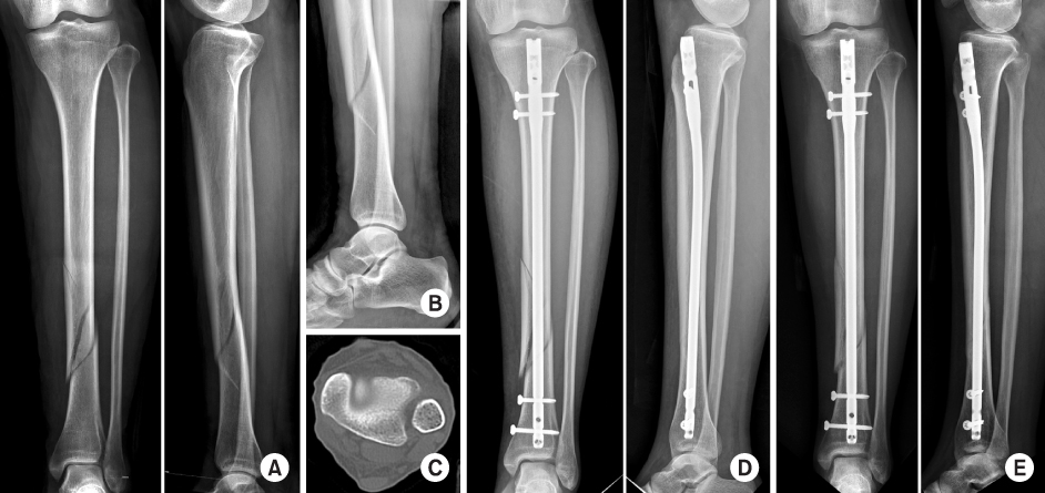
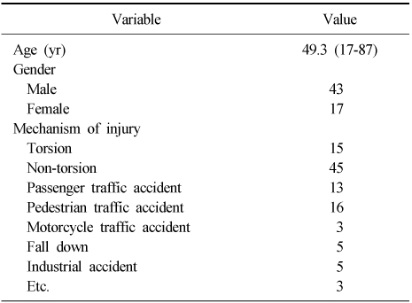
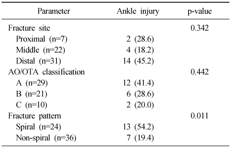
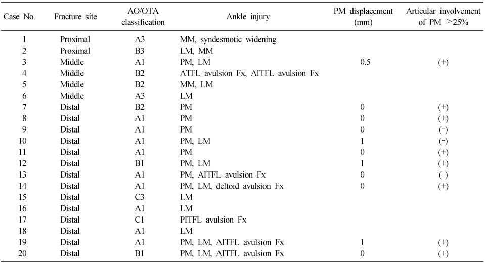
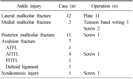
Figure & Data
REFERENCES
Citations
Citations to this article as recorded by 
- Usefulness of Computed Tomography on Distal Tibia Intra-Articular Fracture Associated with Spiral Tibia Shaft Fracture
Seong-Eun Byun, Sang-June Lee, Uk Kim, Young Rak Choi, Soo-Hong Han, Byong-Guk Kim
Journal of the Korean Fracture Society.2016; 29(2): 114. CrossRef
 Cite
CiteAnkle Fracture Associated with Tibia Shaft Fractures


Fig. 1
(A) Initial tibia anteroposterior (AP) and lateral image of a 63-year-old female showing a spiral fracture at the distal third of the tibia shaft. (B) Initial ankle lateral image showing lateral and posterior malleolar fracture. (C) Axial image of ankle computed tomography showing a minimally displaced posterior malleolar fracture. (D) Postoperative tibia AP and lateral image demonstrating articular congruency after screw fixation for a posterior malleolar fragment.
Fig. 2
(A) Initial tibia anteroposterior (AP) and lateral image of a 41-year-old female showing a spiral fracture at the distal third of the tibia shaft. (B) Initial ankle lateral image showing no evidence of any articular disruption of the ankle. (C) Ankle axial computed tomography image showing a non-displaced posterior malleolar fracture. (D) Postoperative tibia AP and lateral image showing articular congruency without fixation for a posterior malleolar fragment after intramedullary nailing. (E) The tibia AP and lateral image at postoperative six months demonstrating fracture healing and no displacement of the posterior malleolar fracture.
Fig. 1
Fig. 2
Ankle Fracture Associated with Tibia Shaft Fractures
Demographics and Mechanism of Injury
Values are presented as median (range) or number.
Incidence Rates of Ankle Injury Combined with Tibial Shaft Fractures
Values are presented as number (%).
Summary of Tibial Shaft Fractures Combined with Ankle Injury
MM: Medial malleolus, LM: Lateral malleolus, PM: Posterior malleolus, ATFL: Anterior talofibular ligament, Fx: Fracture, AITFL: Anteroinferior tibiofibular ligament, PITFL: Posterior inferior tibiofibular ligament.
Distribution of Ankle Injury and Applied Operation
ATFL: Anterior talofibular ligament, AITFL: Anteroinferior tibiofibular ligament, PITFL: Posterior inferior tibiofibular ligament.
Table 1
Demographics and Mechanism of Injury
Values are presented as median (range) or number.
Table 2
Incidence Rates of Ankle Injury Combined with Tibial Shaft Fractures
Values are presented as number (%).
Table 3
Summary of Tibial Shaft Fractures Combined with Ankle Injury
MM: Medial malleolus, LM: Lateral malleolus, PM: Posterior malleolus, ATFL: Anterior talofibular ligament, Fx: Fracture, AITFL: Anteroinferior tibiofibular ligament, PITFL: Posterior inferior tibiofibular ligament.
Table 4
Distribution of Ankle Injury and Applied Operation
ATFL: Anterior talofibular ligament, AITFL: Anteroinferior tibiofibular ligament, PITFL: Posterior inferior tibiofibular ligament.

