 E-submission
 TOTA
TOTA TOTS
TOTS
Articles
- Page Path
- HOME > J Musculoskelet Trauma > Volume 25(2); 2012 > Article
-
Case Report
- Delayed Foreign-body Reaction of Ankle Fracture Treated with a Biodegradable Plate and Screws: A Case Report
- Chul-Hyun Park, M.D., Dae-Hyun Song, M.D., Jae Ho Cho, M.D.
-
Journal of the Korean Fracture Society 2012;25(2):142-145.
DOI: https://doi.org/10.12671/jkfs.2012.25.2.142
Published online: April 17, 2012
Department of Orthopaedic Surgery, Inje University Seoul Paik Hospital, Inje University College of Medicine, Seoul, Korea.
*Department of Pathology, The Armed Forces Capital Hospital, Seongnam, Korea.
†Department of Orthopedic Surgery, The Armed Forces Capital Hospital, Seongnam, Korea.
- Address reprint requests to: Jae Ho Cho, M.D. Department of Orthopedic Surgery, Armed Forces Capital Hospital, Yul-dong, Bundang-gu, Sungnam 463-040, Korea. Tel: 82-31-725-6222, Fax: 82-31-706-0987, hohotoy@nate.com
• Received: January 8, 2012 • Accepted: February 17, 2012
Copyright © 2012 The Korean Fracture Society
- 981 Views
- 5 Download
Abstract
- Biodegradable implants made of co-polymers composed of L-lactide, D-lactide, and trimethylene carbonate were used in the present case. To our knowledge, only one reported tissue reaction has been associated with ankle fracture treated with third-generation implants internationally and none yet domestically. We report a delayed foreign-body reaction of ankle fracture treated with a third-generation biodegradable plate and screws. We suggest that ankle fracture patients treated with biodegradable implants should be advised of this possible complication and should be followed for at least 2 years.
CASE REPORT
DISCUSSION
- 1. Böstman O, Pihlajamäki H. Clinical biocompatibility of biodegradable orthopaedic implants for internal fixation: a review. Biomaterials, 2000;21:2615-2621.Article
- 2. Cho JY, Kim JW, Kim SE, Jung KC, Choi SH. Surgical fixation with biodegradable plate for the treatment of ankle fractures. J Korean Fract Soc, 2008;21:31-36.Article
- 3. Kukk A, Nurmi JT. A retrospective follow-up of ankle fracture patients treated with a biodegradable plate and screws. Foot Ankle Surg, 2009;15:192-197.Article
- 4. Laughlin RM, Block MS, Wilk R, Malloy RB, Kent JN. Resorbable plates for the fixation of mandibular fractures: a prospective study. J Oral Maxillofac Surg, 2007;65:89-96.Article
- 5. Losken HW, van Aalst JA, Mooney MP, et al. Biodegradation of Inion fast-absorbing biodegradable plates and screws. J Craniofac Surg, 2008;19:748-756.Article
- 6. Mavrogenis AF, Kanellopoulos AD, Nomikos GN, Papagelopoulos PJ, Soucacos PN. Early experience with biodegradable implants in pediatric patients. Clin Orthop Relat Res, 2009;467:1591-1598.Article
- 7. Nieminen T, Rantala I, Hiidenheimo I, et al. Degradative and mechanical properties of a novel resorbable plating system during a 3-year follow-up in vivo and in vitro. J Mater Sci Mater Med, 2008;19:1155-1163.ArticlePDF
- 8. Rokkanen PU, Böstman O, Hirvensalo E, et al. Bioabsorbable fixation in orthopaedic surgery and traumatology. Biomaterials, 2000;21:2607-2613.Article
- 9. Wood GD. Inion biodegradable plates: the first century. Br J Oral Maxillofac Surg, 2006;44:38-41.Article
REFERENCES
Fig. 1
(A) Preoperative radiograph showing a lateral malleolar fracture.
(B) Radiograph made one week after open reduction and internal fixation with biodegradable implants.
(C) Biodegradable plate and screws (Inion OTPS™, Inion Oy, Tampere, Finland).

Fig. 2The patient noted a gradually enlarging soft-tissue mass adjacent to the previous surgical scar.

Fig. 3
(A) On radiographs, the fracture had healed but osteolytic change had occurred.
(B) Magnetic resonance imaging showed a healed lateral malleolar fracture accompanied by an oval mass with an accumulation of fluid in the sinus formation.

Fig. 4
(A) Intraoperative clinical photographs showing a collection of fluid accompanied by several fragments of whitish material.
(B) Granulomatous tissue and foreign-body fragments.

Fig. 5
(A) Photomicrograph showing some variably shaped, hyaline foreign-body material with a rhomboid shape (H&E stain, ×100).
(B) Photomicrograph showing fragments surrounded by a foreign-body reaction with mononuclear cells, foamy histiocytes, foreign body-type multinucleated giant cells, and granulation tissue in the stroma (H&E stain, ×400).
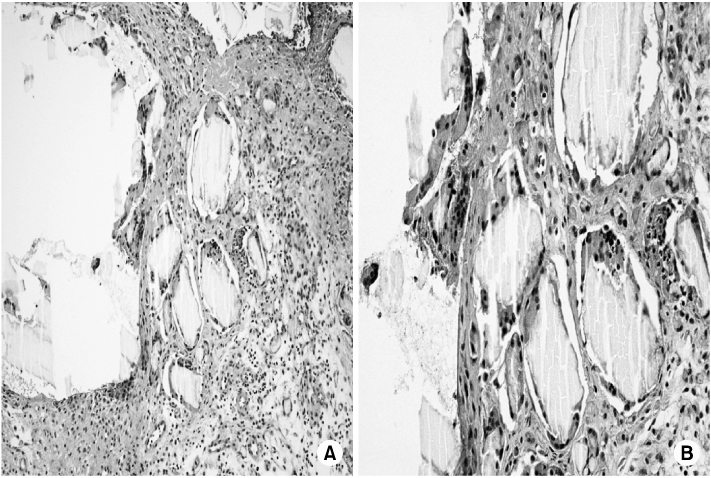
Figure & Data
REFERENCES
Citations
Citations to this article as recorded by 
 Cite
CiteDelayed Foreign-body Reaction of Ankle Fracture Treated with a Biodegradable Plate and Screws: A Case Report
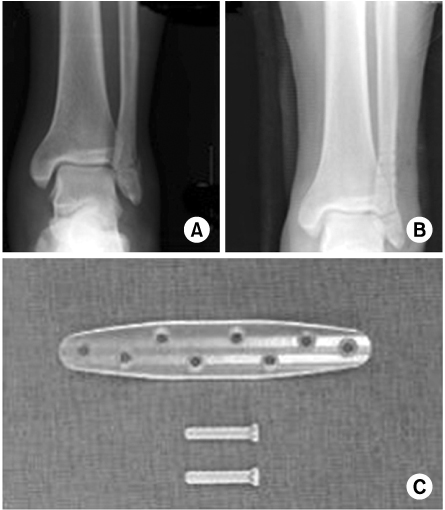
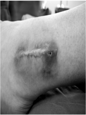
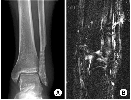
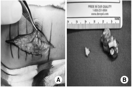

Fig. 1
(A) Preoperative radiograph showing a lateral malleolar fracture.
(B) Radiograph made one week after open reduction and internal fixation with biodegradable implants.
(C) Biodegradable plate and screws (Inion OTPS™, Inion Oy, Tampere, Finland).
Fig. 2
The patient noted a gradually enlarging soft-tissue mass adjacent to the previous surgical scar.
Fig. 3
(A) On radiographs, the fracture had healed but osteolytic change had occurred.
(B) Magnetic resonance imaging showed a healed lateral malleolar fracture accompanied by an oval mass with an accumulation of fluid in the sinus formation.
Fig. 4
(A) Intraoperative clinical photographs showing a collection of fluid accompanied by several fragments of whitish material.
(B) Granulomatous tissue and foreign-body fragments.
Fig. 5
(A) Photomicrograph showing some variably shaped, hyaline foreign-body material with a rhomboid shape (H&E stain, ×100).
(B) Photomicrograph showing fragments surrounded by a foreign-body reaction with mononuclear cells, foamy histiocytes, foreign body-type multinucleated giant cells, and granulation tissue in the stroma (H&E stain, ×400).
Fig. 1
Fig. 2
Fig. 3
Fig. 4
Fig. 5
Delayed Foreign-body Reaction of Ankle Fracture Treated with a Biodegradable Plate and Screws: A Case Report

