 E-submission
 TOTA
TOTA TOTS
TOTS
Search
- Page Path
- HOME > Search
Original Article
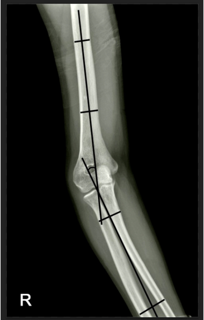
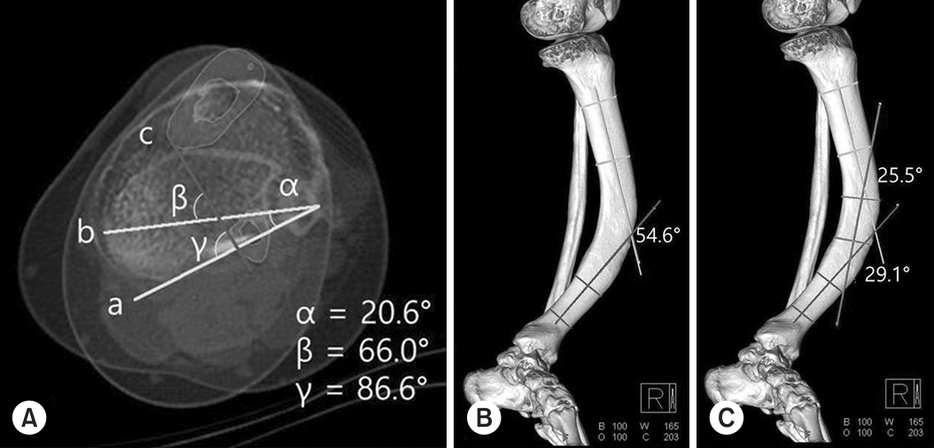
- Reverse V step-cut osteotomy for the correction of cubitus varus in adults: a retrospective study
- Jinyoung Bang, Hyung Jun Koo
- J Musculoskelet Trauma 2025;38(2):102-108. Published online April 25, 2025
- DOI: https://doi.org/10.12671/jmt.2025.00045
-
 Abstract
 PDF
PDF - Background
Cubitus varus deformity in adults most commonly occurs as a late complication resulting from malunion of distal humeral fractures sustained during childhood. This deformity can cause cosmetic problems and anatomical deformities that hinder normal sports activities and potentially lead to long-term complications. Although various surgical techniques exist for correcting cubitus varus, this study investigated the clinical and functional outcomes of reverse V step-cut osteotomy.
Methods
In total, 15 patients underwent surgical treatment with reverse V step-cut osteotomy between 2012 and 2023. The mean age of the patients at the time of surgery was 46.3 years (range, 20–65 years). The preoperative carrying angle was ‒11.09° of varus, which was corrected to +12.81° of valgus postoperatively. The mean preoperative lateral prominence index (LPI) was ‒10.03, and the mean postoperative LPI improved to ‒4.48. A comparison to the unaffected side showed a P-value of 0.978, indicating similarity.
Results
Preoperatively, eight patients exhibited signs of posterolateral rotatory instability, and among them, three underwent concomitant lateral ulnar collateral ligament reconstruction. Seven patients reported ulnar nerve symptoms, and all underwent concurrent ulnar nerve release. Postoperatively, improvements in elbow pain, instability, and ulnar nerve symptoms were observed. One patient required reoperation due to malunion and insufficient correction, but no other complications were noted.
Conclusions
These outcomes demonstrate that reverse V step-cut osteotomy can be an effective treatment method for cubitus varus deformity in adults. Level of evidence: IV.
- 5,393 View
- 101 Download

Case Reports
- Acute on Chronic Stress Fracture of a Varus Deformed Distal Tibia - A Case Report -
- Seong Kee Shin, Ki Chun Kim, Eli Schmidt, Seung Yeon Cho, Ki Chul Park
- J Musculoskelet Trauma 2024;37(4):184-189. Published online October 25, 2024
- DOI: https://doi.org/10.12671/jmt.2024.37.4.184
-
Abstract
PDF
- A severe post-traumatic distal tibia vara deformity is an uncommon condition in orthopedics. Typical symptoms include intractable recurrent pain, fragility related to stress fractures over the tensile area, and a limping gait caused by leg length discrepancy. Surgical management should be performed on acute fractures extending from a stress fracture gap. For successful surgical results, deformity correction is important for sustaining axial load bearing for standing and walking. Procedures to manage this condition have been proposed, but there is a high risk of complications, including metal failure, nonunion, and weakness caused by a long period of rehabilitation. In this case, the authors report a successful result using a modified clamshell osteotomy combined with a proximal and distal wedge bone resection in a single stage.
- 2,299 View
- 45 Download
- Two-Year Follow-Up Results after Tendon Graft and Corrective Osteotomy for the Delayed Rupture of the 2nd-5th Flexor Tendons due to a Malunion of a Distal Radius Fracture - A Case Report -
- Jeung-Hwan Seo, Hyun-Gon Gwak, Jae Hoon Lee
- J Korean Fract Soc 2022;35(2):63-67. Published online April 30, 2022
- DOI: https://doi.org/10.12671/jkfs.2022.35.2.63
-
Abstract
PDF
- The delayed rupture of the flexor tendons is a rare complication of malunited distal radius fractures after nonoperative management. The known cause of a flexor tendon rupture is attrition between the palmarly displaced ulnar head and the involved tendons. Sharp bony spurs on the volar side of the malunited distal radius can also cause flexor tendon rupture. About 30 cases have been reported in literature. There were only four case reports about the delayed rupture of the 2nd, 3rd, 4th, and 5th flexor tendons. In this case, we experienced flexor digitorum superficialis and flexor digitorum profundus tendon ruptures of the index, middle, ring, and little fingers, after 8 months following the malunion of a distal radius fracture. At two years follow-up after tendon graft and corrective osteotomy, the range of motion and motor weakness of the 2nd, 3rd, 4th, and 5th fingers improved.
- 1,256 View
- 2 Download
Review Articles
- A Correction of Malunion or Deformity in the Lower Extremity
- Kyeong Hyeon Park, Joon Woo Kim, Chang Wug Oh
- J Korean Fract Soc 2017;30(4):219-227. Published online October 31, 2017
- DOI: https://doi.org/10.12671/jkfs.2017.30.4.219
-
Abstract
PDF
- The incidence of malunion in the long bone with has been reduced because of the advancements in surgical technique. However, nonunion or malunion are still observed in mechanical axis deformation of the lower limb, resulting in the overload of cartilage and instability of the joint, requiring surgical correction. Preoperative planning for malunion is very important, and accurate evaluation of the deformity is essential. Herein, we describe the indications of corrective osteotomy, choice of patients, and various surgical methods for the treatment of malunion of the long bone.
- 1,979 View
- 38 Download
- Osteotomy Selection: Advantages, Disadvantages, and Indication
- Ki Chul Park, Hyun Uk Kim, Young Sik Song
- J Korean Fract Soc 2017;30(3):167-172. Published online July 31, 2017
- DOI: https://doi.org/10.12671/jkfs.2017.30.3.167
-
Abstract
PDF
- Malunion causes not only cosmetic problems, but also degenerative osteoarthritis due to changes in the anatomical and mechanical axes. Corrective osteotomy may be required in some cases to prevent these complications. The corrective osteotomy is divided into two types: Straight and dome. The straight type is divided into open and closed wedge, in accordance with the correction method. Surgeons should understand the indication, surgical procedure, as well as the advantages and disadvantages of each osteotomy method. Deciding on the method of corrective osteotomy depends on the degree of angulation, soft tissue condition, approximate with joint, implant type, and the experience of the surgeon.
- 1,813 View
- 31 Download
Original Articles
- Cannulated Screw and Wire Fixation with Predrilling for Olecranon Osteotomy in Intra-articular Comminuted Distal Humerus Fractures
- Soo Hong Han, Ho Jae Lee, Woo Hyun Kim, Yong Gil Jo, Won Tae Song
- J Korean Fract Soc 2015;28(2):118-124. Published online April 30, 2015
- DOI: https://doi.org/10.12671/jkfs.2015.28.2.118
-
Abstract
PDF
- PURPOSE
The olecranon osteotomy in intra-articular comminuted distal humerus fractures is a suggested technique for excellent exposure of articular fractures. However, complications including delayed union, nonunion of osteotomy site have been reported. Authors have applied predrilling for cannulated screw before osteotomy for achievement of rapid and accurate reposition of separation part and added wire fixation for secure stability. The purpose of this study is to evaluate the efficacy of this fixation procedure following the olecranon osteotomy during the internal fixation of intra-articular fracture of the distal humerus.
MATERIALS AND METHODS
This study retrospectively analyzed 14 cases (9 women and 5 men) of intra-articular distal humerus fractures in which the olecranon osteotomy was applied. The mean age of patients was 53.4 years (range, 25 to 83 years), and the average follow-up period was 15.9 months. Eleven cases were classified as AO 13-C3, and the other 3 cases were AO 13-C2. Reduction accuracy, union period of osteotomy site on follow-up radiographs and postoperative complications related to olecranon osteotomy were evaluated.
RESULTS
All osteotomized parts showed no position change and solid union with normal alignment at the last follow-up. The mean period of bony union was 3.5 months (range, 2 to 5 months). There were no complications related to olecranon osteotomy except one case of non-displaced fracture of the proximal ulnar shaft at the level of cannulated screw tip caused by forceful passive physical therapy. It was managed by conservative treatment without further problem.
CONCLUSION
Predrilled cannulated screw and wire fixation following the olecranon osteotomy during internal fixation of intra-articular comminuted distal humerus fractures showed satisfactory results in the union of osteotomy site and it could be a recommendable procedure when fractures require olecranon osteotomy. -
Citations
Citations to this article as recorded by
- Comparison of two fixation techniques of olecranon osteotomy after reconstruction of intra-articular distal humerus fractures
Faaiz Ali Shah, Aimal Sattar, Javed Iqbal
World Journal of Orthopedics.2025;[Epub] CrossRef
- Comparison of two fixation techniques of olecranon osteotomy after reconstruction of intra-articular distal humerus fractures
- 924 View
- 7 Download
- 1 Crossref
- A Retrospective Comparative Study of Internal Fixation with Reconstruction Plate Versus Anatomical Locking Compression Plate in Displaced Intercondylar Fractures of Humerus
- Tong Joo Lee, Young Tae Kim, Dae Gyu Kwon, Ju Yong Park
- J Korean Fract Soc 2014;27(4):294-300. Published online October 31, 2014
- DOI: https://doi.org/10.12671/jkfs.2014.27.4.294
-
Abstract
PDF
- PURPOSE
To analyze the clinical result of a conventional reconstruction plate (CRP) fixation and locking compressive plate (LCP) fixation on the surgical treatment of an adult's displaced intercondylar fracture of humerus.
MATERIALS AND METHODS
A total of 40 patients enrolled in the study were treated between August 2002 and May 2012. Fixation with a CRP was performed in 20 patients (group A) and anatomical locking compression plate fixation was performed in 20 patients (group B). The clinical and functional evaluation was performed according to the Mayo elbow performance score and Cassebaum classification of elbow range of motion (ROM), disabilities of the arm, shoulder and hand score.
RESULTS
The Mayo elbow functional evaluation scores, eight cases were excellent, 10 cases were good, and two cases were fair in group A, and 12 cases were excellent, seven cases good, and one case fair in group B; both groups showed satisfactory results. The durations of attaining 90 to 120 degrees of the ROM of joints postoperatively were 8.3 days on average (6 to 15 days) in group A and 5.5 days on average (5 to 9 days) in group B, demonstrating a significant difference between the two groups (p=0.04). Although the correlations of clinical results according to the difference of bone mineral densities (BMDs) were not statistically significant between the two groups (p=0.35), loss of fixation occurred due to loosening of screws in two patients with low BMDs in whose operations reconstruction plates were used.
CONCLUSION
The use of locking compressive plate on the surgical treatment of an diaplaced intercondylar fracture of humerus have a good clinical results because that permits early rehabilitation through good fixation and reduces the complications such as loosening of screws.
- 712 View
- 0 Download
- The Clinical Results of Opening Wedge Osteotomy in the Volarly Malunited Distal Radius
- Seoung Joon Lee, Jin Ho Choi
- J Korean Fract Soc 2014;27(1):29-35. Published online January 31, 2014
- DOI: https://doi.org/10.12671/jkfs.2014.27.1.29
-
Abstract
PDF
- PURPOSE
To report the clinical results of opening wedge osteotomy graft in the volarly malunited distal radius.
MATERIALS AND METHODS
Ten patients with volarly malunited distal radius fractures treated by opening wedge osteotomy were included in this study. Grip power, range of motion of the wrist, radiographic parameter and Mayo wrist scores were retrospectively evaluated.
RESULTS
At the final follow-up, the rotation of the forearm, the range of motion of wrist, and the grip power were improved. The average radial inclination improved to 22.2degrees, the average volar tilting improved to 5.6degrees, and the average ulnar variance improved to 0.8 mm. The average Mayo wrist score was improved to 85.6.
CONCLUSION
Opening wedge osteotomy for volarly malunited distal radius was considered as one of the good treatments to restore anatomy of the distal radius and distal radioulnar joint and also to improve the function of the wrist joint.
- 928 View
- 6 Download
Case Report
- Bilateral Malunion and Distal Radioulnar Joint Dislocation after Operative Treatment of Bilateral Galeazzi Fractures in Child: A Case Report
- Sang Jin Cheon, Dong Joon Kang, Nam Hoon Moon, Seung Han Cha, He Myung Cho
- J Korean Fract Soc 2009;22(4):292-296. Published online October 31, 2009
- DOI: https://doi.org/10.12671/jkfs.2009.22.4.292
-
Abstract
PDF
- Galeazzi fractures in child is rare and seldom necessary of operative treatment because the result of conservative treatment is good. We present the patient who was a 11-year-old male and fell onto his both hands during a hundred-meter dash. His diagnosis was bilateral Galeazzi fractures and limited open reduction and internal fixation with Kirschner pins was initial treatment at local hospital. After 4 weeks postoperatively, Kirschner pins were removed and rehabilitating exercise was started. After 4 months postoperatively, he was transferred to our hospital due to malunion with severe angular deformities and distal radioulnar joint (DRUJ) dislocation. He was treated with corrective osteotomy. Thus, as in this case, we suggest more careful treatment and observation if conservative method of Galeazzi fracture in child is chosen and consider operative method as treatment according to age and pattern of fracture.
- 954 View
- 1 Download
Original Articles
- Operative Treatment of Displaced Intercondylar Fracture of the Distal Humerus with Reconstruction Plate
- Ryuh Sup Kim, Tong Joo Lee, Kyoung Ho Moon, Seung Rim Park, Moon Lee
- J Korean Fract Soc 2007;20(2):172-177. Published online April 30, 2007
- DOI: https://doi.org/10.12671/jkfs.2007.20.2.172
-
Abstract
PDF
- PURPOSE
To evaluate the therapeutic effects of chevron olecranon osteotomy and bilateral reconstruction plate as operative treatment for distal humerus intercondylar fracture.
MATERIALS AND METHODS
Among patients operated for distal humerus intercondylar fracture in our hospital from June, 1997 to October, 2005, 26 patients were selected who could be followed-up for more than one year. The average follow-up period was 15 months. All olecranon osteotomies were chevron osteotomy and all fractures were treated with internal fixation using bilateral reconstruction plate. The ulnar nerve was checked in all cases. Three patients in which case the plate might irritate the ulnar nerve, received with ulnar nerve anterior transposition. Cassebaum's classification and Mayo elbow performance score were used to evaluate at three, six and twelve months.
RESULTS
Mean bone union period was 11.7 weeks. There were 9 excellent cases, 11 good cases, 4 fair cases and 2 poor cases. Mean flexion contracture was 11° and further flexion was 126° at last follow-up.
CONCLUSION
Bilateral reconstruction plate internal fixation using chevron olecranon osteotomy showed strong fixation and good clinical results and it is possible for early rehabilitation treatment.
- 888 View
- 3 Download
- Modified Step-cut Osteotomy of Distal Humerus for the Correction of Cubitus Varus Deformity in Children
- Yeo Hon Yun, Jun Gyu Moon, Duk Moon Chung
- J Korean Fract Soc 2004;17(3):287-294. Published online July 31, 2004
- DOI: https://doi.org/10.12671/jkfs.2004.17.3.287
-
Abstract
PDF
- PURPOSE
evaluate the radiologic and clinical results of modified step-cut osteotomy for correction of cubitus varus deformity in children.
MATERIALS AND METHODS
We analysed 16 children who had varus deformity preoperatively and received modified step-cut osteotomy. The results were evaluated by final follow-up radiographs and clinical results, which were humeral-elbow-wrist angle, lateral prominence, range of motion and complications.
RESULTS
The average preoperative humeral-elbow-wrist (HEW) angle was -15.8degrees and average last follow-up HEW angle was +6.7degrees Lateral prominence under 5 mm occurred in 3 cases and one children showed limited motion and transient ulna neuropathy.
CONCLUSION
The results demonstrate that modified step-cut osteotomy achieve good correction of cubitus varus without lateral bony prominence or complications.
- 724 View
- 3 Download
- Surgical Treatment for the Non-union of the Lateral Humeral Condyle Fracture using Closing Wedge Osteotomy and Bone Graft
- Sang Ho Ha, Hong Moon Sohn, Jun Young Lee, Sun Jong Oh
- J Korean Soc Fract 2003;16(3):379-384. Published online July 31, 2003
- DOI: https://doi.org/10.12671/jksf.2003.16.3.379
-
Abstract
PDF
- PURPOSE
To evaluate the clinical results of the surgical treatment for established nonunion of lateral humeral condyle fracture using closing wedge osteotomy and bone graft.
MATERIALS AND METHODS
Six patients diagnosed as symptomatic established nonunion of lateral humeral condyle fracture and cubitus valgus deformity were reviewed retrospectively. The average age was 23 years old and mean follow up period was 32 months. We investigate the changes of the symptoms and radiographic findings, and determine the results by Oppenheim's criteria.
RESULTS
According to Oppenheim's criteria, 3 patients showed excellent, 2 good, 1 poor. Carrying angle is improved to 10.2 degrees and range of motion was decreased by mean 9 degrees. All of the patients' muscle weakness and pain were improved, and was achieved solid union at the last follow up.
CONCLUSION
In the treatment of symptomatic established lateral humeral fracture and cubitus valgus deformity, better functional and cosmetic results are anticipated by a closing wedge osteotomy and bone graft. -
Citations
Citations to this article as recorded by- In SituLate Metaphyseal Osteosynthesis for the Fractures of the Lateral Humeral Condyle in Children
Kun Bo Park, Seung Whan Lee, Hyun Woo Kim, Hui Wan Park, Ki Seok Lee
Journal of the Korean Fracture Society.2008; 21(2): 151. CrossRef
- In SituLate Metaphyseal Osteosynthesis for the Fractures of the Lateral Humeral Condyle in Children
- 940 View
- 3 Download
- 1 Crossref
- The Lateral Closing Osteotomy using Threaded Steinmann Pin for the Cubitus Varus Deformity Followed by Supracondylar Fracture around the Elbow
- Jin Hak Kim, Song Lee, Byung Ki Kwon, Hyun Soo Kim, Soon Young Jeong, Dea Jung Choi
- J Korean Soc Fract 2003;16(3):370-378. Published online July 31, 2003
- DOI: https://doi.org/10.12671/jksf.2003.16.3.370
-
Abstract
PDF
- PURPOSE
To investigate the usefulness of closing wedge osteotomy with threaded steinmann pin and wiring for the treatment of cubitus varus deformity after elbow fracture during childhood.
MATERIALS AND METHODS
From February 1994 to February 2002. We performed closing wedge osteotomy with threaded steinmann pin and wiring in 16 elbows with cubitus varus deformity. There are 11 men and 5 women. Mean age was 21.6 years and mean follow-up was 19.2 months. Mean deformed carrying angle was varus 21.7 degree. Mean period from initial injury to treatment was 16.5 years.
RESULTS
Mean angle that was corrected by above operation methods was valgus 12 degree. Average periods of immobilization was 27.8 days. One tardy ulnar nerve syndrome before surgery was solved at 8 weeks after operation. 2 cases with superficial infection was treated easily. 14 cases of all were estimated as good with Oppenheim's criteria.
CONCLUSION
Closing wedge osteotomy with threaded steinmann pin and wiring makes early range of motion exercise being possible as rigid fixation. The supracondylar closing wedge osteotomy with threaded Steinmann pin and wiring is thought to be the useful method.
- 650 View
- 1 Download
- Treatments of the Malunited Tibial Shaft Fracture
- Taik Seon Kim, Jae Ik Shim, Sung Jong Lee, Suk Ha Lee, Yeon Sik Yu, Young Bae Kim, Kwang Yeol Park
- J Korean Soc Fract 2000;13(4):897-904. Published online October 31, 2000
- DOI: https://doi.org/10.12671/jksf.2000.13.4.897
-
Abstract
PDF
- PURPOSE
The malunited diaphyseal tibia fractures result in tibial shortening, angular deformities, gait disturbance, development of joint pain, etc. The authors analyzed the results of treatment consist of corrective osteotomy for diaphyseal malunion with internal or external fixation.
MATERIALS AND METHODS
The authors reviewed 18 cases of tibial diaphyseal malunion treated in Korea Veterans Hospital between January 1992 and December 1998. Mean follow-up period was 4.2 years. The preoperative deformities were varus, anterior or posterior bowing and shortening. The preoperative symptoms were knee joint pain, ankle joint pain, and gait disturbance. Corrective osteotomy was done on the site of malunion in all cases. Fixation were done with IM nailings(13 cases), plates(3 cases) and Ilizarov external fixator. We analyzed the unions radiologically and the knee pains with HSS score.
RESULTS
All malunions were successfully corrected. Mean duration of union was 4.5 month. In the coronal plane, preoperative varus deformity(mean 16.5degrees varus) was corrected to 3degrees of valgus. In the saggital plane, anterior and posterior bowing was corrected to neutral. In 15 cases of the patient with knee joint pain, the mean HSS score was improved from 69 preopertively to 82 postoperatively.
CONCLUSION
The correction of tibia diaphyseal malunion had good results by osteotomy at the malunited site and firm internal or external fixation. And it also improved knee joint pain significantly.
- 713 View
- 5 Download
Case Report
- Treatment of rotational malalignment after interlocking intramedullary nailing of femur : Report of a case
- Suk Kyu Choo, Byung Jik Kim, Byung Hee Min
- J Korean Soc Fract 1999;12(4):833-836. Published online October 31, 1999
- DOI: https://doi.org/10.12671/jksf.1999.12.4.833
-
Abstract
PDF
- Rotational malalignment after IM nail of femur is a common problem and if the deformity is great, may cause pain, limitation of motion, even require corrective osteotolny later. The rotational malalignment of femur is not easy to find out during operation because prominant landmarks is lack. We experienced 25 years old male patient with 45 of internal malalignment of femur after IM nail at other hospital. The patient was treated by corrective osteotomy at the fracture site and exchange nailing, but we confronted the obstacle that was remained rotatonal unstability after slotted nailing, and we have to use additional plate fixation. This problem can be prevented by using rigid unslotted nail.
- 587 View
- 0 Download
Original Articles
- Tardy Ulnar Nerve Palsy Caused by Post-Traumatic Elbow deformities
- Seung koo Rhee, seok Whan Song, Hwa Sung Lee, Ho Tae Kim
- J Korean Soc Fract 1998;11(2):420-426. Published online April 30, 1998
- DOI: https://doi.org/10.12671/jksf.1998.11.2.420
-
Abstract
PDF
- Thirty-five patients with tardy ulnar nerve palsy caused by cubitus valgus (33 cases0 and varus (2 cases) deformities were retrospectively studied. All patients had a history of old fracture on the distal humerus during childhood. The mean interval between the previous fractures and the onset of ulnar neuropathy was 19 years. The severity of nerve palsy was classified as McGowan's grade I in 24 patients, grade II in 8 patients, and grade III in 3 patients. The mean carrying angle was average 29 degrees in 33 cases with cubitus valgus and it was decreased to average 11 degrees postoperatively, but the angle was average -23 degrees preoperatively in 2 cases with cubitus varus and it was corrected to average 9 degrees postoperatively. the cause of palsy was analysed by mechanical stetching in 11 cases, compression by a fibrous band between the two heads of flexor carpi ulnaris in 8 cases, and diffuse fibrous adhesion around the ulnar tunnel in 5 cases. All patients was treated with supracondylar closing wedge osteotomy accompanied with anterior ulnar nerve transposition in 13 patients, corrective osteotomy only in 12 patients, and anterior ulnar nerve transposition only in 10 patients. Their end results were analysed as good in 24 cases, fair in 8 cases, and poor in 3 cases within average 6 months after the operations (4 to 13 months). The poor results was obtained in 3 cases out of 9 cases with corrective osteotomy group (33.3%). Conclusively, a tardy ulnar nerve palsy caused by post-traumatic elbow deformities should be corredcted with anterior ulnar nerve transposition with or without corrective closing wedge osteotomy but not by corrective osteotomy only, because of compressive neuropathy by diffuse fibrous adhesion or bands of two heads of FCU around the ulnar tunnel in elbow.
- 1,359 View
- 8 Download
- Treatment of Cubitus Varus Using Tension Band Wiring after the Supracondylar Osteotomy
- Dne Yong Han, Hui Wan Park, Dong Eun Shin, Ki Won Suh
- J Korean Soc Fract 1997;10(3):678-684. Published online July 31, 1997
- DOI: https://doi.org/10.12671/jksf.1997.10.3.678
-
Abstract
PDF
- Cubitus varus is the most common angular deformity that results from supracondylar fractures in children. Although this deformity rarely limit elbow function, the correction is frequently requested due to cosmetic problem. This paper was prepared to describe the operative method and to evaluate the clinical results of tension band wirinB after distal humeral supracondylar osteotomy for cubitus varus. The result was evaluated using Oppenheim criteria. Excellent or good cases were 94.4%. We concluded that tension band wiring is a satisfactory method of treatment for cubitus varus.
-
Citations
Citations to this article as recorded by- Evaluation of tension-band osteosynthesis for cubitus varus deformity in pediatric patients: A retrospective review
Man Duc Minh Phan, Terry Richard Light, Tiep Van Phan, Phi Duong Nguyen
Journal of Hand and Microsurgery.2025; 17(2): 100213. CrossRef
- Evaluation of tension-band osteosynthesis for cubitus varus deformity in pediatric patients: A retrospective review
- 766 View
- 2 Download
- 1 Crossref
- Treatment of Intercondrlar Fracture of Elbow using Y-Plate through Extraartieular Olecranon Osteotomy in the Posterior Approach
- Ho Guen Chang, Sang Su Lee, Eung Ju Lee, Jun Dong Chang, Won Ho Cho, Chang Ju Lee
- J Korean Soc Fract 1996;9(4):1118-1124. Published online October 31, 1996
- DOI: https://doi.org/10.12671/jksf.1996.9.4.1118
-
Abstract
PDF
- Iniercondylar fractures of the distal humerus in adults are rare and notoriously difficult to treat. We treated 8 patients by open reduction and internal fixation using Y-plate with extraarticular olecranon osteotomy in posterior approach. L-shaped osteotomy was performed at the extraarticular portion of olecranon with triceps tendon insertian remained to proximal portion of it, using air-saw. The follow-up period ranged from 12 to 30 months with average of 19.7 months. The fractures were dassified according to AO classification. The results were evaluated using Jupiter et al grading system. 1) There was no loosening of fixation Cevice of humeral condyle and olecranon. 2) Niether delayed union nor nonunion of olecranon and humeral condyle were found. 3) Exellent grade was achieved in 4 patients(50%), three(38%) had good, and one(12%) poor. Flexion-extension arc ranged from 60 degree to 120 degree with mean of 98.8 degree. 4) Complications included postoperatile neuritis in one, myositis ossificance in one, and heterotopic bone in one patient. Authors would introduce the method and result of extraarticular olecranon osteotomy in posterior approach for the intercondylar fracture of distal humerus, as a new technique.
- 735 View
- 0 Download
- Supracondylar Closing Wedge Osteotomy for Posttraumatic Angular Deformity of Distal Humerus: Methods for Reducing Secondary Deformity
- Sung Soo Kim, Sung Keun Sohn, Chul Hong Kim
- J Korean Soc Fract 1996;9(3):706-714. Published online July 31, 1996
- DOI: https://doi.org/10.12671/jksf.1996.9.3.706
-
Abstract
PDF
- The angular deformity of distal humerus is one of the most frequent complication of supracondylar fracture in growing children. The deformity rarely limits function, but corrected by patients request due to cosmetic problem. Many orthopedic surgeons have suggested various operation methods but with high incidence of complications related to these operations, also we often experience secondary deformity after inaccurate osteotomy. Therefore to identify desirable operative method to reduce secondary deformity, a retrospective study of 17 patients operated with angular deformity following distal humerus fracture was carried out in which replanning with isosceles triangle method was done in all cases. The following results were obtained. 1. The complications were two cases of metal failure and one of non union. 2. The basic requirement of closing wedge osteotomy without secondary deformity was that:the center line of isosceles triangle whose apex angle should be identical to the deformity angle and be placed on the concave apex of deformity, should overlap the transverse bisector of hurnerusforearm axes. In inevitable cases, the disparity should be minimized to alleviate secondary deformity. 3. The translation was calculated by the equation of T=Dxsin α(T:translation, D:proximal or distal migration of the point of contact of humerus-forearm axes, α:angle of the deformity). In conclusion, we think that the deformity may be corrected safely and easily using minute preoperative planning with application of above principle.
- 736 View
- 3 Download
- Treatment for the Malunion of the Distal Radius
- Hyoun Oh Cho, Kyoung Duck Kwak, Sung Do Cho, Cheol Soo Ryoo, Woo Keun Jung
- J Korean Soc Fract 1996;9(2):290-294. Published online April 30, 1996
- DOI: https://doi.org/10.12671/jksf.1996.9.2.290
-
Abstract
PDF
- Malunited fractures of the distal radius may result in adequate function of the wrist with absence of pain in elderly patients. However, posttraumatic dedormity in younger, active patients is less well toterated, especially in those engaged in heavy manual work or who require a normal range of motion of the wrist. surgical correction of the malunion of the distal radius should be considered for this group of patients. Operation for the malunited fractures of the distal radius was performed in ten cases during the periods between January, 1990 and December, 1993, who were followed for an average of 15 months.The procedures included radial osteotomy(RO) in four malunions of short duration, radial osteotomy with ulnar shortening (RO & US) in these malunions of long duration and ulnar shortening(US) in three cases. We reviewed these cases retrospectively with respect to the clinical findings(pain, grip strength, range of motion of the wrist) and radiograpic changes(volar tilt, radial articular inclination and radiul shortening). Symptoms(radioulnar or radiocarpal pain) were improved in all cases. By compairing with the opposite sides, resedual loss of grip strength was 35% in RO group, 40% in RO & US and 31% in & US group. Residual loss of motion in flexion and extension or in deviation was similar in all groups, whill loss in rotation was less in RO or RO & US group than in US group. Inclination of the radial articular surface (radial inclination and volar tilt) was restored up to the degree similar to the opposite wrist in RO or US group, while was not in US group. Radial length was restored up to the dgegrees similar to the opposite wrist in all groups. The overall results were good or very good in five among the seven cases of RO group(with or without ulnar shortening), while good only in one among the cases of US group.
- 673 View
- 4 Download
- The Fractures of the Femoral Neck in Children
- Ki Soo Kim, Seung Hee Ko, Chang Mun Seo, Yong Su Choi, Kyung Ho Kim, Dong Myung Lee
- J Korean Soc Fract 1994;7(2):562-570. Published online November 30, 1994
- DOI: https://doi.org/10.12671/jksf.1994.7.2.562
-
Abstract
PDF
- The femoral neck fracture in childhood is rare and occurred by severe trauma. Its treatment method and prognosis are different from adult, and(it was) difficult to treat due to frequent complication. So the femoral neck fractures in children are called as unsolved fracture. Eleven cases of childrens femoral neck fracture were treated by closed reduction and internal fixation using cancellous screws or Knowles pins at Kwang lu Christian Hospital from January 1986 to January 1992, and were analysed clinically and radiologically. According to classification of Delbet and Colona, the transcervical fracture were 6 cases and the cervicotrochanteric fracture f cases. 10 of the eleven cases were displaced fractures. Avascular necrosis was evident in 6 cases(54.5%) and all of them were displaced fracture initially. 2 cases of avascular necrosis were treated with Intertrochanteric varus osteotomy with angle blade plate.
- 677 View
- 0 Download
- Supracondylar Osteotomy for Cubitus Varus and Valgus
- Duk Seop Shin, Jong Chul Ahn, Se Dong Kim, Yong Seok Choi
- J Korean Soc Fract 1994;7(1):49-57. Published online May 31, 1994
- DOI: https://doi.org/10.12671/jksf.1994.7.1.49
-
Abstract
PDF
- Between December 1989 and january 1944, 17 corrective supracondylar osteotomy of humerus for cubitus varus and valgus were performed at department of orthopaedic surgery in Yeugnam University. Supracondylar fracture was the most common cause of deformity Average age at operation was 18.6 years old and average follow up period was 14 months. The operation was done under the comprehensive preoperative plan, and Internal fixation was done with K-w,res ,n younger patients, and with plates and screws in elder ones(77%). Period for external fixation could be shortened by firm internal fixation. The result was exllent in nine cases, good in four, and poor in four. No ulna and radial nerve palsy were found in operation of cubitus varus. There were three tardy ulna nerve palsy In cubitus valgus, then anterior transposition of ulna nerve was done.
- 718 View
- 3 Download
- Supracondylar Osteotomy for Cubitus Vnrus Deformity by Using Plate in Adults
- Hyun Ki Yoon, Sung Seok Soe, Young Ku Lee
- J Korean Soc Fract 1992;5(2):319-324. Published online November 30, 1992
- DOI: https://doi.org/10.12671/jksf.1992.5.2.319
-
Abstract
PDF
- Cubitus varus deformity is the most common complication of supracondylar fractures of the humerus in children. For the correction of this deformity, three basic types of osteotomies were known. Among them, the lateral closing wedge osteotomy is the easiest, safest and the most stable method. After osteotomy, the methods of fixation are plate fixation, crossed kirschner wires, staple, and French techniques. Between 1987 and 1991, 15 corrective supracondylar osteotomy of the humerus in adults were perromed at department of orthopaedic surgery inje University, Paik hoshpital Pusan, Korea. All were fixed with plate and screws. From this small series of retrospective study, the authors concluded that plate fixation is good method for the prevention of complication after osteotomy and results are satisfactory.
- 586 View
- 2 Download
- The problem associated with tibia fractures with intact fibula
- Joo Chul Ihn, Jong Chul Ahn, Se Dong Kim, Jae Sung Seo, Kyung Ho Shin
- J Korean Soc Fract 1991;4(1):85-93. Published online May 31, 1991
- DOI: https://doi.org/10.12671/jksf.1991.4.1.85
- 1,206 View
- 13 Download
- Supracondylar osteotomy for cubitus varus deformity in adult
- Ik Dong Kim, Poong Taek Kim, Byung Chul Park, Young Goo Lyu, Il Hyung Park, Byung Guk Min
- J Korean Soc Fract 1991;4(1):22-29. Published online May 31, 1991
- DOI: https://doi.org/10.12671/jksf.1991.4.1.22
- 969 View
- 2 Download
- The Supracondylar Osteotomy for the Angular Dformity followed by a Fracture Around the Elbow
- Soo Ill Kang, Kang Hyung Lee, Chan Su Park, Myung Ku Kim, Myung Seon Kim
- J Korean Soc Fract 1990;3(1):103-109. Published online May 31, 1990
- DOI: https://doi.org/10.12671/jksf.1990.3.1.103
-
Abstract
PDF
- The fracture around the elbow is frequent in the children. The cubitus varus and cubitus valgus deformities are the common late complications of the elbow fracture. The reason of correction for these problem is not the elbow fuction, but the cosmetic problem of tardy ulnar nerve palsy. We performed five supracondylar osteotomies that are modification of Milch osteotomy from Mar, 1988 to Jan, 1989 of which four cases were cubitus varus and one case was cubitus valgus at In Chon Christian Hospital. The results were as follows; 1. the cubitus varus deformities were four cases and the cubitus balus deformity was one. 2. The injuries were supracondylar fractures of the humerus for the cubitus varus and lateral condyle fracture of the humerus for the cubitus valgus. 3. The result was excellent by modified Milch osteotomy with derotation in the case of rotational deformity. 4. In adult, the bone healing was promoted by deepening the triangular shaped notch in the Milch osteotomy by widening the contact surface.
- 672 View
- 2 Download
 First
First Prev
Prev

