 E-submission
 TOTA
TOTA TOTS
TOTS
Articles
- Page Path
- HOME > J Musculoskelet Trauma > Volume 22(4); 2009 > Article
-
Case Report
- Bilateral Malunion and Distal Radioulnar Joint Dislocation after Operative Treatment of Bilateral Galeazzi Fractures in Child: A Case Report
- Sang Jin Cheon, M.D., Dong Joon Kang, M.D., Nam Hoon Moon, M.D., Seung Han Cha, M.D., He Myung Cho, M.D.
-
Journal of the Korean Fracture Society 2009;22(4):292-296.
DOI: https://doi.org/10.12671/jkfs.2009.22.4.292
Published online: October 30, 2009
Department of Orthopedic Surgery, College of Medicine, Pusan National University, Busan, Korea.
- Address reprint requests to: Sang Jin Cheon, M.D. Department of Orthopaedic Surgery, Pusan National University Hospital, 10, Ami-dong 1-ga, Seo-gu, Busan 602-739, Korea. Tel: 82-51-240-7248, Fax: 82-51-240-7248, scheon@pusan.ac.kr
• Received: May 4, 2009 • Accepted: August 12, 2009
Copyright © 2009 The Korean Fracture Society. All rights reserved.
This is an Open Access article distributed under the terms of the Creative Commons Attribution Non-Commercial License (http://creativecommons.org/licenses/by-nc/3.0/) which permits unrestricted non-commercial use, distribution, and reproduction in any medium, provided the original work is properly cited.
- 954 Views
- 1 Download
Abstract
- Galeazzi fractures in child is rare and seldom necessary of operative treatment because the result of conservative treatment is good. We present the patient who was a 11-year-old male and fell onto his both hands during a hundred-meter dash. His diagnosis was bilateral Galeazzi fractures and limited open reduction and internal fixation with Kirschner pins was initial treatment at local hospital. After 4 weeks postoperatively, Kirschner pins were removed and rehabilitating exercise was started. After 4 months postoperatively, he was transferred to our hospital due to malunion with severe angular deformities and distal radioulnar joint (DRUJ) dislocation. He was treated with corrective osteotomy. Thus, as in this case, we suggest more careful treatment and observation if conservative method of Galeazzi fracture in child is chosen and consider operative method as treatment according to age and pattern of fracture.
- 1. Campbell RM Jr. Operative treatment of fractures and dislocations of the hand and wrist region in children. Orthop Clin North Am, 1990;21:217-243.Article
- 2. Green NE, Swiontkowski MF. Galeazzi fractures. In: Browner BD, Jupiter JB, Levine AM, Trafton PG, editors. Skeletal Trauma in Children. 3rd ed. Philadelphia: WB Saunders Ins; 2003. p. 229-234.
- 3. Herring JA. Fractures of the forearm. In: Hering HA, editor. Tachdjian's Pediatric Orthopaedics. 3rd ed. Philadelphia: WB Saunders Ins; 2002. p. 2218-2246.
- 4. Mikić ZD. Galeazzi fracture-dislocations. J Bone Joint Surg Am, 1975;57:1071-1080.ArticlePubMed
- 5. Mino DE, Palmer AK, Levinsohn EM. The role of radiography and computerised tomography in the diagnosis of subluxation and dislocation of the distal radioulnar joint. J Hand Surg Am, 1983;8:23-31.PubMed
- 6. Reckling FW. Unstable fracture-dislocations of the forearm (Monteggia and Galeazzi Lesions). J Bone Joint Surg Am, 1982;64:857-863.ArticlePubMed
- 7. Walsh HP, McLaren CA, Owen R. Galeazzi fractures in children. J Bone Joint Surg Br, 1987;69:730-733.ArticlePubMedPDF
REFERENCES
Fig. 1Initial radiographs after the trauma showed bilateral distal radius fractures and bilateral distal radioulnar joint dislocations.
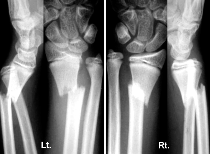
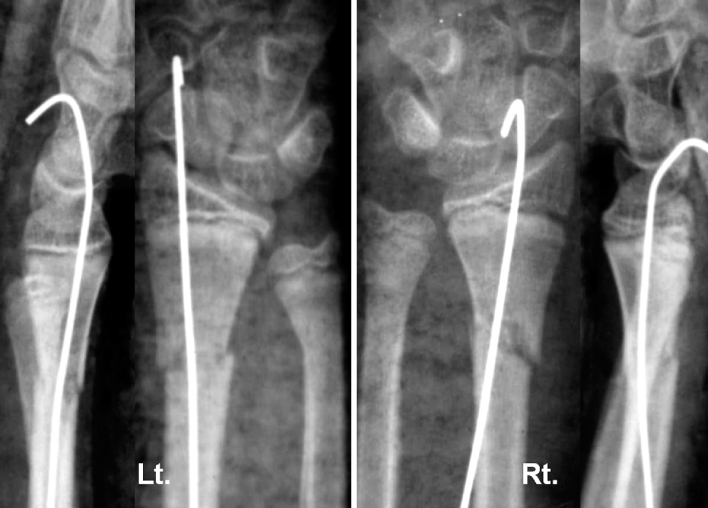
Fig. 3Radiographs taken at 4 weeks postoperatively after removal of K-wire and cast showed callus formation of fractures of distal radius, but showed angulation of distal radius and volar subluxation of ulnar head.

Fig. 4Radiographs and photographs taken at the presentation to our hospital 4 months after trauma. Radiographs showed 40 degree-angulated distal radius and dorsal dislocation of ulnar head.
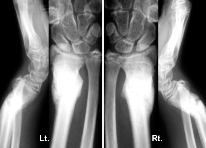
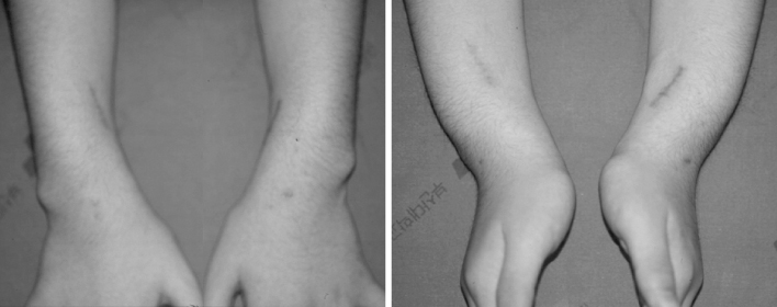
Fig. 6Radiographs after corrective osteotomy and temporary K-wire fixation of distal radioulnar joint showed correction of angular deformity and reduction of ulnar head.
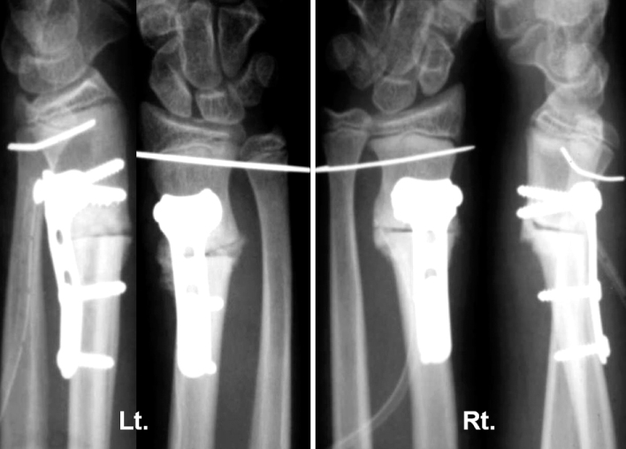
Fig. 7Radiographs taken at 7 months after corrective osteotomy showed well-healed distal radial osteotomy site and reduced distal radioulnar joint.


Figure & Data
REFERENCES
Citations
Citations to this article as recorded by 
 Cite
CiteBilateral Malunion and Distal Radioulnar Joint Dislocation after Operative Treatment of Bilateral Galeazzi Fractures in Child: A Case Report


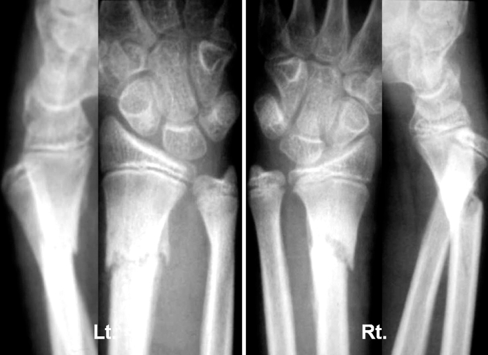


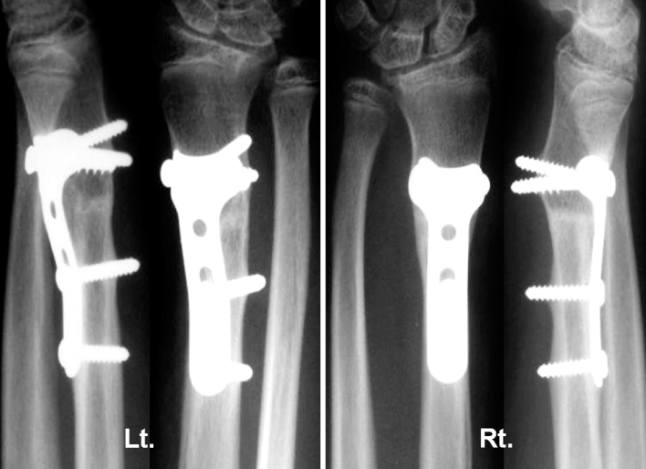
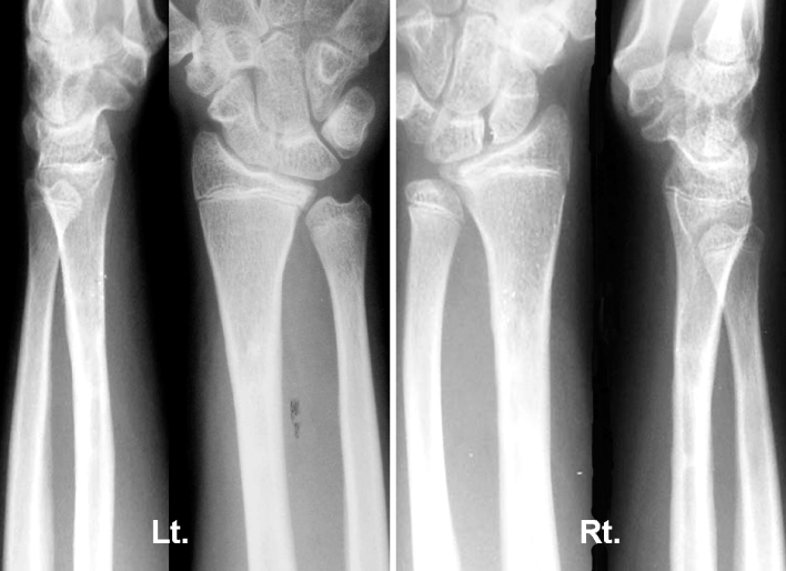
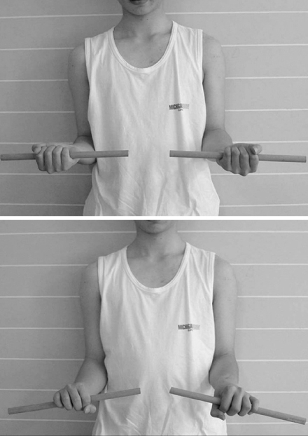
Fig. 1
Initial radiographs after the trauma showed bilateral distal radius fractures and bilateral distal radioulnar joint dislocations.
Fig. 2
Radiographs after limited open reduction and pinning and cast immobilization.
Fig. 3
Radiographs taken at 4 weeks postoperatively after removal of K-wire and cast showed callus formation of fractures of distal radius, but showed angulation of distal radius and volar subluxation of ulnar head.
Fig. 4
Radiographs and photographs taken at the presentation to our hospital 4 months after trauma. Radiographs showed 40 degree-angulated distal radius and dorsal dislocation of ulnar head.
Fig. 5
Photographs showed bowing of both distal forearms.
Fig. 6
Radiographs after corrective osteotomy and temporary K-wire fixation of distal radioulnar joint showed correction of angular deformity and reduction of ulnar head.
Fig. 7
Radiographs taken at 7 months after corrective osteotomy showed well-healed distal radial osteotomy site and reduced distal radioulnar joint.
Fig. 8
Radiographs and photographs taken at 2 years after corrective osteotomy. Radiographs showed correction of angular deformities and maintenance of reduction of distal radioulnar joints.
Fig. 9
Photographs showed normal range of motion of supination and pronation.
Fig. 1
Fig. 2
Fig. 3
Fig. 4
Fig. 5
Fig. 6
Fig. 7
Fig. 8
Fig. 9
Bilateral Malunion and Distal Radioulnar Joint Dislocation after Operative Treatment of Bilateral Galeazzi Fractures in Child: A Case Report

