 E-submission
 TOTA
TOTA TOTS
TOTS
Search
- Page Path
- HOME > Search
Case Report
- Paradoxical hypertrophy as a cause of femoral insufficiency fractures analyzed through differences in force application in Korea: three case reports
- Yong-Uk Kwon, Dae-Hyun Park, Hyoung-Gu Kang
- J Musculoskelet Trauma 2026;39(2):174-180. Published online April 23, 2026
- DOI: https://doi.org/10.12671/jmt.2025.00388
-
 Abstract
 PDF
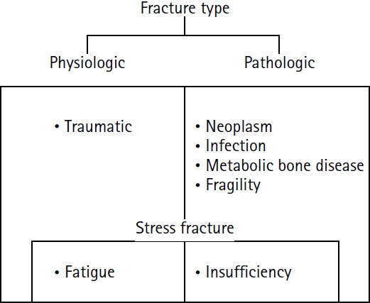
PDF - Previous studies have extensively examined the association between femoral insufficiency fractures and prolonged bisphosphonate therapy. However, alternative etiologies remain insufficiently characterized. This study aimed to analyze nonpharmacologic factors associated with femoral insufficiency fractures, with particular emphasis on paradoxical cortical hypertrophy and altered biomechanical load distribution. We reviewed three cases of femoral insufficiency fracture that were surgically treated at our institution between January 2018 and January 2022. None of the patients had a history of bisphosphonate use. Clinical histories—including underlying comorbidities, prior surgical procedures, and radiographic findings—were evaluated. Serial radiographs obtained before and after fracture occurrence were analyzed to characterize fracture morphology and associated cortical changes. Case 1 involved a patient with posttraumatic hip synostosis; case 2 involved a patient with osteogenesis imperfecta; and case 3 involved a patient who had previously undergone intramedullary nailing for an intertrochanteric fracture. Lateral femoral bowing and cortical hypertrophy preceded fracture development in two cases, whereas focal cortical hypertrophy at the distal locking screw site was observed in the third case. No history of bisphosphonate therapy was identified in any patient. Fractures developed at sites characterized by increased cortical remodeling and abnormal load concentration. Femoral insufficiency fractures can occur in the absence of bisphosphonate therapy. Paradoxical cortical hypertrophy and altered biomechanical force distribution appear to be important contributing factors. Level of evidence: IV.
- 257 View
- 10 Download

Technical Note
- Operative Positioning Technique for an Intertrochanteric Fracture in a Patient with an Ipsilateral Above-the-Knee Amputation - Technical Note -
- Dae-Hyun Park, Yong-Uk Kwon, Dong-Seok Kim
- J Korean Fract Soc 2021;34(4):137-141. Published online October 31, 2021
- DOI: https://doi.org/10.12671/jkfs.2021.34.4.137
-
Abstract
PDF
- A 45-year-old man with a remote history of a left above-the-knee amputation presented to the emergency department with left hip pain after a mechanical fall. This case was an operative challenge because commonly used intraoperative traction methods could not be applied to a patient with an above-the-knee amputation. We describe a rarely utilized surgical technique of applying traction to an amputated extremity via a Steinmann pin during closed reduction and internal fixation of an intertrochanteric fracture.
-
Citations
Citations to this article as recorded by
- Periprosthetic Femur Fractures in Osseointegration Amputees
Jason Shih Hoellwarth, S. Robert Rozbruch
JBJS Case Connector.2022;[Epub] CrossRef
- Periprosthetic Femur Fractures in Osseointegration Amputees
- 1,337 View
- 8 Download
- 1 Crossref
Review Article
- Surgical Treatment for Displaced Intra-Articular Calcaneal Fractures
- Chul Hyun Park, Oog Jin Shon
- J Korean Fract Soc 2016;29(3):221-231. Published online July 31, 2016
- DOI: https://doi.org/10.12671/jkfs.2016.29.3.221
-
Abstract
PDF
- Calcaneal fractures are the most common type of tarsal fracture, and comminuted and bursting fractures are common due to the anatomic characteristics of the calcaneus. Assessment and treatment of calcaneal fractures has improved significantly over time. Despite advancements in surgical techniques and equipment, these fractures remain difficult to treat. In this review article, the physiopathology, classification, and surgical treatments of displaced intra-articular calcaneal fractures are updated.
-
Citations
Citations to this article as recorded by- Current Treatment of Calcaneal Fractures and Dislocation
Dae Jin Nam, Sung Hyun Lee
Journal of the Korean Fracture Society.2022; 35(2): 74. CrossRef
- Current Treatment of Calcaneal Fractures and Dislocation
- 1,934 View
- 20 Download
- 1 Crossref
Case Reports
- Multiple Stress Fractures Related to Low-dose Adefovir Dipivoxil Treatment in a Patient with Chronic Hepatitis B: A Case Report
- Chul Hyun Park, Hyo Sae Ahn, Dong Chul Lee
- J Korean Fract Soc 2014;27(4):327-331. Published online October 31, 2014
- DOI: https://doi.org/10.12671/jkfs.2014.27.4.327
-
Abstract
PDF
- Stress fractures typically result from repeated abnormal mechanical loading to the bones. In particular, multiple stress fractures may occur in patients with systemic disease, such as rheumatoid arthritis, osteoporosis, or osteoarthritis. Adefovir dipivoxil (ADV), a nucleotide analogue of adenosine monophosphate, very rarely causes severe hypophosphatemia when using a low dosage of 10 mg daily for treatment of chronic hepatitis B. To the best of our knowledge, in English literature, this is the first report of multiple stress fractures in a chronic hepatitis B patient who has been treated with a low dosage of ADV. We think it is important to consider that use of ADV in a patient with chronic hepatitis B could be a risk factor for stress fractures.
- 609 View
- 0 Download
- Acute Rupture of Subclavian Artery Pseudoaneurysm after Delayed Osteosynthesis of Clavicular Fracture: A Case Report
- Oog Jin Shon, Jee Hoon Kim, Kang Hyun Park
- J Korean Fract Soc 2014;27(1):82-87. Published online January 31, 2014
- DOI: https://doi.org/10.12671/jkfs.2014.27.1.82
-
Abstract
PDF
- Subclavian vessels are well protected by muscles, fascia and sheaths, so vascular complications associated with clavicular fractures are rare. Pseudoaneurysms after clavicular fractures have been reported, and the occurrence or rupture of pseudoaneurysm has been reported rarely as a late complication. However, cases of pseudoaneurysm after rupture of the clavicular fracture following delayed osteosynthesis of the clavicular fracture have not been reported. A 58-year-old female that presented with a right clavicular shaft fracture obtained conservative treatment. Surgery was performed after 4 months because of non-union in the local medical center. After operation, rupture of the subclavian pseudoaneurysm occurred following osteosynthesis of the clavicular shaft fracture. We report this case here with a review of the literature.
- 759 View
- 1 Download
Original Articles
- Treatment and Prognosis of Femoral Insufficiency Fracture Associated with Prolonged Bisphosphonate Use
- Ki Chan An, Dae Hyun Park, Guemin Gong, Ju Young Kim, Sang Bum Kim, Seung Yeob Sakong
- J Korean Fract Soc 2014;27(1):10-16. Published online January 31, 2014
- DOI: https://doi.org/10.12671/jkfs.2014.27.1.10
-
Abstract
PDF
- PURPOSE
This study was conducted to investigate and identify an appropriate fracture treatment method by analyzing patients in whom a femoral incompetence fracture occurred after receiving a long-term bisphosphonate administration.
MATERIALS AND METHODS
The subjects of this study were 13 cases out of ten patients among those who had a history of receiving bisphosphonate for more than five years and had a fracture or an imminent fracture with a characteristic radiological finding in the femoral subtrochanteric region and the interfemoral region. The period of the drug administration, bone density, the existence of a prodromal symptom, and bilateral fracture were investigated.
RESULTS
In seven out of the 13 cases, the patients complained of painat the femoral and pelvic parts as a prodrome (53.8%), and three of them showed a bilateral fracture (30%). An imminent fracture with a prodrome was observed in six cases (46.2%); for three of these cases, a prophylactic fixture pexis was performed by inserting a metal nail into the medullary cavity, and in two out of these three, a complete fracture was found within 11 months on average (3 to 19 months). In the three prophylactic fixture pexis performed cases, no postoperative complications were found, and a radiological finding of concrescence was seen within one year after the operation. Among the nine operation performed cases after the fracture, non-union was found in two.
CONCLUSION
In the patients who have received bisphosphonate for a long periodof time, a prodome may be a useful indicator of a fracture in the femoral subtrochanteric region and the interfemoral region; therefore, a careful observation is necessary. A prophylactic internal fixation is recommended for patients with imminent fracture with a prodome since they have a high risk of a complete fracture is high in them. -
Citations
Citations to this article as recorded by- Subtrochanteric Fracture Reduction during Intramedullary Nailing: Technical Note
Gyu Min Kong
Journal of the Korean Fracture Society.2019; 32(2): 107. CrossRef
- Subtrochanteric Fracture Reduction during Intramedullary Nailing: Technical Note
- 951 View
- 0 Download
- 1 Crossref
- Fixation of the Femoral Subtrochanteric Fracture with Minimally Invasive Reduction Techniques
- Chul Hyun Park, Chul Wung Ha, Sang Jin Park, Min Su Ko, Oog Jin Shon
- J Korean Fract Soc 2013;26(2):112-117. Published online April 30, 2013
- DOI: https://doi.org/10.12671/jkfs.2013.26.2.112
-
Abstract
PDF
- PURPOSE
To evaluate the results of using minimally invasive reduction techniques in patients with femoral subtrochanteric fracture.
MATERIALS AND METHODS
We retrospectively analyzed 40 patients (41 cases) with subtrochanteric fracture who underwent using minimally invasive reduction techniques. The mean age was 61.4 years (15-89 years), and the mean follow-up period was 32.7 months (12-66 months). Clinical results were assessed using the Parker-Palmer mobility score and the Salvati-Wilson hip functional score. Radiographic results were evaluated using bone union time and femur neck-shaft angle.
RESULTS
No significant difference was observed in the pre- and postoperative Parker-Palmer mobility score. Salvati-Wilson hip functional score showed more than good grade in 37 cases (90%) at the last follow-up. Union was achieved in all 41 cases at an average of 22.5 weeks (18-30 weeks). The mean femoral neck-shaft angle immediately postoperatively was 128.8 degrees (120-140 degrees), and the mean difference versus contralateral sides was 2.5 degrees varus (-6-13 degrees).
CONCLUSION
Fixation of femoral subtrochanteric fracture using minimally invasive reduction techniques showed excellent clinical and radiographic results and low complication rate. -
Citations
Citations to this article as recorded by- Effects of Yuhyangjeongtong-san on Fracture Healing in Rats
Ki-Tae Kim, Na-Young Jo
Journal of Korean Medicine.2019; 40(4): 61. CrossRef - Factors Affecting Time to Bony Union of Femoral Subtrochanteric Fractures Treated with Intramedullary Devices
Jung-Yoon Choi, Yerl-Bo Sung, Jin-Hee Yoo, Sung-Jae Chung
Hip & Pelvis.2014; 26(2): 107. CrossRef
- Effects of Yuhyangjeongtong-san on Fracture Healing in Rats
- 1,164 View
- 9 Download
- 2 Crossref
Case Report
- Tension Band Plating for a Stress Fracture of the Anterior Tibial Cortex in a Basketball Player: A Case Report
- Chul Hyun Park, Woo Chun Lee
- J Korean Fract Soc 2012;25(4):323-326. Published online October 31, 2012
- DOI: https://doi.org/10.12671/jkfs.2012.25.4.323
-
Abstract
PDF
- Stress fractures of the anterior tibial cortex are prone to complete fracture because these stress fractures occur on the tension side of the bone. Recently, surgical treatments are preferred in high-performance athletes requiring rapid return to sports. We report our experience of a case in which stress fracture of the anterior tibial cortex was treated using anterior tension band plating in a male athlete and successful bony union and rapid return to sports were achieved.
-
Citations
Citations to this article as recorded by- Stress fractures of the tibia
Jung Min Park, Ki Sun Sung
Arthroscopy and Orthopedic Sports Medicine.2015; 2(2): 95. CrossRef
- Stress fractures of the tibia
- 1,345 View
- 6 Download
- 1 Crossref
Original Article
- Comparison of Results of Minimally Invasive Plate Osteosynthesis according to Types of Locking Plate in Distal Femoral Fractures
- Oog Jin Shon, Moon Soo Kwon, Chul Hyun Park
- J Korean Fract Soc 2012;25(4):269-276. Published online October 31, 2012
- DOI: https://doi.org/10.12671/jkfs.2012.25.4.269
-
Abstract
PDF
- PURPOSE
To compare results of minimally invasive plate osteosynthesis using a locking compression plate and a periarticular locking plate in distal femur fractures.
MATERIALS AND METHODS
We retrospectively reviewed 31 consecutive femoral fractures who treated by minimally invasive plate osteosynthesis from April 2006 to May 2009. Sixteen patients were treated using a locking compression plate (group A) and 15 patients were treated using a periarticular locking plate (group B).
RESULTS
The mean operation time was 78 minutes and 76 minutes (p=0.273), and the mean radiation exposure time was 1.9 minutes and 2.3 minutes (p=0.001) in the group A and B, respectively. The plate bending during operation was performed in 4 cases of group A. The knee range of motion was 117.5degrees and 118.2degrees (p=0.825), and the Lysholm score was 81.3 and 81.8 (p=0.723) in the group A and B, respectively. Schazker criteria showed more than good grade in 93.8% of group A and in 93.3% of group B (p=1.0).
CONCLUSION
No significant differences in clinical results were observed between the two groups. However, a lower anatomical compliance was showed in the locking compression plate, and a higher risk of radiation exposure was showed in the periarticular locking plate. -
Citations
Citations to this article as recorded by- Incidence of nonunion after surgery of distal femoral fractures using contemporary fixation device: a meta‐analysis
Byung-Ho Yoon, In Keun Park, Youngwoo Kim, Hyoung-Keun Oh, Suk Kyu Choo, Yerl-Bo Sung
Archives of Orthopaedic and Trauma Surgery.2021; 141(2): 225. CrossRef - The Mid-Term Result after Osteosynthesis of Intra-Articular Fractures of Distal Femur
Sam Guk Park, Jeong Jae Moon, Oog Jin Shon
Journal of the Korean Fracture Society.2016; 29(4): 242. CrossRef
- Incidence of nonunion after surgery of distal femoral fractures using contemporary fixation device: a meta‐analysis
- 1,190 View
- 0 Download
- 2 Crossref
Case Reports
- Acute Compartment Syndrome of the Thigh Caused by Contusion: 4 Cases Report
- Oog Jin Shon, Gi Beom Kim, Chul Hyun Park
- J Korean Fract Soc 2012;25(3):215-218. Published online July 31, 2012
- DOI: https://doi.org/10.12671/jkfs.2012.25.3.215
-
Abstract
PDF
- Acute compartment syndrome of the thigh, which usually occurs in the anterior compartment, is a rare condition. It can have various causes including femur fractures, vessel injury, pseudoaneurysm of the femoral or popliteal artery, and use of anticoagulant. However, there have been few reports of acute compartment syndrome of the thigh without fracture caused by blunt trauma. We report 4 cases of acute compartment syndrome of the thigh without fracture caused by blunt trauma, in which three patients were treated with fasciotomy and a Vacuum-Assisted wound Closure system and the other one had a delayed diagnosis, and eventually underwent above-knee amputation.
-
Citations
Citations to this article as recorded by- A Clinical Case Study of Residual Symptoms after Decompression of Traumatic Compartment Syndrome
Min Jung Ji, Seong Chul Lim, Jae Soo Kim, Hyun Jong Lee, Yun Kyu Lee
The Acupuncture.2015; 32(3): 197. CrossRef - Clinical Outcomes of Fasciotomy for Acute Compartment Syndrome
Ji Yong Park, Young Chang Kim, Ji Wan Kim
Journal of the Korean Fracture Society.2015; 28(4): 223. CrossRef
- A Clinical Case Study of Residual Symptoms after Decompression of Traumatic Compartment Syndrome
- 1,135 View
- 2 Download
- 2 Crossref
- Delayed Foreign-body Reaction of Ankle Fracture Treated with a Biodegradable Plate and Screws: A Case Report
- Chul Hyun Park, Dae Hyun Song, Jae Ho Cho
- J Korean Fract Soc 2012;25(2):142-145. Published online April 30, 2012
- DOI: https://doi.org/10.12671/jkfs.2012.25.2.142
-
Abstract
PDF
- Biodegradable implants made of co-polymers composed of L-lactide, D-lactide, and trimethylene carbonate were used in the present case. To our knowledge, only one reported tissue reaction has been associated with ankle fracture treated with third-generation implants internationally and none yet domestically. We report a delayed foreign-body reaction of ankle fracture treated with a third-generation biodegradable plate and screws. We suggest that ankle fracture patients treated with biodegradable implants should be advised of this possible complication and should be followed for at least 2 years.
- 983 View
- 5 Download
Original Articles
- Comparison of Plate Versus Threaded K-wire for Fixation of Midshaft Clavicular Fractures
- Young Jin Ko, Chul Hyun Park, Oog Jin Shon, Jae Sung Seo
- J Korean Fract Soc 2012;25(2):123-128. Published online April 30, 2012
- DOI: https://doi.org/10.12671/jkfs.2012.25.2.123
-
Abstract
PDF
- PURPOSE
To compare clinical outcomes of the plate and threaded K-wire for fixation of midshaft clavicular fractures.
MATERIALS AND METHODS
From 2005 Jan to 2009 May, medical records of 18 patients who underwent open reduction and internal fixation with plate (group 1) and 13 others who underwent intramedullary fixation with threaded K-wire (group 2) were reviewed. The mean follow up periods were 21.9 and 18.9months. The Functional results were evaluated with The Disabilities of the Arm, Shoulder and Hand (DASH) score and Constant shoulder score. The statistical evaluation was assessed with Paired T-test, Chi-square test.
RESULTS
The DASH score were 11.5+/-2.7 in group 1 and 12.4+/-4.3 in group 2. The constant shoulder score were 92.0+/-3.1 in group 1 and 87.1+/-2.8 in group 2. Length of surgical wound (cm) were 10.6+/-3.4 in group 1 and 4.8+/-1.5 in group 2. Postoperative pain and range of motion change were superior in group 1.
CONCLUSION
There was no significant difference between the two groups in functional and radiological results. But, there were patient's complaints about length of surgical wound in group 1 and hardware irritation in group 2. -
Citations
Citations to this article as recorded by- A Comparison between Minimally Invasive Percutaneous Plate Osteosynthesis and Plate Fixation in the Treatment of Clavicle Midshaft Fracture
Seong-Ho Yoo, Suk-Woong Kang, Bu-Hwan Kim, Moo-Ho Song, Yeong-Joon Kim, Gyu-Taek Park, Chang-Hun Kwack
Journal of the Korean Orthopaedic Association.2017; 52(1): 1. CrossRef - Plate fixation versus intramedullary fixation for midshaft clavicle fractures: Meta-analysis of complications and functional outcomes
Hao Xiao, Hengbo Gao, Tuokang Zheng, Jianhui Zhao, Yingping Tian
Journal of International Medical Research.2016; 44(2): 201. CrossRef - Meta-analysis of plate fixation versus intramedullary fixation for the treatment of mid-shaft clavicle fractures
Bing Zhang, Yanbin Zhu, Fei Zhang, Wei Chen, Ye Tian, Yingze Zhang
Scandinavian Journal of Trauma, Resuscitation and Emergency Medicine.2015;[Epub] CrossRef
- A Comparison between Minimally Invasive Percutaneous Plate Osteosynthesis and Plate Fixation in the Treatment of Clavicle Midshaft Fracture
- 1,072 View
- 3 Download
- 3 Crossref
- Minimally Invasive Anterior Plating of Humeral Shaft Fractures
- Hyun Joo Lee, Chang Wug Oh, Do Hyung Kim, Kyung Hyun Park
- J Korean Fract Soc 2011;24(4):341-346. Published online October 31, 2011
- DOI: https://doi.org/10.12671/jkfs.2011.24.4.341
-
Abstract
PDF
- PURPOSE
We evaluated the efficacy and results of minimally invasive anterior plating for humeral shaft fracture.
MATERIALS AND METHODS
Twenty-two cases of humeral shaft fracture were reviewed, including 8 cases of type A, 8 of type B and 6 of type C (AO/OTA classification). There were three open fractures. The fracture was fixed with MIPO (minimally invasive plate osteosynthesis) technique under C-arm guide. A locking compression plate was located in anterior aspect of the humerus with at least three screws fixed in each fragment. Radiologic and functional results were evaluated.
RESULTS
In 20 of 22 cases, bony union was achieved with the mean period of 17.5 weeks, including 2 cases of delayed union. There were 2 cases of nonunion, which needed the further operative procedure. Except one case of distal 1/3 fracture, all cases showed satisfactory elbow and shoulder function with the mean Mayo elbow score of 17.4 and mean UCLA shoulder score of 97.3. In complication, there was one case of radial nerve palsy due to improper traction, but it was completely improved after 3 months. Otherwise, there was no complication including infection.
CONCLUSION
Anterior MIPO for humeral shaft fracture may be another option of operative methods with high union and low complication rate. -
Citations
Citations to this article as recorded by- Minimal Invasive Plate Osteosynthesis versus Conventional Open Plating in Simple Humeral Shaft Fracture (AO Type A, B1, B2)
Boseon Kim, GwangChul Lee, Hyunwoong Jang
Journal of the Korean Fracture Society.2017; 30(3): 124. CrossRef - Clinical and Radiographical Follow-up for Residual Displacement of Fracture Fragments after Interlocking Intramedullary Nailing in Humeral Shaft Fractures
Jae-Kwang Yum, Dong-Ju Lim, Eui-Yub Jung, Su-Een Sohn
The Journal of the Korean Shoulder and Elbow Society.2013; 16(2): 107. CrossRef - Operative Treatment of Humerus Shaft Fracture: Conventional Open Plating or Minimally Invasive Plate Osteosynthesis
Hyun-Joo Lee, Chang-Wug Oh
Journal of the Korean Fracture Society.2012; 25(2): 155. CrossRef
- Minimal Invasive Plate Osteosynthesis versus Conventional Open Plating in Simple Humeral Shaft Fracture (AO Type A, B1, B2)
- 1,301 View
- 22 Download
- 3 Crossref
- Relationship between Lamina Fractures and Dural Tear in Low Lumbar Burst Fractures
- Ki Chan An, Dae Hyun Park, Yong Wook Kwon
- J Korean Fract Soc 2011;24(3):256-261. Published online July 31, 2011
- DOI: https://doi.org/10.12671/jkfs.2011.24.3.256
-
Abstract
PDF
- PURPOSE
To investigate the relationship between the greenstick laminar fractures and the dural tear in low lumbar burst fractures and their optimal treatment.
MATERIALS AND METHODS
We enrolled 51 patients (52 cases) who had been diagnosed with low lumbar burst fracture from June 2003 to May 2007. The average age was 39 years (range, 22 to 58), 30 male patients (58.8%), and 21 female patients (41.2%). Average follow-up periods was 19 months (range, 11 to 45). Lumbar CT scan were taken 1 mm slices in precision for all patients. We judged it incomplete fracture if lumbar CT scans show loss of cortical continuity over 3 slices if there is an aggrement of two among one radiologist and two orthopaedic surgeons reached a consensus. Dural tear and entrapment of nerve root were confirmed intraoperatively by the senior surgeon.
RESULTS
In 52 burst fractures, complete lamina fractures occurred in 21 cases and there were green stick laminar fractures in 14 cases. Neurologic defect has been found in 12 cases, 5 (63%) from complete laminar fractures and 3 (37%) from green stick laminar fractures. Dural tears has been detected in 9 cases (26%), 4 (19%) from complete laminar fractures and 5 (36%) from green stick laminar fractures.
CONCLUSION
Dural tear and nerve root entrapment can be accompanied in patients with green stick fracture. There is necessary to consider the possibility of dural tear and nerve root entrapment before operation and to indentify carefully to the presence of nerve root entrapment during operation. -
Citations
Citations to this article as recorded by- Risk factors for damage to the dura mater in thoracic and lumbar spine injury
A. G. Martikyan, A. A. Grin, A. E. Talypov, S. L. Arakelyan
Hirurgiâ pozvonočnika (Spine Surgery).2022; 19(1): 31. CrossRef - Clinical Efficacy of Large-Channel Percutaneous Lumbar Endoscopic Decompression in the Treatment of Lumbar Spinal Stenosis Secondary to Old Compression Fractures
Junlin Liu, Qingquan Kong, Walter Munesu Chirume, Pin Feng, Bin Zhang, Junsong Ma, Yuan Hu
World Neurosurgery.2022; 166: e118. CrossRef - Diagnostics, pathogenesis and treatment of damage to the dura mater in spinal injury
A. G. Martikyan, A. A. Grin
Russian journal of neurosurgery.2018; 20(2): 74. CrossRef
- Risk factors for damage to the dura mater in thoracic and lumbar spine injury
- 1,152 View
- 2 Download
- 3 Crossref
- Comparison of Results of Tension Band Wire and Hook Plate in the Treatment of Unstable Fractures of the Distal Clavicle
- Chul Hyun Park, Oog Jin Shon, Jae Sung Seo
- J Korean Fract Soc 2011;24(1):55-59. Published online January 31, 2011
- DOI: https://doi.org/10.12671/jkfs.2011.24.1.55
-
Abstract
PDF
- PURPOSE
To compare the clinical and radiological outcomes of two surgical methods with tension band wire and Hook plate for unstable distal clavicle fractures.
MATERIALS AND METHODS
Thirty patients with type II distal clavicle fractures were evaluated, who were operated with tension band wire (Group I) and Hook plate (Group II) fixation, from June 2005 to June 2009, and could be followed-up for more than 1 year after operation. The reduction and union were evaluated by the immediate post-operative and final radiographs. The functional outcome was evaluated by Kona's system and Constant-Murley scoring system.
RESULTS
All 30 cases showed bony union. By Kona's functional evaluation, there were 16 cases with excellent and good results in Group I and 14 cases in Group II. The average Constant score was 88.3 (71~100) in Group I and 89.6 (72~100) in Group II, but there was no significant difference in both groups. As complications, there were 2 case with subacromial impingement, and 1 case showed subacromial erosion. There was no K-wire migration, deep infection and acromioclavicular joint arthritis.
CONCLUSION
Tension band and Hook plate fixation technique gave satisfactory clinical and radiological results in patients with type II distal clavicle fractures. These results suggest that tension band wire and Hook plate fixation technique seems to be an effective method for type II distal clavicle fracture. But we think thal early removal of plate is necessary due to risks for subacromial impingement and erosion in Hook plate fixation. -
Citations
Citations to this article as recorded by- Hook Plate Fixation for Unstable Distal Clavicle Fractures: A Prospective Study
Kyung-Cheon Kim, Hyun-Dae Shin, Soo-Min Cha, Yoo-Sun Jeon
The Journal of the Korean Shoulder and Elbow Society.2011; 14(1): 6. CrossRef
- Hook Plate Fixation for Unstable Distal Clavicle Fractures: A Prospective Study
- 1,166 View
- 6 Download
- 1 Crossref
Case Report
- Ipsilateral Femoral Segmental and Tibial Fractures : A Case Report
- Oog Jin Sohn, Chul Hyun Park, Sang Keun Bae
- J Korean Fract Soc 2009;22(3):193-196. Published online July 31, 2009
- DOI: https://doi.org/10.12671/jkfs.2009.22.3.193
-
Abstract
PDF
- The ipsilateral femoral segmental and tibial fractures seldom occur such as traffic accidents needed high energy mechanisms. For these fractures, surgical stabilization and early mobilization of joint produce can be the best clinical outcomes. We have experienced a case of ipsilateral femoral segmental and tibial fracture and gained good clinical results with surgical treatment. We have reported here on this case and included a review of the relevant literature.
-
Citations
Citations to this article as recorded by- Clinical Outcome after Treatment of Tibia Segmental Fracture with Intramedullary Nailing and Minimal Invasive Plate Osteosynthesis
Jun Young Lee, Hyung Seok Park, Dong Hyuk Cha
Journal of the Korean Fracture Society.2020; 33(3): 142. CrossRef
- Clinical Outcome after Treatment of Tibia Segmental Fracture with Intramedullary Nailing and Minimal Invasive Plate Osteosynthesis
- 1,316 View
- 11 Download
- 1 Crossref
Original Articles
- Treatment of Femoral Shaft Fractures with External Fixators in Children
- Ha Yong Kim, Jong Hyun Park, Seung Hun Lee, Kap Jung Kim, Kwang Won Lee, Byung Sung Kim, Won Sik Choy
- J Korean Soc Fract 2002;15(1):36-44. Published online January 31, 2002
- DOI: https://doi.org/10.12671/jksf.2002.15.1.36
-
Abstract
PDF
- PURPOSE
This study was to assess the amount of overgrowth and convenience after external fixation of pediatric femoral fracture.
MATERIALS AND METHODS
Followed-up more than 18 months were 20 childrens treated with external fixator for femoral fracture(mean follow-up periods: 25.5 months). Mean age was 7.15 years(range: 4-11 years). End to end apposition was done on the closed reduction. Evaluation of the result was done with five parameters; clinical results, radiological results, parents`satisfaction with questionnaire, hospital fee and complications.
RESULTS
Clincal results were not any disability in all cases. No angulation deformity was estimated in all cases, and overgrowth was estimated average 4.8mm (range: -1 ~ 13mm). Answer for questionnaire was revealed satisfactory result. Total hospital fee was average 831 thousand won in external fixator group, and average 289 thousand won in treated group with cast.
CONCLUSION
We propose that external fixation in closed femoral shaft fractures of children could be a rational alternative mode of therapy, because it has excellent clinical & radiological results and parents were satisfied with its convenience & final results. Total hospital fee was statistically higher in external fixator group. -
Citations
Citations to this article as recorded by- Comparison of Flexible Intramedullary Nailing with External Fixation in Pediatric Femoral Shaft Fractures
Do-Young Kim, Sung-Ryong Shin, Un-Seob Jeong, Yong-Wook Park, Sang-Soo Lee, Keun-Min Park
The Journal of the Korean Orthopaedic Association.2008; 43(1): 30. CrossRef - WDM-PON upstream transmission using Fabry–Perot laser diodes externally injected by polarization-insensitive spectrum-sliced supercontinuum pulses
Yang Jing Wen, Chang-Joon Chae
Optics Communications.2006; 260(2): 691. CrossRef
- Comparison of Flexible Intramedullary Nailing with External Fixation in Pediatric Femoral Shaft Fractures
- 964 View
- 5 Download
- 2 Crossref
- Clinical Study on Operative Technique and Management of Tibial condylar Fractures
- Su Chan Lee, Beom Koo Lee, Do Hyun Moon, Jin Hong Ko, Young Kyu Kim, Hong Ki Park, Jun MO Jung, Hyun Park
- J Korean Soc Fract 1998;11(3):533-539. Published online July 31, 1998
- DOI: https://doi.org/10.12671/jksf.1998.11.3.533
-
Abstract
PDF
- We reviewed fifteen cases of tibial condylar fractures, especially, Schatzker type VI treated with hybrid method from January 1995 to May 1997. We attained satisfactory bony union in all cases. There were not serious complications such as deep wound infection and severe angular deformity, but partial ankylosis. After operation, the patients could do knee motion exercise immediately and had no difficulty in getting maintenance of reduction and fracture healing. In conclusions, the hybrid method is an excellent treatment in soft tissue care, maintenance of reduction, and early ambulation and fracture healing in the cases of tibial condylar fractures.
- 613 View
- 0 Download
- Fracture around Hip Joint Combined with Ipsilateral Femoral Shaft Fracture
- Sung Kwan Whang, Jung Ho Rha, Jin Rok Oh, Young Hyun Park
- J Korean Soc Fract 1997;10(4):746-754. Published online October 31, 1997
- DOI: https://doi.org/10.12671/jksf.1997.10.4.746
-
Abstract
PDF
- Fracture around hip joint combined with ipsilateral femoral shaft fracture is relatively uncommon injury and usually result from high-energy trauma. In many of cases, this fracture accompanies with multiple fractures and may be unrecognized the fracture around hip joint. The mechanism of this fracture is that the force on knee joint breaks femoral shaft, and then, when adductive position of femoral shaft, the remained force dislocates hip joint or breaks acetabulum, when abductive position of the shaft, the force breaks femoral neck or intertrochanter. The treatment methods of this fracture are many, but there is no choice of treatment. So, when we select method of treatment, we must consider patients all situations(patients age, pattern of fracture, qulality of bone, ability of surgeon, etc.). The purpose of this study is to make the algorithim of the selection of treatment method for rracture around hip joint combined with ipsilateral femoral shaft fracture. We reviewed 37 cases of fracture around hip joint combined with iplilateral femoral shaft fracture from Febrary 1978 to June 1996. The minimal follow-up period was 1 year. From the review, we made the algorithm of the selection of treatment method for fracture around hip joint combined with ipsilaterl femoral shaft fracture.
- 567 View
- 2 Download
- Management of Unstable Proximal Tibial Fractures Using the Ilizarov
- Hong Gee Park, Beom Goo Lee, Soo Chan Lee, Do Hyun Moon, Jin Hong Ko, Ki Dong Kang, Hyun Park
- J Korean Soc Fract 1997;10(2):332-337. Published online April 30, 1997
- DOI: https://doi.org/10.12671/jksf.1997.10.2.332
-
Abstract
PDF
- We reviewed fifteen cases of unstable tibial fractures treated with Ilizarov method from May 1995 to May 1996. We attained satisfactory bony union in all cases without bone graft(Average time 19 weeks). There were numbers of complications, such as pin tract infection, angular deformity and joint ankylosis but its were soluble and careful management & numbers of minor surgery were needed to prevent & solve such complications. Post-op immediate weight bearing and ROM exercise were possible and showed no difficulty in getting mainteance of reduction & fracture healing, and serious joint ankylosis waa not developed. In conclusions, Ilizarov method is an excellent treatment in getting reduction, maintenance of reduction, early ambulation and fracture healing in the cases of unstable tibia fractures.
- 616 View
- 3 Download
- Chronic isolated distal radioulnar dislocation in child a case report
- Choong Gil Lee, Jin Woo Kwon, Soo Yong Kim, Jae Hyun Park
- J Korean Soc Fract 1993;6(1):165-168. Published online May 31, 1993
- DOI: https://doi.org/10.12671/jksf.1993.6.1.165
- 663 View
- 1 Download
- Clinical analysis of fixation failure in the treatment of intertro chanteric fracture of the femur
- Jong Deuk Rha, Young Hoon Kim, Sung Il Yoon, Jun Soon Kang, Duck Hyun Park
- J Korean Soc Fract 1991;4(2):298-305. Published online November 30, 1991
- DOI: https://doi.org/10.12671/jksf.1991.4.2.298
- 565 View
- 0 Download
 First
First Prev
Prev

