 E-submission
 TOTA
TOTA TOTS
TOTS
Articles
- Page Path
- HOME > J Musculoskelet Trauma > Volume 27(1); 2014 > Article
-
Original Article
- Treatment and Prognosis of Femoral Insufficiency Fracture Associated with Prolonged Bisphosphonate Use
- Ki Chan An, M.D., Dae Hyun Park, M.D., Guemin Gong, M.D., Ju-Young Kim, M.D., Sang-Bum Kim, M.D., Seung-Yeob Sakong, M.D.
-
Journal of the Korean Fracture Society 2014;27(1):10-16.
DOI: https://doi.org/10.12671/jkfs.2014.27.1.10
Published online: January 17, 2014
Department of Orthopedic Surgery, Busan Paik Hospital, Inje University College of Medicine, Busan, Korea.
- Address reprint requests to: Dae Hyun Park, M.D. Department of Orthopedic Surgery, Busan Paik Hospital, Inje University College of Medicine, 75 Bokji-ro, Busanjin-gu, Busan 614-735, Korea. Tel: 82-51-890-6257, Fax: 82-51-892-6619, ronin211@naver.com
• Received: April 10, 2013 • Revised: July 2, 2013 • Accepted: September 15, 2013
Copyright © 2014 The Korean Fracture Society. All rights reserved.
This is an Open Access article distributed under the terms of the Creative Commons Attribution Non-Commercial License (http://creativecommons.org/licenses/by-nc/3.0/) which permits unrestricted non-commercial use, distribution, and reproduction in any medium, provided the original work is properly cited.
- 949 Views
- 0 Download
- 1 Crossref
Abstract
-
Purpose
- This study was conducted to investigate and identify an appropriate fracture treatment method by analyzing patients in whom a femoral incompetence fracture occurred after receiving a long-term bisphosphonate administration.
-
Materials and Methods
- The subjects of this study were 13 cases out of ten patients among those who had a history of receiving bisphosphonate for more than five years and had a fracture or an imminent fracture with a characteristic radiological finding in the femoral subtrochanteric region and the interfemoral region. The period of the drug administration, bone density, the existence of a prodromal symptom, and bilateral fracture were investigated.
-
Results
- In seven out of the 13 cases, the patients complained of painat the femoral and pelvic parts as a prodrome (53.8%), and three of them showed a bilateral fracture (30%). An imminent fracture with a prodrome was observed in six cases (46.2%); for three of these cases, a prophylactic fixture pexis was performed by inserting a metal nail into the medullary cavity, and in two out of these three, a complete fracture was found within 11 months on average (3 to 19 months). In the three prophylactic fixture pexis performed cases, no postoperative complications were found, and a radiological finding of concrescence was seen within one year after the operation. Among the nine operation performed cases after the fracture, non-union was found in two.
-
Conclusion
- In the patients who have received bisphosphonate for a long periodof time, a prodome may be a useful indicator of a fracture in the femoral subtrochanteric region and the interfemoral region; therefore, a careful observation is necessary. A prophylactic internal fixation is recommended for patients with imminent fracture with a prodome since they have a high risk of a complete fracture is high in them.
- 1. Arkan SSN, Bakir KK, Göran OS. Bisphosphonate-induced femoral fragility fractures: what do we know? Orthop Res Rev, 2010;2:27-34.
- 2. Banffy MB, Vrahas MS, Ready JE, Abraham JA. Nonoperative versus prophylactic treatment of bisphosphonate-associated femoral stress fractures. Clin Orthop Relat Res, 2011;469:2028-2034.ArticlePubMedPMC
- 3. Boivin G, Meunier PJ. Changes in bone remodeling rate influence the degree of mineralization of bone. Connect Tissue Res, 2002;43:535-537.Article
- 4. Currey JD. Effects of differences in mineralization on the mechanical properties of bone. Philos Trans R Soc Lond B Biol Sci, 1984;304:509-518.ArticlePubMedPDF
- 5. Fowler JR, Craig MR. Association of low-energy femoral shaft fractures and bisphosphonate use. Orthopedics, 2012;35:e38-e40.ArticlePubMed
- 6. Gates BJ, Sonnett TE, Duvall CA, Dobbins EK. Review of osteoporosis pharmacotherapy for geriatric patients. Am J Geriatr Pharmacother, 2009;7:293-323.Article
- 7. Girgis CM, Sher D, Seibel MJ. Atypical femoral fractures and bisphosphonate use. N Engl J Med, 2010;362:1848-1849.Article
- 8. Isaacs JD, Shidiak L, Harris IA, Szomor ZL. Femoral insufficiency fractures associated with prolonged bisphosphonate therapy. Clin Orthop Relat Res, 2010;468:3384-3392.Article
- 9. Kwek EB, Goh SK, Koh JS, Png MA, Howe TS. An emerging pattern of subtrochanteric stress fractures: a long-term complication of alendronate therapy? Injury, 2008;39:224-231.Article
- 10. Leslie WD, O'Donnell S, Jean S, et al. Osteoporosis Surveillance Expert Working Group. Trends in hip fracture rates in Canada. JAMA, 2009;302:883-889.Article
- 11. Luckman SP, Hughes DE, Coxon FP, Graham R, Russell G, Rogers MJ. Nitrogen-containing bisphosphonates inhibit the mevalonate pathway and prevent post-translational prenylation of GTP-binding proteins, including Ras. J Bone Miner Res, 1998;13:581-589.ArticlePDF
- 12. Morris CD, Einhorn TA. Bisphosphonates in orthopaedic surgery. J Bone Joint Surg Am, 2005;87:1609-1618.Article
- 13. Odvina CV, Zerwekh JE, Rao DS, Maalouf N, Gottschalk FA, Pak CY. Severely suppressed bone turnover: a potential complication of alendronate therapy. J Clin Endocrinol Metab, 2005;90:1294-1301.Article
- 14. Park JG, Song KS, Jung HJ, Lee JS, Lee TJ, Kim KS. Bilateral femoral subtrochanteric insufficiency fractures after long-term bisphosphonate therapy. J Korean Orthop Assoc, 2010;45:146-150.Article
- 15. Puah KL, Tan MH. Bisphosphonate-associated atypical fracture of the femur: spontaneous healing with drug holiday and re-appearance after resumed drug therapy with bilateral simultaneous displaced fractures--a case report. Acta Orthop, 2011;82:380-382.Article
- 16. Rodan GA, Reszka AA. Osteoporosis and bisphosphonates. J Bone Joint Surg Am, 2003;85:Suppl 3. 8-12.Article
- 17. Russell RG, Watts NB, Ebetino FH, Rogers MJ. Mechanisms of action of bisphosphonates: similarities and differences and their potential influence on clinical efficacy. Osteoporos Int, 2008;19:733-759.ArticlePDF
- 18. Shane E, Burr D, Ebeling PR, et al. Atypical subtrochanteric and diaphyseal femoral fractures: report of a task force of the American Society for Bone and Mineral Research. J Bone Miner Res, 2010;25:2267-2294.
- 19. Wasserman N, Yerramshetty J, Akkus O. Microcracks colocalize within highly mineralized regions of cortical bone tissue. Eur J Morphol, 2005;42:43-51.Article
REFERENCES
Fig. 1A 79-year-old woman was diagnosed as subtrochanteric fracture of the right femur after slipping (A). Internal fixation using intramedullary nail (B). Nine months after the surgery, non-union was confirmed (C). Bone grafting (D) union was observed 1 year after the surgery (E).


Fig. 3An 86-year-old woman was diagnosed with left femoral shaft fracture and received internal fixation using intramedullary nail. On arrival, incomplete fracture of femoral shaft and prodromal symptom was confirmed on the other side (A) prophylactic intramedullary nailing was done (B). Bone union was observed 1 year after the surgery (C).
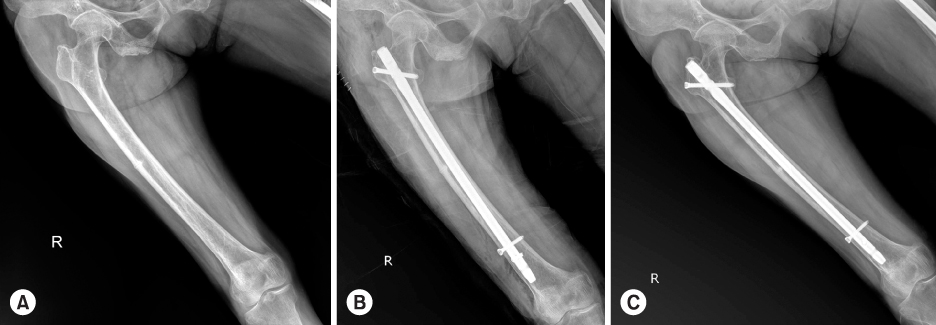
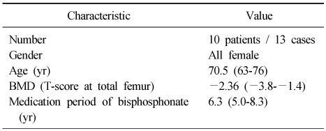
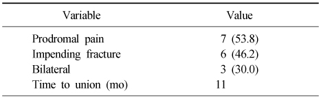
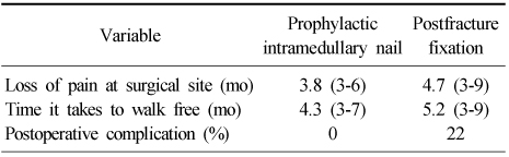
Figure & Data
REFERENCES
Citations
Citations to this article as recorded by 
- Subtrochanteric Fracture Reduction during Intramedullary Nailing: Technical Note
Gyu Min Kong
Journal of the Korean Fracture Society.2019; 32(2): 107. CrossRef
 Cite
CiteTreatment and Prognosis of Femoral Insufficiency Fracture Associated with Prolonged Bisphosphonate Use
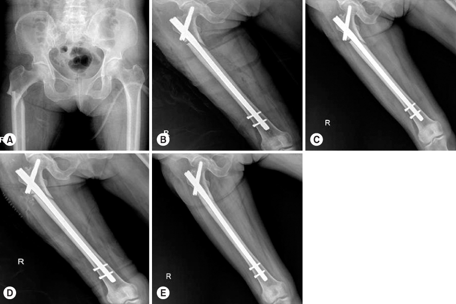
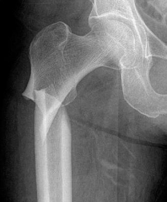

Fig. 1
A 79-year-old woman was diagnosed as subtrochanteric fracture of the right femur after slipping (A). Internal fixation using intramedullary nail (B). Nine months after the surgery, non-union was confirmed (C). Bone grafting (D) union was observed 1 year after the surgery (E).
Fig. 2
Specific radiologic finding.
Fig. 3
An 86-year-old woman was diagnosed with left femoral shaft fracture and received internal fixation using intramedullary nail. On arrival, incomplete fracture of femoral shaft and prodromal symptom was confirmed on the other side (A) prophylactic intramedullary nailing was done (B). Bone union was observed 1 year after the surgery (C).
Fig. 1
Fig. 2
Fig. 3
Treatment and Prognosis of Femoral Insufficiency Fracture Associated with Prolonged Bisphosphonate Use
Baseline Characteristics of the Patient
Values are presented as median (range). BMD: Bone mineral density.
Character of Insufficiency Fracture
Values are presented as number (%) or only number.
Comparison of Prophylactic Fixation and Postfracture Fixation
Values are presented as median (range) or percent.
Table 1
Baseline Characteristics of the Patient
Values are presented as median (range). BMD: Bone mineral density.
Table 2
Character of Insufficiency Fracture
Values are presented as number (%) or only number.
Table 3
Comparison of Prophylactic Fixation and Postfracture Fixation
Values are presented as median (range) or percent.

