 E-submission
 TOTA
TOTA TOTS
TOTS
Articles
- Page Path
- HOME > J Musculoskelet Trauma > Volume 27(1); 2014 > Article
-
Case Report
- Acute Rupture of Subclavian Artery Pseudoaneurysm after Delayed Osteosynthesis of Clavicular Fracture: A Case Report
- Oog-Jin Shon, M.D., Jee-Hoon Kim, M.D., Kang-Hyun Park, M.D.
-
Journal of the Korean Fracture Society 2014;27(1):82-87.
DOI: https://doi.org/10.12671/jkfs.2014.27.1.82
Published online: January 17, 2014
Department of Orthopedic Surgery, Yeungnam University College of Medicine, Daegu, Korea.
- Address reprint requests to: Kang Hyun Park, M.D. Department of Orthopedic Surgery, Yeungnam University College of Medicine, 170 Hyeonchung-ro, Nam-gu, Daegu 705-717, Korea. Tel: 82-53-620-3640, Fax: 82-53-628-4240, propulsing@naver.com
• Received: August 1, 2013 • Revised: September 23, 2013 • Accepted: December 19, 2013
Copyright © 2014 The Korean Fracture Society. All rights reserved.
This is an Open Access article distributed under the terms of the Creative Commons Attribution Non-Commercial License (http://creativecommons.org/licenses/by-nc/3.0/) which permits unrestricted non-commercial use, distribution, and reproduction in any medium, provided the original work is properly cited.
- 657 Views
- 1 Download
Abstract
- Subclavian vessels are well protected by muscles, fascia and sheaths, so vascular complications associated with clavicular fractures are rare. Pseudoaneurysms after clavicular fractures have been reported, and the occurrence or rupture of pseudoaneurysm has been reported rarely as a late complication. However, cases of pseudoaneurysm after rupture of the clavicular fracture following delayed osteosynthesis of the clavicular fracture have not been reported. A 58-year-old female that presented with a right clavicular shaft fracture obtained conservative treatment. Surgery was performed after 4 months because of non-union in the local medical center. After operation, rupture of the subclavian pseudoaneurysm occurred following osteosynthesis of the clavicular shaft fracture. We report this case here with a review of the literature.
The patient's information of this case report was published in Journal of Cardiothoracic Surgery (JCTS). This article received permission from both JCTS and Journal of the Korean Fracture Society.
- 1. Altamimi SA, McKee MD. Canadian Orthopaedic Trauma Society. Nonoperative treatment compared with plate fixation of displaced midshaft clavicular fractures. Surgical technique. J Bone Joint Surg Am, 2008;90:Suppl 2. (Pt 1):1-8.
- 2. Coulier B, Mairy Y, Etienne PY, Joris JP. Late diagnosis of a traumatic pseudo-aneurysm of the subclavian artery. J Belge Radiol, 1996;79:26-28.
- 3. Ding M, Hu J, Ni J, Lv H, Song D, Shu C. Iatrogenic subclavian arteriovenous fistula: rare complication of plate osteosynthesis of clavicle fracture. Orthopedics, 2012;35:e287-e289.Article
- 4. Graham JM, Feliciano DV, Mattox KL, Beall AC Jr, DeBakey ME. Management of subclavian vascular injuries. J Trauma, 1980;20:537-544.Article
- 5. Howard FM, Shafer SJ. Injuries to the clavicle with neurovascular complications. A study of fourteen cases. J Bone Joint Surg Am, 1965;47:1335-1346.
- 6. Johnson B, Thursby P. Subclavian artery injury caused by a screw in a clavicular compression plate. Cardiovasc Surg, 1996;4:414-415.Article
- 7. Pairolero PC, Walls JT, Payne WS, Hollier LH, Fairbairn JF 2nd. Subclavian-axillary artery aneurysms. Surgery, 1981;90:757-763.
- 8. Serrano JA, Rodríguez P, Castro L, Serrano P, Carpintero P. Acute subclavian artery pseudoaneurysm after closed fracture of the clavicle. Acta Orthop Belg, 2003;69:555-557.
- 9. Shackford SR, Connolly JF. Taming of the screw: a case report and literature review of limb-threatening complications after plate osteosynthesis of a clavicular nonunion. J Trauma, 2003;55:840-843.Article
REFERENCES

Fig. 2Preoperative radiograph showing nonunion of the clavicle mid-shaft fracture at 4 months after injury.
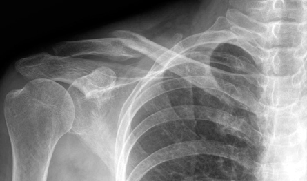
Fig. 3Preoperative T2 magnetic resonance imaging showing a suspicious lesion of the subclavian pseudoaneurysm (white arrows).
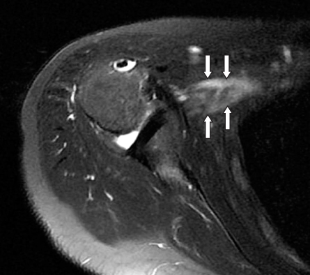
Fig. 4Postoperative radiograph showing nonunion of the clavicle mid-shaft fracture treated using an anatomical plate.

Fig. 5
(A, B) Angio computed tomography showing subclavian pseudoaneurysm (white arrow) and arteriovenous fistula (black arrow). (C) Three-dimensional angio computed tomography showing subclavian pseudoaneurysm (white arrow).
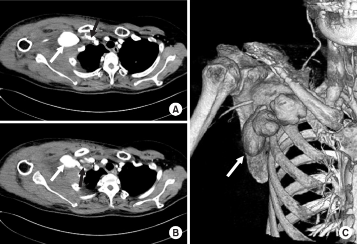
Fig. 6Intraoperative photograph showing the status of clavicular osteotomy. The subclavian artery pseudoaneurysmal sac was removed and an arterial opening (black arrow) was observed.

Fig. 7Postoperative radiograph showing osteosynthesis of the clavicular osteotomy after vessel repair.


Figure & Data
REFERENCES
Citations
Citations to this article as recorded by 
 Cite
CiteAcute Rupture of Subclavian Artery Pseudoaneurysm after Delayed Osteosynthesis of Clavicular Fracture: A Case Report
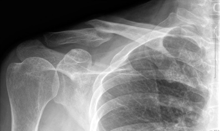


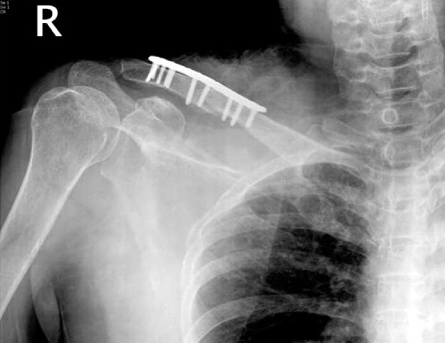

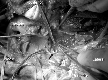
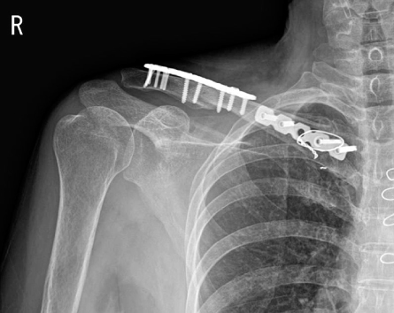
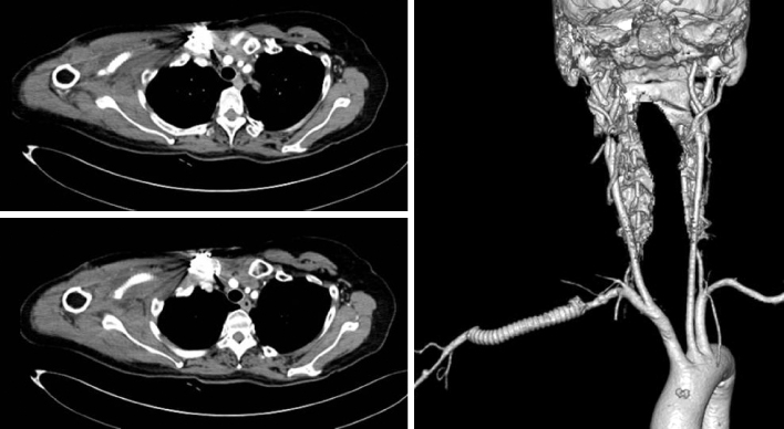
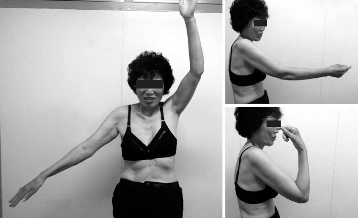
Fig. 1
Initial radiograph showing clavicular mid-shaft fracture.
Fig. 2
Preoperative radiograph showing nonunion of the clavicle mid-shaft fracture at 4 months after injury.
Fig. 3
Preoperative T2 magnetic resonance imaging showing a suspicious lesion of the subclavian pseudoaneurysm (white arrows).
Fig. 4
Postoperative radiograph showing nonunion of the clavicle mid-shaft fracture treated using an anatomical plate.
Fig. 5
(A, B) Angio computed tomography showing subclavian pseudoaneurysm (white arrow) and arteriovenous fistula (black arrow). (C) Three-dimensional angio computed tomography showing subclavian pseudoaneurysm (white arrow).
Fig. 6
Intraoperative photograph showing the status of clavicular osteotomy. The subclavian artery pseudoaneurysmal sac was removed and an arterial opening (black arrow) was observed.
Fig. 7
Postoperative radiograph showing osteosynthesis of the clavicular osteotomy after vessel repair.
Fig. 8
Follow-up 3-dimensional computed tomography angiogram showing good distal blood flow to the axillary artery through a subclavian artery graft.
Fig. 9
Photographs showing recovery state of upper extremities, but with limitation of motion.
Fig. 1
Fig. 2
Fig. 3
Fig. 4
Fig. 5
Fig. 6
Fig. 7
Fig. 8
Fig. 9
Acute Rupture of Subclavian Artery Pseudoaneurysm after Delayed Osteosynthesis of Clavicular Fracture: A Case Report

