 E-submission
 TOTA
TOTA TOTS
TOTS
Articles
- Page Path
- HOME > J Musculoskelet Trauma > Volume 25(4); 2012 > Article
-
Case Report
- Tension Band Plating for a Stress Fracture of the Anterior Tibial Cortex in a Basketball Player: A Case Report
- Chul Hyun Park, M.D., Ph.D., Woo Chun Lee, M.D., Ph.D.
-
Journal of the Korean Fracture Society 2012;25(4):323-326.
DOI: https://doi.org/10.12671/jkfs.2012.25.4.323
Published online: October 19, 2012
Department of Orthopedic Surgery, Seoul Paik Hospital, Inje University College of Medicine, Seoul, Korea.
*Department of Orthopedic Surgery, Yeungnam University College of Medicine, Daegu, Korea.
- Address reprint requests to: Woo Chun Lee, M.D., Ph.D. Department of Orthopedic Surgery, Inje University Seoul Paik Hospital, 9, Mareunnae-ro, Jung-gu, Seoul 100-032, Korea. Tel: 82-2-2270-0028, Fax: 82-2-2270-0023, leewoochun@gmail.com
• Received: January 5, 2012 • Revised: February 16, 2012 • Accepted: April 24, 2012
Copyright © 2012 The Korean Fracture Society
- 1,415 Views
- 7 Download
- 1 Crossref
Abstract
- Stress fractures of the anterior tibial cortex are prone to complete fracture because these stress fractures occur on the tension side of the bone. Recently, surgical treatments are preferred in high-performance athletes requiring rapid return to sports. We report our experience of a case in which stress fracture of the anterior tibial cortex was treated using anterior tension band plating in a male athlete and successful bony union and rapid return to sports were achieved.
- 1. Barrick EF, Jackson CB. Prophylactic intramedullary fixation of the tibia for stress fracture in a professional athlete. J Orthop Trauma, 1992;6:241-244.Article
- 2. Batt ME, Kemp S, Kerslake R. Delayed union stress fractures of the anterior tibia: conservative management. Br J Sports Med, 2001;35:74-77.Article
- 3. Baublitz SD, Shaffer BS. Acute fracture through an intramedullary stabilized chronic tibial stress fracture in a basketball player: a case report and literature review. Am J Sports Med, 2004;32:1968-1972.ArticlePubMedPDF
- 4. Beals RK, Cook RD. Stress fractures of the anterior tibial diaphysis. Orthopedics, 1991;14:869-875.ArticlePubMed
- 5. Boden BP, Osbahr DC. High-risk stress fractures: evaluation and treatment. J Am Acad Orthop Surg, 2000;8:344-353.Article
- 6. Borens O, Sen MK, Huang RC, et al. Anterior tension band plating for anterior tibial stress fractures in high-performance female athletes: a report of 4 cases. J Orthop Trauma, 2006;20:425-430.
- 7. Burrows HJ. Fatigue infraction of the middle of the tibia in ballet dancers. J Bone Joint Surg Br, 1956;38-B:83-94.ArticlePDF
- 8. Choo SK, Oh HK, Choi HW, Song JG. Anterior knee pain after intramedullary nailing for tibial shaft fractures. J Korean Fract Soc, 2011;24:28-32.Article
- 9. Court-Brown CM, Gustilo T, Shaw AD. Knee pain after intramedullary tibial nailing: its incidence, etiology, and outcome. J Orthop Trauma, 1997;11:103-105.Article
- 10. Harmon KG. Lower extremity stress fractures. Clin J Sport Med, 2003;13:358-364.Article
- 11. Rettig AC, Shelbourne KD, McCarroll JR, Bisesi M, Watts J. The natural history and treatment of delayed union stress fractures of the anterior cortex of the tibia. Am J Sports Med, 1988;16:250-255.ArticlePDF
REFERENCES
Fig. 1
(A) Preoperative plain radiographs and (B) computed tomograph of the 13-year-old basketball player show a distinct radiolucency and periosteal thickening in the anterolateral cortex.

Fig. 2Intraoperative photo shows that multiple drilling was performed with a 2.5 mm drill bit after corticotomy.


Figure & Data
REFERENCES
Citations
Citations to this article as recorded by 
- Stress fractures of the tibia
Jung Min Park, Ki Sun Sung
Arthroscopy and Orthopedic Sports Medicine.2015; 2(2): 95. CrossRef
 Cite
CiteTension Band Plating for a Stress Fracture of the Anterior Tibial Cortex in a Basketball Player: A Case Report
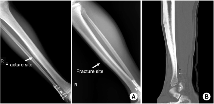
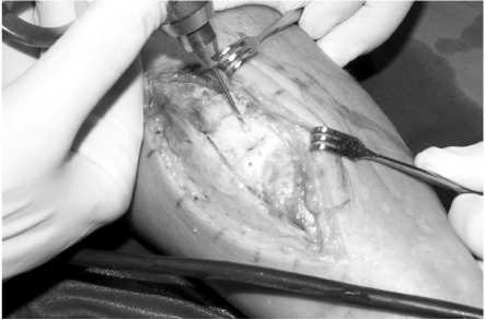
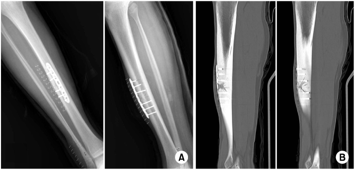
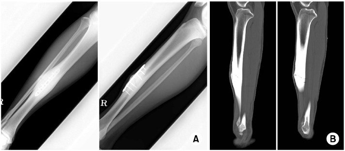
Fig. 1
(A) Preoperative plain radiographs and (B) computed tomograph of the 13-year-old basketball player show a distinct radiolucency and periosteal thickening in the anterolateral cortex.
Fig. 2
Intraoperative photo shows that multiple drilling was performed with a 2.5 mm drill bit after corticotomy.
Fig. 3
(A) Plain radiographs and (B) computed tomograph show the plate fixation with corticotomy and multiple drilling of the fracture site.
Fig. 4
(A) Plain radiographs and (B) computed tomograph at 15 months after surgery show the bony union.
Fig. 1
Fig. 2
Fig. 3
Fig. 4
Tension Band Plating for a Stress Fracture of the Anterior Tibial Cortex in a Basketball Player: A Case Report

