 E-submission
 TOTA
TOTA TOTS
TOTS
Search
- Page Path
- HOME > Search
Original Articles
- Association between decreased bone mineral density and Pauwels angle in femoral neck fractures: a cross-sectional study
- Soo-Hwan Jung, Yong-Uk Kwon, Ji-Hun Park
- J Musculoskelet Trauma 2026;39(1):20-29. Published online January 25, 2026
- DOI: https://doi.org/10.12671/jmt.2025.00269
-
 Abstract
 PDF
PDF Supplementary Material
 - Background
Progressive osteoporosis reduces the trabecular structures of the proximal femur, whereas the primary compression trabeculae (PCTs) are relatively preserved. We hypothesize that the loss of the vertically oriented PCTs in osteoporosis, which act as a mechanical barrier, affects fracture line propagation and influences the Pauwels angle. This study investigated the association between bone mineral density (BMD) and Pauwels angles in low-energy femoral neck fractures (FNFs).
Methods
This cross-sectional study included 150 patients (mean age, 75.3 years; range, 50–94 years) diagnosed with intracapsular FNFs between May 2019 and May 2023. BMD was measured within 1 month of the injury date using dual-energy X-ray absorptiometry, and modified Pauwels angles were assessed using a computed tomography-based multiplanar reconstruction program. Multiple linear regression analysis was performed to evaluate the factors influencing the Pauwels angles. The dependent variable was the Pauwels angle, while the independent variables included sex, age, height, body weight, body mass index, American Society of Anesthesiologists score, Charlson comorbidity index score, smoking status, alcohol use, preinjury walking ability, and femoral neck BMD T-scores.
Results
Higher femoral neck BMD T-scores were significantly associated with increased Pauwels angles (β=3.449, P<0.001). Greater body weight was independently associated with increased Pauwels angles (β=0.213, P=0.007).
Conclusions
The Pauwels angle demonstrated a significant association with BMD, with lower BMD associated with less steep Pauwels angles. In the absence of BMD measurement, the Pauwels angle may indicate osteoporosis severity in patients with low-energy FNFs. Level of evidence: III.
- 778 View
- 21 Download

- Computed tomography plane reformatting to reduce projection error in measuring Pauwels angle of femoral neck fractures: a cross-sectional study
- Gyu Min Kong, Jae-Young Lim, Se-Lin Jeong, Gu-Hee Jung
- J Musculoskelet Trauma 2026;39(1):38-47. Published online January 25, 2026
- DOI: https://doi.org/10.12671/jmt.2025.00038
-
Abstract
PDF
- Objectives
This study aimed to assess fracture verticality in both coronal and axial planes after eliminating projection error in femoral neck fractures among non-older adults, and to demonstrate its clinical utility using computed tomography (CT)-based modeling at actual size.
Methods
This retrospective observational study enrolled 57 patients (30 males and 27 females), aged 20–65 years, with displaced femoral neck fractures. Based on CT images, an actual-size fracture model was constructed. The CT scanning plane was reformatted with the neck-shaft fragment realigned vertically to the ground and parallel to the femoral neck axis. Three consecutive images were used to generate coronal reformats at the centerline and posterior border to measure central and posterior coronal plane verticality as Pauwels’ angle (PA). The central image of the reformatted axial plane was used to assess axial plane verticality. Differences in verticality were analyzed using analysis of variance.
Results
Three coronal morphology types were identified: linear (n=30), concave (n=25), and convex (n=2). Two axial morphology types were observed: cephalad (n=35) and trochanteric (n=22). The mean central PA, posterior PA, and axial verticality were 55.43°±13.79°, 51.44°±11.13°, and 85.74°±18.41°, respectively. Only the central PA showed a significant difference (P<0.001). The PA was significantly higher in the linear coronal type between images (P<0.05) and in the trochanteric axial type (P<0.05).
Conclusions
After reformatting the scanning plane, the central PA showed significant variation between images. Femoral neck fractures of the linear type in the coronal plane and the trochanteric type in the axial plane demonstrated greater verticality than other morphological types. Level of evidence:
- 518 View
- 12 Download
- Comparative results of the femoral neck system versus the dynamic hip screw for stable femoral neck fractures in older adults in Korea: a retrospective cohort study
- Byung-Chan Choi, Byung-Woo Min, Kyung-Jae Lee, Jun-Sik Hong
- J Musculoskelet Trauma 2025;38(4):203-211. Published online October 24, 2025
- DOI: https://doi.org/10.12671/jmt.2025.00276
-
Abstract
PDF
- Background
This study aimed to compare the clinical and radiological outcomes of the femoral neck system (FNS) and the dynamic hip screw (DHS) for the internal fixation of stable femoral neck fractures in older adults.
Methods
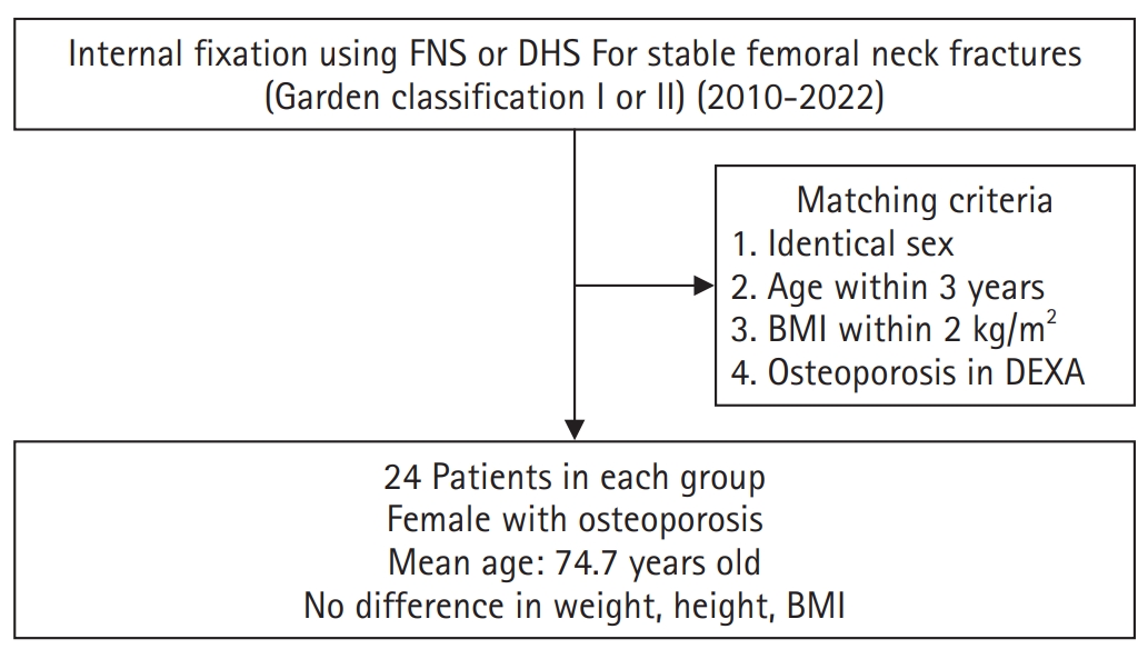
This retrospective cohort study included 48 matched older adult patients based on sex, age, BMI, and osteoporosis status, who had undergone internal fixation with either FNS or DHS for stable femoral neck fractures between January 2010 and December 2022. To minimize selection bias, a 1:1 case-control matching was performed based on sex, age, body mass index (BMI), and the presence of osteoporosis. A total of 48 patients (24 in each group) were included. We compared perioperative data (operation time, hemoglobin change, transfusion rate), functional outcomes using the Koval score, and radiological outcomes, including union rate, femoral neck shortening, and complication rates.
Results
The mean operation time was significantly shorter in the FNS group than in the DHS group (60.9 minutes vs. 70.8 minutes; P=0.007). There were no statistically significant differences between the two groups in the union rate (87.5% in FNS vs. 95.8% in DHS), femoral neck shortening, final Koval score distribution, or overall complication rates (12.5% in both groups).
Conclusions
For treating stable femoral neck fractures in older adults, the FNS demonstrated comparable clinical and radiological outcomes to the DHS, with the distinct advantage of a shorter operation time. While these findings suggest that the FNS is a promising and safe alternative that may reduce the surgical burden, definitive conclusions are precluded by the small sample size, warranting further research to corroborate these results. Level of evidence: IV.
- 2,318 View
- 27 Download
- Risk factors of surgical complications after use of the femoral neck system: a random forest analysis
- Chul-Ho Kim, Hyun-Chul Shon, Han Soul Kim, Ji Wan Kim, Eic Ju Lim
- J Musculoskelet Trauma 2025;38(3):160-167. Published online July 23, 2025
- DOI: https://doi.org/10.12671/jmt.2025.00157
-
Abstract
PDF
- Background
The femoral neck system (FNS), a novel fixation device for managing femoral neck fractures (FNFs), has gained popularity in recent years. However, analyses of the surgical complications and reoperation risks associated with the use of FNS remain limited.
Methods
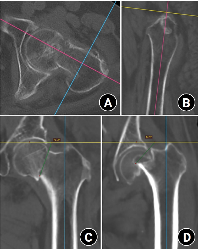
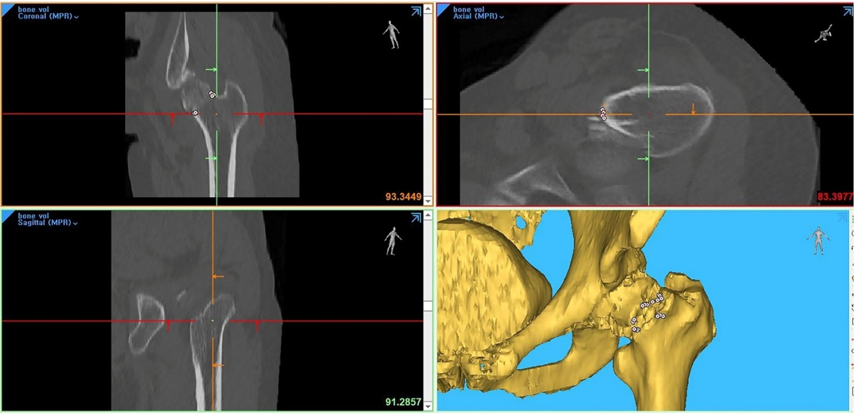
This retrospective observational study analyzed 57 patients who had undergone FNS fixation for FNF at two university hospitals between July 2019 and February 2024. Demographic, perioperative, and outcome variables, including age, sex, fracture classification (Garden, Pauwels, and AO), implant characteristics, tip-apex distance (TAD), neck shortening, and neck-shaft alignment, were analyzed. In addition to univariate analysis, a machine learning analysis was conducted using a random forest classifier with stratified sampling (80% training, 20% testing). The accuracy, precision, recall, F1-score, and area under the receiver’s operating curve were calculated to assess model performance.
Results
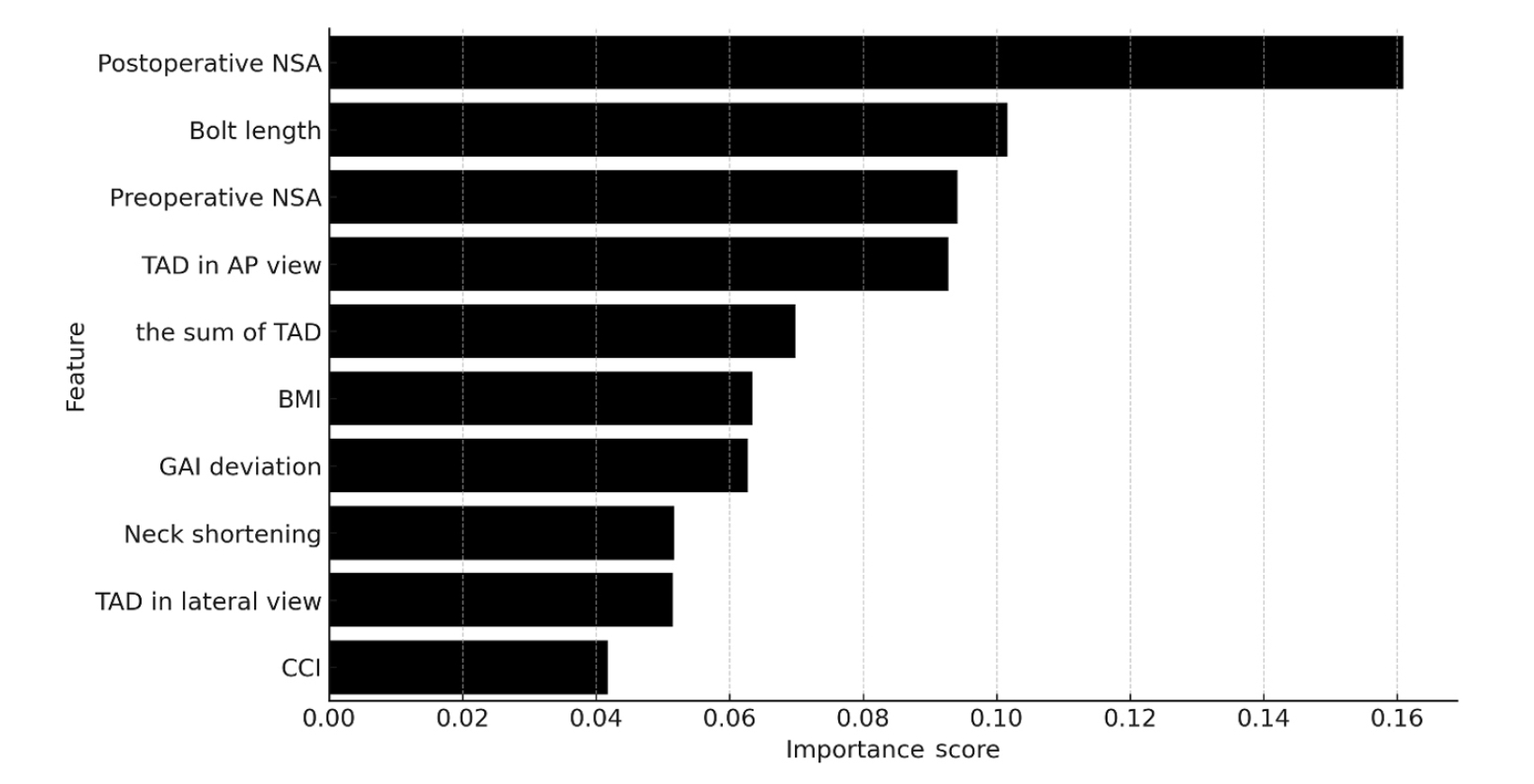
Ten patients experienced osteonecrosis of the femoral head (n=6), implant cut-out or penetration (n=3), and peri-implant fracture (n=1). Univariate analysis revealed that the TAD in the complication group was significantly shorter than that in the control group (12.1 vs. 16.7 mm; P=0.012). Additionally, neck shortening in the complication group was greater than that in the control group (4.9 vs. 2.3 mm; P=0.011). The random forest model achieved an accuracy of 83.3% and identified postoperative neck-shaft angle (NSA) as the most important predictor of complications (feature importance, 0.161), followed by bolt length (0.102) and preoperative NSA (0.094).
Conclusions
Risk factor analysis conducted using a random forest model identified postoperative NSA as the most important feature associated with postoperative complications following FNS. Therefore, care should be taken to normalize the postoperative NSA during FNF surgery. Level of Evidence: III. -
Citations
Citations to this article as recorded by
- Length-stable fixation reduces femoral neck shortening in unstable femoral neck fractures: A retrospective comparative study of length-stable dynamic hip screw versus femoral neck system fixation
Seonghyun Kang, Wonseok Choi, Jeong Seok Choi, Eic Ju Lim, SungJin Ahn, Jong-Keon Oh, William T. Kent, Whee Sung Son, Jae-Woo Cho
Journal of Orthopaedic Surgery.2026;[Epub] CrossRef
- Length-stable fixation reduces femoral neck shortening in unstable femoral neck fractures: A retrospective comparative study of length-stable dynamic hip screw versus femoral neck system fixation
- 1,739 View
- 53 Download
- 1 Crossref
Review Article
- Osteoporotic Hip Fracture: How We Make Better Results?
- Byung-Chan Choi, Kyung-Jae Lee
- J Korean Fract Soc 2024;37(1):52-59. Published online January 31, 2024
- DOI: https://doi.org/10.12671/jkfs.2024.37.1.52
-
Abstract
PDF
- The prevalence of osteoporosis and incidence of osteoporotic fractures is increasing gradually as life expectancy is prolonged and the aged population increases. Osteoporotic hip fractures (femoral neck fractures and femoral intertrochanteric fractures) have high mortality because the patients with these fractures are elderly and have several comorbidities. Thorough preparation and a multidisciplinary approach in the preoperative period are critical, and early surgery is recommended. There are also several principles to treat osteoporotic hip fractures and prevent fixation failures. Many studies have suggested various treatment methods for femoral neck fractures and femoral intertrochanteric fractures. Functional recovery treatment is essential based on the patient’s health and activity levels. Finally, aggressive management of osteoporosis and the prevention of falling is needed to treat osteoporotic hip fractures successfully.
- 878 View
- 26 Download
Original Articles
- Risk Factors of Fixation Failure in Femoral Neck Fractures
- Sung Hyun Yoon, Kyu Beom Kim, Hyung Jun Lee, Kyung Wook Kim
- J Korean Fract Soc 2023;36(4):118-124. Published online October 31, 2023
- DOI: https://doi.org/10.12671/jkfs.2023.36.4.118
-
Abstract
PDF
- Purpose
Internal fixation after a femoral neck fracture (FNF) is one of the conventional treatment options for the young and active elderly patients. However, fixation failure of internal fixation is a probable complication. The treatment of fixation failure after a primary internal fixation of the FNF remains a challenge.
Materials and Methods
Between July 2002 and March 2017, 83 patients who underwent internal fixation after FNF were retrospectively analyzed. Radiological assessments, including Pauwels’ angle, fracture level, reduction quality, and bone union, were measured, preoperatively and postoperatively. Moreover, intraoperative variables such as time to surgery, surgical time, and estimated blood loss were also evaluated.
Results
The patients were divided into the fixation failure and the non-failure groups. Among the 83 patients, 17 cases (20.5%) of fixation failure after the primary internal fixation of the FNF were identi-fied. When comparing the two groups according to the radiographic data, Pauwels’ angle and the reduction quality based on Garden’s angle showed significant differences (p<0.001). Moreover, when comparing the intraoperative variables, unlike the surgical time and estimated blood loss, significant differences were noted in the time interval from injury to surgery and specifically in whether the surgery was performed within 12 hours after injury (p<0.001).
Conclusion
Pauwels’ angle, reduction quality, and time to surgery are the major factors that can predict the possibility of internal fixation failure of the FNF. Early and accurate anatomical reduction is needed to decrease complications after the internal fixation of the FNF.
- 3,217 View
- 45 Download
- Comparison of Clinical Outcomes for Femoral Neck System and Cannulated Compression Screws in the Treatment of Femoral Neck Fracture
- Jae Kwang Hwang, KiWon Lee, Dong-Kyo Seo, Joo-Yul Bae, Myeong-Geun Song, Hansuk Choi
- J Korean Fract Soc 2023;36(3):77-84. Published online July 31, 2023
- DOI: https://doi.org/10.12671/jkfs.2023.36.3.77
-
Abstract
PDF
- Purpose
This study compared the clinical and radiological results of the femoral neck system (FNS) and cannulated compression screws (CCS) for the fixation of femoral neck fractures.
Materials and Methods
Patients who underwent FNS or CCS internal fixation for femoral neck fractures between January 2016 and January 2022 were analyzed retrospectively. The hip joint function using the Harris hip score (HHS) was evaluated three months and one year after surgery. The operation time, fracture healing time, and associated surgical complications in the two groups were compared and analyzed statistically.
Results
Seventy-nine patients were categorized into 38 FNS and 41 CCS groups. The FNS group had a longer operation time and higher postoperative HHS at three months (p<0.01). Femoral neck shortening was lower in the FNS group (p=0.022). There were no significant differences in the fracture healing time and other complications.
Conclusion
There were no differences in most clinical outcomes and complications between the two groups except for the three-month HHS and femoral neck shortening. This study suggests that FNS could be an alternative to CCS for treating femoral neck fractures.
- 1,360 View
- 26 Download
- Computational Simulation of Femoral Neck System and Additional Cannulated Screws Fixation for Unstable Femoral Neck Fractures and the Biomechanical Features for Clinical Applications
- Ju-Yeong Kim
- J Korean Fract Soc 2023;36(1):1-9. Published online January 31, 2023
- DOI: https://doi.org/10.12671/jkfs.2023.36.1.1
-
Abstract
PDF
- Purpose
To identify the biomechanical features for clinical applications through a computational simulation of the fixation of the Femoral Neck System (FNS) with additional cannulated screws for a Pauwels type III femoral neck fractures.
Materials and Methods
Thirty cadaveric femurs underwent computed tomography, and the images were transferred to the Mimics ® program, resulting in three-dimensional proximal femur models. A three-dimensional scan of the FNS and 6.5 mm and 7.0 mm cannulated screws was performed to enable computerized virtual fixation of FNS with additional cannulated screws for unstable femoral neck fractures. Furthermore, the cannulated screw used for additional fixation was modeled and used as a cylinder within the Ansys program. The biomechanical characteristics of these models were investigated by applying a physiological load virtually.
Results
The maximum von Mises stress value at bone was 380.14 MPa in FNS and 297.87 MPa in FNS+7.0 mm full-thread cannulated screw. The maximum von Mises stress value at FNS was 786.83 MPa in FNS and 435.62 MPa in FNS+7.0 mm full-thread cannulated screw. The FNS group showed the highest maximum von Mises stress values at bone and FNS. For total deformation, the maximum deformation value was 10.0420 mm in FNS and 9.2769 mm in FNS+7.0 mm full-thread cannulated screws. The FNS group represented the highest maximum deformation compared to the other groups.
Conclusion
Considering the anatomical spatiality and biomechanical characteristics of the FNS in unstable femoral neck fractures, when one 7.0 mm full thread cannulated screw was also fixed to the anterosuperior portion of the FNS, significant biomechanical stability was demonstrated.
- 1,051 View
- 12 Download
- Mortality-Related Risk Factors in Total Hip Arthroplasty for Femoral Neck Fractures in Elderly Patients
- Jae Sung Suh, Hyung Gon Ryu, Young Ju Roh, Dae Won Shin
- J Korean Fract Soc 2022;35(2):51-56. Published online April 30, 2022
- DOI: https://doi.org/10.12671/jkfs.2022.35.2.51
-
Abstract
PDF
- Purpose
Total hip arthroplasty (THA) using dual mobility components (DMC) is a reasonable surgical option for displaced femoral neck fractures in elderly patients, resulting in lower dislocation rates and improved stability. The purpose of this study was to investigate the clinical outcomes and risk factors responsible for mortality in elderly patients who were diagnosed with a displaced femoral neck fracture and had undergone DMC-THA.
Materials and Methods
Out of 147 cases of THA from December 2018 to June 2020, a total of 79 cases were enrolled in this study, with the following characteristics: (1) Garden stage III or IV, (2) over 75 years of age, and (3) over 1 year of follow-up. All the patients received DMC-THA surgery using the anterolateral approach.
Results
The mean follow-up period was 15.0±8.43 months and a total of one dislocation case was observed. The mortality rate was 17.7% (14/79), and it was especially higher in patients with a past medical history of malignancy (odds ratio [OR]=7.18, p=0.03) or a cognitive disorder such as dementia (OR=5.48, p=0.03). Preoperative low initial hemoglobin levels (OR=0.65, p=0.04) and low UCLA (Uni-versity of California at Los Angeles) score (OR=0.47, p=0.02) were also associated with mortality.
Conclusion
When considering THA as a treatment approach in elderly patients with a displaced femoral neck fracture, a high mortality rate is expected in patients with low preoperative hemoglobin levels or a history of malignancy or cognitive disorders. Hence, thorough monitoring and management should be undertaken before and after surgery. -
Citations
Citations to this article as recorded by- Comparison of Operation Time, Vital Signs, Bleeding Tendency, and Recovery Time Based on Anesthesia Methods in Patients Undergoing Hip Fracture Surgery
Je Bog Yoo, Woo Young In, Chang Ok Pyo, Jeung Hee Kwon, Min Ji Lee, Kwang Hee Kim, Kyoung Ok Kim, Mi Yu
Journal of PeriAnesthesia Nursing.2025;[Epub] CrossRef
- Comparison of Operation Time, Vital Signs, Bleeding Tendency, and Recovery Time Based on Anesthesia Methods in Patients Undergoing Hip Fracture Surgery
- 602 View
- 29 Download
- 1 Crossref
Review Article
- Pediatric Femoral Neck Fracture
- Joo Hyung Han, Hoon Park
- J Korean Fract Soc 2021;34(1):34-43. Published online January 31, 2021
- DOI: https://doi.org/10.12671/jkfs.2021.34.1.34
-
Abstract
PDF
- Pediatric femoral neck fracture is an uncommon injury with a high complication rate, regardless of the appropriate diagnosis and management. The bony anatomy and blood supply of the proximal femur in a skeletally immature patient differ from those in adult patients. Generally, these fractures result from high-energy trauma, but pathologic hip fractures also occur, usually from low-energy trauma. Pediatric femoral neck fractures are categorized using the Delbet classification system. This classification guides management and aids clinicians in determining the risk of avascular osteonecrosis. The ideal surgical treatment is determined by the fracture type and the age of the patient. Reduction, which is achieved using a closed or open procedure, combined with stable fixation and/or cast immobilization, is recommended for most of these fractures. Anatomical reduction within 24 hours from the injury may result in a good surgical outcome. Although the effects of capsular decompression after reduction and fixation have not been established, decompression is easy to perform and may reduce the risk of avascular necrosis. Despite appropriate management, osteonecrosis can occur after all types of pediatric femur neck fractures. Other complications include coxa vara, nonunion, and premature physeal arrest.
- 1,944 View
- 39 Download
Case Report
- Rare Experience of Bilateral Femoral Neck and Shaft Fractures - A Case Report -
- DaeHyun Choe, Jae-Ho Lee, Ki-Chul Park
- J Korean Fract Soc 2020;33(3):154-158. Published online July 31, 2020
- DOI: https://doi.org/10.12671/jkfs.2020.33.3.154
-
Abstract
PDF
- Ipsilateral fractures of the femoral neck and shaft are relatively common injuries and accompany 2% to 9% of all femoral shaft fractures. On the other hand, it is extremely rare for these injuries to occur bilaterally. This paper reports the authors’ experience of a case with bilateral femoral neck and shaft fractures. The patient sustained multiple injuries, including liver laceration with hemoperitoneum, bilateral open fractures of the tibia, and bilateral femoral neck, and shaft fractures caused by a high-speed motor vehicle accident. Under the circumstances, damage-control orthopedic principles were applied, and external fixators were initially placed. After the patient’s general condition showed improvement, both femurs were fixed with a reconstruction nail. Fracture healing was achieved without complications, such as avascular necrosis of the femoral head. Despite the rare occurrence, this paper describes this case because these injuries must be managed with meticulous attention.
- 719 View
- 8 Download
Original Articles
- Computational Simulation of Multiple Cannulated Screw Fixation for Femoral Neck Fractures and the Anatomic Features for Clinical Applications
- Jin Hoon Jeong, Gu Hee Jung
- J Korean Fract Soc 2018;31(2):37-44. Published online April 30, 2018
- DOI: https://doi.org/10.12671/jkfs.2018.31.2.37
-
Abstract
PDF
- PURPOSE
To identify the anatomic features for clinical applications through a computational simulation of the fixation of three cannulated screws for a femoral neck fracture.
MATERIALS AND METHODS
Thirty cadaveric femurs underwent computed tomography and the images were transferred to the Mimics® program, resulting in three-dimensional proximal femur models. A three-dimensional scan of the 7.0 mm cannulated screw was performed to enable computerized virtual fixation of multiple cannulated screws for femoral neck fractures. After positioning the screws definitively for cortical support, the intraosseous position of the cannulated screws was evaluated in the anteroposterior image and axial image direction.
RESULTS
Three cannulated screws located at the each ideal site showed an array of tilted triangles with anterior screw attachment and the shortest spacing between posterior and central screws. The central screw located at the lower side was placed in the mid-height of the lesser trochanter and slightly posterior, and directed toward the junction of femoral head and neck to achieve medial cortical support. All the posterior screws were limited in height by the trochanteric fossa and were located below the vastus ridge, but the anterior screws were located higher than the vastus ridge in 10 cases. To obtain the maximum spacing of the anterior and posterior screws on the axial plane, they should be positioned parallel to the cervical region nearest the cortical bone at a height not exceeding the vastus ridge.
CONCLUSION
The position of cannulated screws for cortical support were irregular triangular arrangements with the anterosuperior apex. The position of the ideal central screw in the anteroposterior view was at the mid-height of the lesser trochanter toward the junction of the femoral head and neck, and the anterior and posterior screws were parallel to the neck with a maximal spread just inferior to the vastus ridge. -
Citations
Citations to this article as recorded by- Computational Simulation of Femoral Neck System and Additional Cannulated Screws Fixation for Unstable Femoral Neck Fractures and the Biomechanical Features for Clinical Applications
Ju-Yeong Kim
Journal of the Korean Fracture Society.2023; 36(1): 1. CrossRef
- Computational Simulation of Femoral Neck System and Additional Cannulated Screws Fixation for Unstable Femoral Neck Fractures and the Biomechanical Features for Clinical Applications
- 899 View
- 0 Download
- 1 Crossref
- Treatment of 5th Metacarpal Neck Fracture Using Percutaneous Transverse Fixation with K-Wires
- Jae Hak Jung, Kwan Hee Lee, Yong Ju Kim, Woo Jin Lee, Sung Hyun Choi
- J Korean Fract Soc 2012;25(4):317-322. Published online October 31, 2012
- DOI: https://doi.org/10.12671/jkfs.2012.25.4.317
-
Abstract
PDF
- PURPOSE
To evaluate the radiologic and clinical results of percutaneous transverse fixation with K-wires for 5th metacarpal neck fracture.
MATERIALS AND METHODS
Between January 2007 and September 2010, 18 patients with a 5th metacarpal neck fracture, who underwent operative treatment, were included in this study. The surgical method was percutaneous transverse fixation using K-wires. We evaluated fracture angulation in oblique radiographs preoperatively, postoperatively, and at final follow-up, and used SPSS to perform statistical analysis. We also performed clinical evaluation using the Disabilities of the Arm, Shoulder and Hand (DASH) score.
RESULTS
All of the 18 cases were completely united, and in the oblique radiographs, the angulation was corrected from 50.69degrees to 11.68degrees. The average difference between postoperative and final follow-up angulations was 0.14degrees, which was statistically insignificant. Clinically, the DASH score was 1.030 and no complications were observed.
CONCLUSION
Percutaneous transverse fixation using K-wires could be one of the best ways to treat a 5th metacarpal neck fracture because of its simple method and low rate of complications.
- 2,063 View
- 55 Download
- Antegrade Intramedullary Prebent K-wire Fixation for the 5th Metacarpal Neck Fracture
- Tae Hyung Kim, Bo Hyeon Kim, In Ho Jung, Dong Hyun Kim
- J Korean Fract Soc 2011;24(1):67-72. Published online January 31, 2011
- DOI: https://doi.org/10.12671/jkfs.2011.24.1.67
-
Abstract
PDF
- PURPOSE
To evaluate radiological and clinical results of the antegrade intramedullary prebent K-wire fixation for the 5th metacarpal neck fracture.
MATERIALS AND METHODS
Between January, 2006 and December, 2009, 31 patients with displaced neck fracture of the fifth metacarpal who received antegrade intramedullary prebent K-wire fixation were included in this study. Radiological and clinical outcome evaluations were performed.
RESULTS
All the fractures were completely united. In the oblique radiographs, the average of preoperative angulation was corrected from 38.9degrees to 4.4degrees. The average difference between postoperative and final follow-up was 1.2degrees. Clinical outcomes were satisfactory except for one patient who had sustained ulnar nerve dorsal branch injury during surgery.
CONCLUSION
Antegrade intramedullary prebent K-wire fixation may be preferentially considered as one of the best ways to fix the displaced neck fractures of the fifth metacarpal. -
Citations
Citations to this article as recorded by- Clinical Outcomes of Customized Staple Fixation Using K-wire in Metacarpal Base or Neck Fractures
Hong-ki Jin, Hyoung Min Kim, Yong Seung Oh, Jihoon Kim
Journal of the Korean Fracture Society.2021; 34(1): 23. CrossRef
- Clinical Outcomes of Customized Staple Fixation Using K-wire in Metacarpal Base or Neck Fractures
- 1,347 View
- 18 Download
- 1 Crossref
- Comparison of Bone Mineral Density in Elderly Patients over 65 Years according to Presence and Types of Hip Fracture
- Myung Ho Kim, Moon Jib Yoo, Joong Bae Seo, Hyun Yul Yoo, Sang Young Moon
- J Korean Fract Soc 2010;23(3):263-269. Published online July 31, 2010
- DOI: https://doi.org/10.12671/jkfs.2010.23.3.263
-
Abstract
PDF
- PURPOSE
We measured the BMD of elderly patients with osteoporotic hip fracture in order to understand the relationship between BMD of each sites and hip fracture occurrence or the types, and also to suggest a reference point for starting an osteoporosis treatment program.
MATERIALS AND METHODS
From July 2007 to February 2010, we investigated total 147 elderly osteoporotic hip fracture patients over 65 years. For control group, 80 patients who were over 65-year-old and did not have any fracture were selected. BMD was compared at each site between each groups statistically.
RESULTS
In the comparison of femur intertrochanter and neck fracture groups, BMD of femur neck and trochanter areas and L2, L3 areas were significantly less in intertrochanteric fracture group. In the analysis according to the classification of intertrochanteric fracture, BMD of intertrochanter and Ward's triangle area were significantly less in unstable fracture group than stable one. Each of the fracture threshold of intertrochanteric and neck fracture group was -1.10 and -1.36 of the T-score in proximal femur, and -1.40 and -1.40 of the T-score in lumbar vertebrae.
CONCLUSION
To examine the BMD of both proximal femur and lumbar vertebrae areas is helpful to predict the hip fracture occurrence and the type of hip fracture. And for the prevention of hip fracture in elderly patients over 65 years, we propose that the aggressive treatment of osteoporosis should be started to prevent fracture for patients with a T-score less than -1.40. -
Citations
Citations to this article as recorded by- Risk factors affecting hip fracture patterns in an elderly Korean patient population
Sug Hun Che, Myung-Rae Cho, Patrick Michael Quinn, Suk-Kyoon Song
Medicine.2023; 102(33): e34573. CrossRef - Does Fracture Severity of Intertrochanteric Fracture in Elderly Caused by Low-Energy Trauma Affected by Gluteus Muscle Volume?
Byung-Kook Kim, Suk Han Jung, Donghun Han
Hip & Pelvis.2022; 34(1): 18. CrossRef
- Risk factors affecting hip fracture patterns in an elderly Korean patient population
- 945 View
- 0 Download
- 2 Crossref
- Factors Predicting Complications after Internal Fixation of Femoral Neck Fractures
- Tae Ho Kim, Jong Oh Kim, Sung Sik Kang
- J Korean Fract Soc 2009;22(2):79-84. Published online April 30, 2009
- DOI: https://doi.org/10.12671/jkfs.2009.22.2.79
-
Abstract
PDF
- PURPOSE
To evaluate the factors predicting complications after internal fixation using multiple cannulated screws in the patients with femoral neck fracture, the authors performed a comparative study of a success group and a failure group and reviewed the literature.
MATERIALS AND METHODS
Sixty-eight patients with intracapsular femoral neck fractures were treated by multiple pinning from January 2000 to July 2007 and followed up more than one year. Relationships between the complications such as failure of union, collapse of femoral head due to osteonecrosis of femoral head and several affecting factors including the degree of displacement by Garden stage, state of reduction, position of screws, patient's age, time interval from injury to operation, anatomical fracture site and two weeks postoperative (99m)Tc-MDP bone scan were analyzed.
RESULTS
Statistically significant factors were the degree of displacement by Garden stage (p<0.001), reduction state (p<0.001) and postoperative two weeks (99m)Tc-MDP bone scan (p<0.001).
CONCLUSION
An accurate anatomical reduction is needed to decrease complications with multiple cannulated screws fixation of femoral neck fracture. Displacement of fracture by Garden stage and (99m)Tc-MDP bone scan are major factors predicting complications.
- 1,096 View
- 2 Download
- Bipolar Hemiarthroplasty for the Femoral Neck Fractures in Elderly Patients
- Woong Kyo Jeong, Sang Won Park, Soon Hyuck Lee, Jong Hoon Park, Suk Ha Lee, Ji Hoon Kang, Gi Won Choi, Won Noh
- J Korean Fract Soc 2008;21(1):8-12. Published online January 31, 2008
- DOI: https://doi.org/10.12671/jkfs.2008.21.1.8
-
Abstract
PDF
- PURPOSE
To evaluate the clinical results of bipolar hemiarthroplasty in elderly patients more than 65 years of age with a femoral neck fracture.
MATERIALS AND METHODS
Forty-six bipolar hemiarthroplasties in 43 patients more than 65 years of age which could be followed more than 3 years were included in this study. The clinical outcomes were evaluated using Harris hip score, pain score and support score. The radiological results were analyzed by femoral stem loosening and bipolar cup migration.
RESULTS
The average Harris hip score was 88.7 (62~96) points. An excellent score was recorded in 34 cases, good in 7 cases, fair in 3 cases and poor in 2 cases. The average pain score was 39.3 points and there were no pain in 20 cases, slight pain in 17 cases, mild pain in 6 cases and moderate pain in 2 cases. The average support score was 9.6 points and 32 patients could walk without the use of any assistive devices. Two cases were converted to total hip arthroplasty due to femoral stem loosening with or without bipolar cup migration.
CONCLUSION
For the early ambulation and functional recovery of elderly patients with femoral neck fracture, bipolar hemiarthroplasty was considered as one of recommendable methods.
- 920 View
- 8 Download
- Bouquet Pin Intramedullary Nail Technique of the 5th Metacarpal Neck Fractures
- Myung Ho Kim, Moon Jib Yoo, Jong Pil Kim, Ju Hong Lee, Jin Won Lee
- J Korean Fract Soc 2007;20(1):64-69. Published online January 31, 2007
- DOI: https://doi.org/10.12671/jkfs.2007.20.1.64
-
Abstract
PDF
- PURPOSE
To evaluate radiologic and clinical results of bouquet pin intramedullary nail technique for the 5th metacarpal neck fracture.
MATERIALS AND METHODS
Between April, 2005 and February, 2006, 17 patients treated by bouquet pin intramedullary nail technique for the 5th metacarpal neck fracture were evaluated. All patients were reviewed clinically and radiologically after operation.
RESULTS
All of 17 cases of fractures were completely united. In the anteroposterior radiographs, the average of preoperative angulation was corrected from 34.4° to 5.2°. Also, in the oblique radiographs, radiographic results of angulation correction were satisfactory which was corrected from 44.2° to 11.7°. Although, the averages of difference between postoperative and final follow-up angulations were 1.5° in the anteroposterior radiographs and 0.9° in the oblique radiographs, they were not statistically different. All patients were excellent clinically except 1 patient who has moderate joint stiffness after operation.
CONCLUSION
Selecting of appropriate patients who is indicated, bouquet pin intramedullary nail technique for the 5th metacarpal neck fracture could be a good treatment method without complications. -
Citations
Citations to this article as recorded by- Percutaneous retrograde intramedullary single wire fixation for metacarpal shaft fracture of the little finger
Soo-Hong Han, Seung-Yong Rhee, Soon-Chul Lee, Seung-Chul Han, Yoon-Sik Cha
European Journal of Orthopaedic Surgery & Traumatology.2013; 23(8): 883. CrossRef - Treatment of 5th Metacarpal Neck Fracture Using Percutaneous Transverse Fixation with K-Wires
Jae-Hak Jung, Kwan-Hee Lee, Yong-Ju Kim, Woo-Jin Lee, Sung-Hyun Choi
Journal of the Korean Fracture Society.2012; 25(4): 317. CrossRef - Antegrade Intramedullary Prebent K-wire Fixation for the 5th Metacarpal Neck Fracture
Tae-Hyung Kim, Bo Hyeon Kim, In-Ho Jung, Dong-Hyun Kim
Journal of the Korean Fracture Society.2011; 24(1): 67. CrossRef - Percutaneous Retrograde Intramedullary Pin Fixation for Isolated Metacarpal Shaft Fracture of the Little Finger
Soo Hong Han, Hyung Ku Yoon, Dong Eun Shin, Seung Chul Han, Young Woong Kim
Journal of the Korean Fracture Society.2010; 23(4): 367. CrossRef
- Percutaneous retrograde intramedullary single wire fixation for metacarpal shaft fracture of the little finger
- 1,311 View
- 22 Download
- 4 Crossref
- The Operative Treatment of Radial Head or Neck Fracture: The Sub-classification of Mason Type II Fracture
- Hyun Dae Shin, Kyung Cheon Kim, Se Min Woo, Yong Bum Joo, Dong Kyu Kim
- J Korean Fract Soc 2006;19(4):449-453. Published online October 31, 2006
- DOI: https://doi.org/10.12671/jkfs.2006.19.4.449
-
Abstract
- PURPOSE
To evaluate the results of treatment according to the sub-classification of the Mason type II fracture.
MATERIALS AND METHODS
From 1999 to 2003, according to the sub-classification of the Mason type II fracture, 33 patients were treated with miniplate in displaced neck fracture (IIa), with compression screw in displaced head fracture (IIb), with miniplate and/or compression screw in displaced head and neck fracture (IIc), with compression screw and miniplate in comminution fracture (III) or excision of head in irreducible state. The clinical results were evaluated by An and Morrey's functional rating index.
RESULTS
Functional rate score averaged 92.7 in type IIa, 88.4 in IIb, 86.4 in IIc, 83.5 in type III with reduced fracture, 75.0 in type III with excised head, and 75.5 in type IV. Complications included heterotopic ossification (2 cases), metal loosening (1 case), malunion (1 case), partial ankylosis of elbow (3 cases), posttraumatic arthritis (1 case).
CONCLUSION
These results supported the recommendation for internal fixation with compression screw in isolated radial head fracture (IIb) and with miniplate in fracuture combined with displaced neck (IIa, IIc, indicated some III). We concluded that sub-classification is useful for dicision making in radial head or neck fracture's treatment.
- 528 View
- 0 Download
Case Report
- Subtrochanteric Fracture after Cannulatd Screw Fixation of Femoral Neck Fracture in a Child: A Case Report
- Moo Sam Seo, Han Seong Park, Dae Won Jeong
- J Korean Fract Soc 2006;19(3):392-395. Published online July 31, 2006
- DOI: https://doi.org/10.12671/jkfs.2006.19.3.392
-
Abstract
- Though femoral neck fractures in adults are usually treated by fixation with multiple screws, subtrochanteric fracture at the insertion site is an uncommon complication, and in children, there has been a few reports about this complication after treatment of slipped capital femoral epiphysis. We report a subtrochanteric fracture at the insertion site of cannulated screws used in femoral neck fracture of a 9-years old boy.
- 545 View
- 0 Download
Original Article
- Analysis of Affecting Factors of Fixation Failure of Femoral Neck Fractures Using Internal Fixation
- Soo Jae Yim, Seung Han Woo, Min Young Kim, Jong Seok Park, Eung Ha Kim, Yoo Sung Seo, Byung Il Lee
- J Korean Fract Soc 2006;19(3):297-302. Published online July 31, 2006
- DOI: https://doi.org/10.12671/jkfs.2006.19.3.297
-
Abstract
- PURPOSE
To evaluate the factors which influence on the fixation failure after internal fixation using multiple cannulated screws in the patients with femoral neck fracture.
MATERIALS AND METHODS
Ninty-six patients (male: 63, female: 33) who underwent closed reduction and internal fixation of femoral neck fracture between Feb. 1994 and Jun. 2002 with use of multiple cannulated screws. The mean age was 68 years (17~90) and mean follow-up period was average 50 months (36 months~6 years). The fixation failure was defined by change in fracture position above 10 mm, change in each screws position above 5%, backing above 20 mm, or perforation of the head, respectively. They were evaluated with the age, gender, fracture type, accuracy of reduction, placement of screws, posterior comminution and also studied the risk factors which influenced nonunion and the development of avascular necrosis.
RESULTS
Twenty-four patients out of 96 patients had radiographic signs of fixation failure. The incidence of nonunion in the fixation failure group was 41% (10/24) and AVN was 33% (8/24). There were statistically significant correlations between fixation failure and nonunion and that posterior comminution, poor reduction and improper placement of the screws were the major factors contributing to nonunion.
CONCLUSION
In case of femoral neck fracture of internal fixation using multiple cannulated screws, posterior comminution, poor reduction and improper placement of the screws were the major factors contributing to nonunion and fixation failure. -
Citations
Citations to this article as recorded by- Clinical Results of Internal Fixation of Subcapital Femoral Neck Fractures
Joon Soon Kang, Kyoung Ho Moon, Joong Sup Shin, Eun Ho Shin, Chi Hoon Ahn, Geon Hong Choi
Clinics in Orthopedic Surgery.2016; 8(2): 146. CrossRef - Internal Fixation for Femoral Neck Fracture in Patients between the Ages of Twenty and Forty Years
Ui-Seoung Yoon, Jin-Soo Kim, Hak-Jin Min, Jae-Seong Seo, Jong-Pil Yoon, Joo-Young Chung
Journal of the Korean Fracture Society.2010; 23(1): 1. CrossRef - Factors Predicting Complications after Internal Fixation of Femoral Neck Fractures
Tae-Ho Kim, Jong-Oh Kim, Sung-Sik Kang
Journal of the Korean Fracture Society.2009; 22(2): 79. CrossRef
- Clinical Results of Internal Fixation of Subcapital Femoral Neck Fractures
- 968 View
- 0 Download
- 3 Crossref
Case Report
- Bilateral Femoral Neck Fractures in a Young Adult: A Case Report
- Eea Sub Chung, Jae Kyu Park
- J Korean Fract Soc 2005;18(4):478-480. Published online October 31, 2005
- DOI: https://doi.org/10.12671/jkfs.2005.18.4.478
-
Abstract
PDF
- Ipsilateral femur shaft and neck fractures are occurred by high energy trauma, usually in motor vehicle accidents or fall from a height. Simultaneous Ipsilateral femur shaft and neck fractures and contralateral femur neck fracture are not yet reported in Korea. Authors report a case of simultaneous bilateral femoral neck fractures combined with a ipsilateral femoral shaft fracture in a young adult treated with anatomical reduction, internal fixation and vascularized bone graft with a review of the literature.
- 562 View
- 2 Download
Original Articles
- Value of Preoperative Bone Scan in Evaluation of Femur Shaft Fracture
- Young Jin Seo, Soon eok Kwon, Jun Dong Chang
- J Korean Fract Soc 2005;18(3):227-231. Published online July 31, 2005
- DOI: https://doi.org/10.12671/jkfs.2005.18.3.227
-
Abstract
PDF
- PURPOSE
To evaluate the availability of bone scan as a preoperative study by analyzing patients who developed ipsilateral femoral neck fractures during intramedullary nailing for femoral shaft fractures.
MATERIALS AND METHODS
Among 28 patients who conducted preoperative bone scan before performing intramedullary nailing for femoral shaft fractures, three patients developed femoral neck fractures during the operation. We analyzed retrospectively the result of bone scan including clinical and radiological findings of three patients.
RESULTS
Among 28 patients, 7 showed hot uptake in femoral neck area compared to the unaffected side in preoperative bone scan; All 3 patients who developed femoral neck fractures during the operaion showed hot uptakein the area. Among 7 patients who showed hot uptake, there were no abnormalities in plain radiograph and computerized tomography of femoral neck area.
CONCLUSION
The risk of femoral neck fracture should be considered during the intramedullary nailing for femoral shaft fracture, if there was hot uptake in femoral neck area in preoperative bone scan.
- 565 View
- 1 Download
- Bipolar Hemiarthroplasty of Displaced Femoral Neck Fractures in Pakinsonism Patients
- Hyung Ku Yoon, Byung Kuk Kim, Dong Eun Shin, Sang Jun Song, Hyung Kun Park, Ji Hoon Chang
- J Korean Fract Soc 2005;18(2):126-130. Published online April 30, 2005
- DOI: https://doi.org/10.12671/jkfs.2005.18.2.126
-
Abstract
PDF
- PURPOSE
To evaluate clinical outcome and functional result after cemented bipolar hemiarthroplasty of displaced neck fracture in parkinsonism patients.
MATERIALS AND METHODS
12 parkinsonism patients treated by cemented bipolar hemiarthroplasty of displaced femur neck fracture from August 1994 to October 2002 were evaluated. Posterolateral approach was performed. Preoperative and postoperative walking ability, activity of daily life and severity of parkinsonism were compared. The effects of parkinsonism on clinical outcome were analyzed retrospectively.
RESULTS
The median difference of walking ability was 1 (p=0.001) and that of ADL scale was -3 (p=0.0005). There was no significant change in the severity of parkinsonism (p=0.5), and the severity and duration of parkinsonism were not correlated with postoperative functional status. 7 cases of voiding difficulty, 5 of temporary delirium, and 2 of temporary respiratory insufficiency were noted as general complications. 2 cases of dislocation and 1 of infection were noted as orthopaedic complications.
CONCLUSION
In parkinsonism patient, walking ability was worsened, activity was more independent, but severity of parkinsonism was not changed after hemiarthroplasty of displaced femur neck fracture. Orthopaedic surgeons should bear in mind that functional outcome is poor and orthopaedic complication rate high in parkinsonism. -
Citations
Citations to this article as recorded by- Failure of Long Spinal Construct and Pseudarthrosis in a Patient with Parkinson Disease for the Treatment of Degenerative Lumbar Spinal Disorder: Case Report
Hong Kyun Kim, Hyun Woo Na, Kook Jin Chung
Journal of Korean Society of Spine Surgery.2014; 21(4): 174. CrossRef
- Failure of Long Spinal Construct and Pseudarthrosis in a Patient with Parkinson Disease for the Treatment of Degenerative Lumbar Spinal Disorder: Case Report
- 729 View
- 1 Download
- 1 Crossref
- Bipolar Hemiarthroplasty for the Femur Neck Fractures in Patients Aged Around Ninety
- Hyung Ku Yoon, Duck Yun Cho, Dong Eu Shin, Jae Haw Kim, Jin Soo Lee, Jae Hyung Kim
- J Korean Fract Soc 2004;17(3):209-213. Published online July 31, 2004
- DOI: https://doi.org/10.12671/jkfs.2004.17.3.209
-
Abstract
PDF
- PURPOSE
To evaluate the functional changes, postop delirium and complications after cemented bipolar hemiarthroplasty for the femur neck fractures in patients aged around ninety.
MATERIALS AND METHODS
Between May 1995 and April 2002, of the twenty seven patients, 17 who follow-up for at least one year were included in this study. Walking ability, activity of daily living, mental status, chronic illness, postoperative delirium and complications were evaluated retrospectively using Yoon's walking class, ADL scale, MMSE-K score, ASA classification, DSM IV respectively.
RESULTS
The walking ability was decreased to 2.4 from 3.3 tendency of reliance in ADL scale was increased to 8.3 from 4.5, MMSE-K score was decreased to 15.9 from 21.7. There was no significant change in status of chronic illness. Postoperative delirium occurred in eight (47%) cases and all of them recovered completely. complications included bladder problem in eleven (66%) cases, temporary respiratory distress in two (12%) cases, hip dislocation in two (12%) cases, infection in one (6%) case. Overall thirteen (78%) cases were able to walk with supports.
CONCLUSION
This study indicates that physicians treation femur neck fractures in patients aged around ninety must anticipate worsening of the functional changes more especially in regard to walking level, activity of daily living and mental status, little changes of chronic disease status, complete recovery of postop delirium and high complication rate
- 504 View
- 1 Download
- Factors Predisposing to Complications After Internal Fixation of Femoral Neck Fracture
- Sang Won Park, Chang Yong Hur, Jong Ryoon Baek, Seong Jun Park
- J Korean Soc Fract 2003;16(4):441-446. Published online October 31, 2003
- DOI: https://doi.org/10.12671/jksf.2003.16.4.441
-
Abstract
PDF
- PURPOSE
To analyze the factors predisposing to complications after internal fixation of femoral neck fracture.
MATERIALS AND METHODS
We reviewed retrospectively the results of percutaneous internal fixation of femoral neck fracture using multiple pinning, in 52 cases who were treated from Jan. 1996 to Dec. 2001. Relationship between the complications and several factors such as the age, sex, time interval from injury to operation, Garden stage, Singh index, internal fixation device and state of redction were analyzed.
RESULTS
The functional results by Lunceford criteria were excellent in 23 cases (44%), good in 15 cases (29%), fair in 2 cases (3.8%) and poor in 12 cases (23.1%). The avascular necrosis of the femoral head were occured in 14 cases (26.9%). Among these, 1 case of non-union, 2 cases of mal-union were accompanied. No stastically significant relationship between the age, sex, time interval from injury to operation, Garden stage, Singh index, internal fixation device, state of redction and complication. However, there was 4 times higher complication rate in Garden stage 3 or 4 group than its rate in Garden stage 1 (odds ratio 3.889), and 3 times higher complication rate in non-anatomical reduction group (odds ratio 3.22).
CONCLUSION
Factors predisposing to complications after internal fixation of femoral neck fracture seemed to closely relate with Garden stage and state of reduction. -
Citations
Citations to this article as recorded by- Bipolar Hemiarthroplasty for the Femoral Neck Fractures in Elderly Patients
Woong-Kyo Jeong, Sang-Won Park, Soon-Hyuck Lee, Jong-Hoon Park, Suk-Ha Lee, Ji-Hoon Kang, Gi-Won Choi, Won Noh
Journal of the Korean Fracture Society.2008; 21(1): 8. CrossRef
- Bipolar Hemiarthroplasty for the Femoral Neck Fractures in Elderly Patients
- 814 View
- 0 Download
- 1 Crossref
Case Report
- Femoral Neck Fracture in Bilateral Above Knee Amputee: A Case Report
- Kye Young Han
- J Korean Soc Fract 2003;16(1):116-119. Published online January 31, 2003
- DOI: https://doi.org/10.12671/jksf.2003.16.1.116
-
Abstract
PDF
- Femoral neck fracture is a common fracture in elderly or osteoporotic women. But femoral neck fracture in previously amputed patients is rare, so the guideline of appropriate treatment is rarely discussed. Especially, femoral neck fracture in patients with above knee amputation was more rare. Hereby I report a case of femoral neck fracture occurred to 58-year-old male bilateral above knee amputee with the review of literatures.
- 507 View
- 3 Download
Original Articles
- Impacted Cancellous Allograft and Quadratus Femoris Pedicle Bone Graft of Femoral Neck Fracture Nonunion
- Soo Jae Yim, Seung Han Woo
- J Korean Soc Fract 2002;15(4):519-525. Published online October 31, 2002
- DOI: https://doi.org/10.12671/jksf.2002.15.4.519
-
Abstract
PDF
- PURPOSE
The aim of this study was attempted to evaluate the effects of impacted cancellous allograft and quadratus femoris pedicle bone graft in the management of nonunion of femur neck fracture.
MATERIALS AND METHODS
Between March 1998 and April 1999, 5 patients, rating from 36 to 45 years of age, were treated with impacted cancellous allograft and quadratus femoris pedicle bone graft and all cases were nonunion with displaced transcervical fracture whose primary treatment had been done with closed reduction and multiple pinning. The duration of follow-up was from 36 months to 48 months and the mean follow-up period was 40 months. Clinical evaluation was done according to Lunceford functional results and radiologically bone union was evaluated by 3 monthly X-ray check.
RESULTS
After follow-up from 36 months to 48 months, all cases resulted in the bone union. Four cases, radiologically bone union was progressed during 14 weeks, and the other, obtained at 6 months. All cases, at 18 months, radiologically complete bone union was obtained. Clinical result was above fair results and no one complaints pain and instability.
CONCLUSION
For patients with nonunion of femoral neck fracture, impacted cancellous allograft and quadratus femoris pedicle bone graft was provide a good result of union.
- 476 View
- 1 Download
- The Significance of Posterior Cortex in Complicated Femoral Neck Fractures which were Internal Fixated
- You Sung Suh, Seok Bong Jung, Soo Jae Yim, Jong Seok Park, Byung Ill Lee
- J Korean Soc Fract 2002;15(4):511-518. Published online October 31, 2002
- DOI: https://doi.org/10.12671/jksf.2002.15.4.511
-
Abstract
PDF
- PURPOSE
When a surgeon carries out an operative treatment on a patient who has fractures of the femoral neck, he decides to do either the internal fixation for bony union or the aggressive treatment according to his experience and preparation, not according to the objective standard. The aim of this retrospective study is to prepare a guideline for the operative method.
MATERIALS AND METHODS
We analyse possible factors of the patient who has nonunion, avascular necrosis and loss of fixation after doing internal fixation in femoral neck fractures RESULTS: In this treated case of femoral neck, the appearance of complications are influenced by the maintenance of internal fixation, shape of fractures, osteoporosis, and the position of fixations; but in the complicated cases without the loss of fixation, the shape of fractures always have posterior cortical communition.
CONCLUSION
When we choose between simple fixation and aggressive treatments in cases of fractures of the femoral neck, we must treat according to the patient 's condition, displacement of the fracture, operative technique and existence of a posterior cortical comminuted fracture.
- 558 View
- 0 Download
- Adverse Effect of the Absorbable Rods in Treatment of the Radial Head & Neck Fractures
- Weon Ik Lee, Jun Dong Chang, Soo Joong Choi, Byeong Kook Lee, Young Jin Seo, Chang Ju Lee
- J Korean Soc Fract 2002;15(3):414-420. Published online July 31, 2002
- DOI: https://doi.org/10.12671/jksf.2002.15.3.414
-
Abstract
PDF
- PURPOSE
We report complications occurred from 6 patients among 14 patients who received the operation for their radial head and neck fractures by using the absorbable rod made by poly-glycolic acid(PGA).
MATERIALS AND METHODS
We analyze the postoperative results of 14 patients who recieved fixation by absorbable rod for the radial head and neck fractures from March 1991 to March 2000. All of the fractures were are reducible and modified Mason 's type II.
RESULTS
After average 15 months follow up, flexion contracture was average 20 degrees and full flexion was average 130 degrees. Complications were occurred in 6 cases. Osteolysis was occurred in 3 cases and in 2 cases among theses 3 cases, radial head excision was performed. Synovitis was occured in other 3 cases and in one case joint fluid was drainaged from operation wound for 2 weeks and in other 2 cases, synovitis was progressed to arthritis.
CONCLUSION
The absorbable rod made of PGA in radial head and neck fracture have relatively high rate of adverse tissue responses. So surgeon should consider adverse tissue response of PGA. Development of more biocompatible absorbable and slow degrading material should be needed.
- 607 View
- 0 Download
- Treatment of Pertrochanteric Fracture with Femoral Neck Fracture
- Weon Yoo Kim, Chang Whan Han, Woo Sung Choi, Jong Hoon Ji, Chang Youn Moon, Jin Young Kim
- J Korean Soc Fract 2002;15(3):307-311. Published online July 31, 2002
- DOI: https://doi.org/10.12671/jksf.2002.15.3.307
-
Abstract
PDF
- OBJECTIVES
To establish the precise diagnosis of a comminuted pertrochanteric fracture with femoral neck fracture in a senile osteoporotic patient and report of a preliminary clinical results of early bipolar hemiarthroplasty. MATERIAL & METHODS: Consecutive seven cases of comminuted pertrochanteric fractures who were suspicious to have combination with femoral neck fracture were evaluated. All cases had routine radiographs and CT scans of proximal femur and performed with bipolar hemiarthroplasties. Observation of the retrieved femoral head to evaluate a fracture and recorded with photograph. Postoperative evaluation was done with Daubine & Postel clinical grading with medical recording and personal telephone. The clinical evaluation was focused on the recovery for preinjured walking distance.
RESULTS
All patients were proved to have combination with pertrochanteric fractures and femoral neck fractures. In addition, all patients were recovered to more than good in clinical grading and pre-injured walking distance.
CONCLUSION
To make a precise diagnosis of pertrochanteric fractures with femoral neck fracture it is recommended to perform the CT scan with prompt reading of the simple radiographs in suspicious case. An early bipolar hemiarthroplasty was also recommended to treat this kind of senile difficult fracture.
- 656 View
- 5 Download
- Complications and Affecting Factors for Intracapsular Femoral Neck Fractures Treated by Multiple Pinning
- Sung Jung Kim, Shin Yoon Kim, Gi Bong Cha, Chang Wug Oh, Il Hyung Park, Joo Chul Ihn
- J Korean Soc Fract 2002;15(2):201-208. Published online April 30, 2002
- DOI: https://doi.org/10.12671/jksf.2002.15.2.201
-
Abstract
PDF
- PURPOSE
To investigate the relationship between the complications of intracapsular femoral neck fractures treated by multiple pinning and several affecting factors.
MATERIALS AND METHODS
Sixty-eight patients with intracapsular femoral neck fractures were treated by multiple pinning from March 1993 to January 2000 and followed at more than one year. Relationship between the complications such as failure of union, collapse of femoral head due to osteonecrosis of femoral head and several affecting factors including displacement of fracture according to Garden stage, state of reduction, position of screws, time interval from injury to operation, and fracture level were analyzed. The Fisher exact test, chi-square test, and multivariate logistic regression analysis were used to find the relevant factors influencing incidence of complications. Statistical significance was set at p < 0.05.
RESULTS
Position of screw was the most important single factor affecting the results of treatment of intracapsular femoral neck fracture (p=0.046). Moreover, the Garden stage and position of screw were revealed affecting the incidence of complications together with other factors (each p value was 0.028 and 0.027).
CONCLUSION
We considered that satisfactory position of screw was important to reduce complications after multiple pinning for intracapsular femoral neck fracture. And the results of operation also seemed to closely relate with multiple factors including Garden stage and status of reduction. -
Citations
Citations to this article as recorded by- Factors Predicting Complications after Internal Fixation of Femoral Neck Fractures
Tae-Ho Kim, Jong-Oh Kim, Sung-Sik Kang
Journal of the Korean Fracture Society.2009; 22(2): 79. CrossRef
- Factors Predicting Complications after Internal Fixation of Femoral Neck Fractures
- 703 View
- 0 Download
- 1 Crossref
- Bone scintigraphy after multiple pinning of femoral neck fractures
- Kee Haeng Lee, Youn Soo Kim, Chang Hoon Jeong, Suk Ku Han, Hyoung Min Kim, Jun Seok Kim
- J Korean Soc Fract 2001;14(4):567-574. Published online October 31, 2001
- DOI: https://doi.org/10.12671/jksf.2001.14.4.567
-
Abstract
PDF
- PURPOSE
To determine the value of bone scintigraphy in predicting avascular necrosis following femoral neck fracture, and to analyze of relationship between pintract sign (increased radioactivity along the pins) and avascular necrosis after multiple pinning of femoral neck fracture.
MATERIALS AND METHODS
We analyzed 20 femoral neck fractures, which were fixed with cannulated screws(14 cases) or Knowles pins(6 cases). The follow-up period was longer than 18 months, and bone scintigraphy was carried out at postoperative 3 weeks, 3 months interval to 1 year, 6 months interval to 2 years, and then every 1 year.
RESULTS
There were 14 cases with positive pin-tract sign and increased uptake of femoral head on bone scintigraphy performed at the postoperative 3 weeks, and I case with positive pin-tract sign and partially decreased uptake of femoral head. None of them developed avascular necrosis. There was I case with negative pin-tract sign and partially decreased uptake of femoral head, which showed increased uptake later and didn't develop avascular necrosis. There were 4 cases with negative pin-tract sign and generally decreased uptake of the femoral head, and all of them developed avascular necrosis.
CONCLUSION
Bone scintigraphy is a useful method predicting the avascular necrosis following femoral neck fracture, and pin-tract sign may be an early postoperative sign indicating that there is little possibility of development of avascular necrosis.
- 550 View
- 2 Download
- The Effectiveness of Bone Scintigraphy of Femur neck fracture
- Sang Won Park, Seung Beom Han, Soon Hyuck Lee, Woong Kyo Chung, Seung Yong Wang
- J Korean Soc Fract 2001;14(3):323-330. Published online July 31, 2001
- DOI: https://doi.org/10.12671/jksf.2001.14.3.323
-
Abstract
PDF
- PURPOSE
To evaluate the effectiveness of bone scintigraphy using 99mTc-methylene diphosphonate(99mTc-MDP) for prediction of viability of femoral head in femur neck fracture that have been treated with osteosynthesis.
MATERIALS AND METHOD
Thirty two patients were included in this study who underwent preoperative and postoperative bone scintigraphy using 99mTc-MDP following femur neck fracture. The uptake of istope was estimated visually as either normal or reduced compared with the opposite side. The complications as avascular necrosis and non-union were checked and compared with the preoperative and postoperative bone scintigraphy and the predictive values of positive and negative scintigraphy were calculated.
RESULTS
Among thirty-two patients, bone union occured in nineteen patients except 12 avascular necrosis and 1 non-union. Average bone union peried was 4.4 months and 50% was occured between 3 and 6 months. In seventeen patients who showed reduced isotope uptake, twelve patients developed complications and predictive value of positive scintigraphy was calculated as 0.76. In fifteen patients shown normal isotope uptake, none developed complications and predictive value of negative scintigraphy was calculated as 1.00.
CONCLUSION
Preoperative bone scintigraphy using 99mTc-MDP was useful method to evaluate the viability of femoral head following femur neck fracture and to choose the treatment modality of displaced femur neck fracture especially in elderly person. -
Citations
Citations to this article as recorded by- Bipolar Hemiarthroplasty for the Femoral Neck Fractures in Elderly Patients
Woong-Kyo Jeong, Sang-Won Park, Soon-Hyuck Lee, Jong-Hoon Park, Suk-Ha Lee, Ji-Hoon Kang, Gi-Won Choi, Won Noh
Journal of the Korean Fracture Society.2008; 21(1): 8. CrossRef
- Bipolar Hemiarthroplasty for the Femoral Neck Fractures in Elderly Patients
- 835 View
- 1 Download
- 1 Crossref
- Complications after Surgical Treatment in Fracture of The Neck of Humerus
- Ho Jung Kang, Sang Jin Shin, Dae Eui Lim, Eung Shick Kang
- J Korean Soc Fract 2001;14(1):91-98. Published online January 31, 2001
- DOI: https://doi.org/10.12671/jksf.2001.14.1.91
-
Abstract
PDF
- PURPOSE
The causes and risk factors of complications following operative treatment of fracuture of neck of humerus were analysis. MATERIALS & METHODS: From 1995 to 1998, 32 cases of fracture of neck of humerus on which operative treatment have been taken were reviewed. The average age was 48.3 years. There were 13 cases of two part fracture, 11 cases of three part fracture and 8 cases of four part fracture, with 4 cases associated with comminution. Closed reduction and pinning was performed in 11 cases. An external fixator was applied in 1 case. Other 18 cases underwent open reduction using various fixation method including 4 K-wires, 2 cannulated screws, 5 plates, 1 Ender nail and 6 tension band wirings combined with screws each. 2 cases were underwent hemiarthroplasty.
RESULTS
Thirteen patients (41%) had postoperative complications. There were 3 nonunion, 2 pin site infection, 2 inferior subluxation of humeral head, 3 impingement syndrome, 1 hardware failure, 1 avascular necrosis of humeral head and 1 glenoid rim erosion. The incidence of postoperative complication was high in ages older than 40 years and the four part and comminuted fractures. The insufficient fixation due to osteoporosis, incomplete reduction, surgical technique and use of inappropriate implant were considered as related causative factures.
CONCLUSION
The patient's age, the quality of bone, severity of fracture and methods of fixation are all important contributing factors for postoperative complications.
- 658 View
- 6 Download
- Treatment of Femoral Neck Fractures with Cannulated Screws
- Chan Hoon Yoo, Hong Tae Kim, Young Soo Byun, Sang Chul Shin, Byung Doo Jang, Kyoung Hoon Hyun
- J Korean Soc Fract 2000;13(3):445-453. Published online July 31, 2000
- DOI: https://doi.org/10.12671/jksf.2000.13.3.445
-
Abstract
PDF
- PURPOSE
This study was performed to evaluate the results of femoral neck fractures in adult treated with cannulated screws and the factors that may affect results.
MATERIALS AND METHODS
From April 1992 to December 1998, the authors analysed 53 cases of femoral neck fracture treated with cannulated screws and followed more than one year. According to Garden's classification and anatomic location, we classified the fracture type. We used Garden alignment index for the accuracy of reduction and Singh index for the degree of osteoporosis. The clinical results were analysed by Lunceford's assessment.
RESULTS
According to Lunceford's assessment, the results were good or excellent in 40 cases(75%). Mean bony union time was 16.3 weeks. There were 10 cases(19%) of avascular necrosis of the femoral head, 6 cases(11%) of nonunion and 2 cases(4%) of malunion. There were significant relationship between complication rate and accuracy of reduction(P<0.01), operative delay more than 7 days(P<0.05).
CONCLUSION
The important factors that may affect the results are accuracy of reduction and interval between injury and time of operation, the others were degree of displacement, anatomic site, degree of osteoporosis. The results of this study indicate that cannulated screw fixation is an effective method for femoral neck fractures in adult.
- 649 View
- 1 Download
- Closed Reduction and Percutaneous Pinning in Displaced Surgical Neck Fracture of the Proximal Humerus
- Ju Hong Lee, Gyu Hyung Kim
- J Korean Soc Fract 2000;13(2):406-413. Published online April 30, 2000
- DOI: https://doi.org/10.12671/jksf.2000.13.2.406
-
Abstract
PDF
- PURPOSE
: to appreciate the effectiveness of th closed reduction and percutaneous pinning(CRPP) in reducible but unstable displaced surgical neck fracture of the humerus.
MATERIALS AND METHODS
: reviewed 30 patients(19 cases in CRPP and 11 cases in ORIF) with at least 1 year follow-up, comparing clinical union time, elapse time for surgery and clinical results using UCLA end-result scoring system in two froups and determining prognostic factors in CRPP.
RESULTS
: Clinical union was seen 8.4 weeks in CRPP and 11.2 weeks in ORIF. The difference between two groups in the clinical results was not significant. Lower UCLA score in CRPP correlated with the increment in age(p<0.05), but not with sex and metaphyseal comminution. Elapse time for surgery was taken average 38minutes in CRPP and average 95 minutes in ORIF. The postoperative complications in CRPP were 1 in nonunion, 4 in stiffness and 4 in pin loosening, most of them were occurred in female over sixty. SUMMARY : CRPP is a useful alternative and may be primarily applicable method in respect of comparable results to ORIF, minimal soft tissue damage and shorter surgical time. However, in cases of female with sixty or more, ORIF would be preferred because of poor bone quality, less compliant, and frequent joint stiffness.
- 620 View
- 0 Download
- Comparison between results of treatment of the Femoral Neck and Intertrochanteric Fractures : Focused on Mortality rate and Complications
- Chung Soo Hwang, Phil Hyun Chung, Suk Kang, Tae Hoon Kim, Han Chul Kim, Yong Soon Kim
- J Korean Soc Fract 1999;12(4):792-802. Published online October 31, 1999
- DOI: https://doi.org/10.12671/jksf.1999.12.4.792
-
Abstract
PDF
- The incidence of femoral neck and intertrochanteric fractures has steadily increased with lengthening of the life span. It is well known that anatomical characteristics of femoral neck may evoke complications such as nonunion and avascular necrosis. And there are many problems in the treatment of femoral intertrochanteric fractures due to osteoporosis, unstable pattern of fracture and poor general condition in elderly patients. The author analyzed 56 cases(56 patients) of femoral neck fractures and 63 cases(61 patients) of femoral intertrochanteric fractures which we have been able to follow up more than 1 year from March 1991 to March 1997. The purpose of this study is 1) to analyze results of treatment, predisposing factors, complications and mortality rates, and so 2) to reduce the mortality rate and complication in these fractures. The results were as follows , 1. The difference in union time between both type of fractures was not significant. 2. The mortality rate during admission was 1.8% in femoral neck fractures and 6.3% in intertrochanteric fractures. 3. The mortality rate during 1 years was 3.6% in femoral neck fractures and 9.5% in femoral intertrochanteric fractures. 4. The predisposing factors associated with postoperative mortality rate were malnutrition. chronic obstructive pulmonary disease, previous contralateral hip fracture, and operation within 3 days.
-
Citations
Citations to this article as recorded by- Anesthetic considerations for surgical treatment of geriatric hip fracture
Dong Kyu Lee, Seunguk Bang, Sangseok Lee
Anesthesia and Pain Medicine.2019; 14(1): 8. CrossRef - A Comparison of Clinical Results between Compression Hip Screw and Proximal Femoral Nail as the Treatment of AO/OTA 31-A2.2 Intertrochanteric Femoral Fractures
Phil Hyun Chung, Suk Kang, Jong Pil Kim, Young Sung Kim, Ho Min Lee, In Hwa Back, Kyeong Soo Eom
Journal of the Korean Orthopaedic Association.2016; 51(6): 493. CrossRef - Postoperative Mortality and the Associated Factors in Elderly Patients with Hip Fracture
You-Sung Suh, Yong-Beom Kim, Hyung-Suk Choi, Hong-Kee Yoon, Gi-Won Seo, Byung-Ill Lee
Journal of the Korean Orthopaedic Association.2012; 47(6): 445. CrossRef - One-Year Mortality Rate of Patients over 65 Years Old with a Hip Fracture
Phil Hyun Chung, Suk Kang, Jong Pil Kim, Young Sung Kim, Ho Min Lee, Young Hwa Choi
Hip & Pelvis.2011; 23(2): 137. CrossRef
- Anesthetic considerations for surgical treatment of geriatric hip fracture
- 1,534 View
- 3 Download
- 4 Crossref
- Cemented Hemiarthroplasty in Femoral Neck Fractures over 70 Years : A Matched-Pair Analysis of unipolar and Bipolar Hemiarthroplasty
- Kang Sup Yoon, Seung Baik Kang, Ji Ho Lee, Jin Soo Tark, Hyeok Rhyou
- J Korean Soc Fract 1999;12(4):773-779. Published online October 31, 1999
- DOI: https://doi.org/10.12671/jksf.1999.12.4.773
-
Abstract
PDF
- PURPOSE
: The goal of treatment in elderly patients with hip fractures is restoration of function We analysed the clinical efficacy of the cemented unipolar hemiarthroplasty and bipolar hemiarthroplalty for the treatment of femoral neck fractures in elderly patients over 70 years. Twenty-four pairs of patients who had a cemented hemiarthroplasty were studied with a retrospective and matched-pair analysis. Half of the patients had received a cemented bipolar hemiarthroplasty and the other half, a cemented unipolar hemiarthroplasty The patients were matched for age, sex, femoral head size, physical status and the ability to walk. At one year follow-up, the frequency of the pain and the limp were 41.7% and 54.2%, respectively, in the unipolar group and 45.8% and 45.8%, respertively, in the bipolar group. The ability to live independently was 66.7% in the unipolar group and 79.2% in the bipolar group. None of these differences were statistically significant. The frequency of the return to the level of function before injury was 37.5% in unipolar group and 45.8% in the bipolar group, which was also not significantly different. Flexion of the hip joint was 96.7+/-6.9 in unipolar group and 101.5+/-7.3 in the bipolar group(p=0.02). Abduction and rotational motion was not significantly different in two groups. There were no revisions in either group. Cemented bipolar hemiarthroplasty did not show better clinical results than cemented unipolar group. -
Citations
Citations to this article as recorded by- Bipolar Hemiarthroplasty for the Femoral Neck Fractures in Elderly Patients
Woong-Kyo Jeong, Sang-Won Park, Soon-Hyuck Lee, Jong-Hoon Park, Suk-Ha Lee, Ji-Hoon Kang, Gi-Won Choi, Won Noh
Journal of the Korean Fracture Society.2008; 21(1): 8. CrossRef
- Bipolar Hemiarthroplasty for the Femoral Neck Fractures in Elderly Patients
- 783 View
- 0 Download
- 1 Crossref
- Ipsilateral Femoral Shaft and Neck fracture
- Dong Chul Lee, Yun Seok Lee, Duk Seop Shin
- J Korean Soc Fract 1999;12(2):245-252. Published online April 30, 1999
- DOI: https://doi.org/10.12671/jksf.1999.12.2.245
-
Abstract
PDF
- The occurrence of ipsilateral hip and femoral shaft fracture is uncommon and this problematic combination occur in 2.5% to 6% of femoral fracture. This combination of fractures result from high energy trauma and occurs in young multiply injured patient. As most orthopedists attention is directed to the shaft fructure of femur, the neck fracture can be commonly missed initially. Ipsilateral femoral shaft and neck fracture has the characteristics that has multiple associated injuries and many complications, such at avascular necrosis and nonunion of the femur neck, coxa vara. We analysed the outcome of treatment to know the results of treatment and its complication in 10 patients who were treated at Orthopedic Department, Yeungnam University Hospital from May 1991 to May 1996. The results were as follows : 1. Sixty percent of femoral neck fracture was basicervical type; Ninty Percent of femoral shaft fracture occured in the middle one third and most of the fractures were comminuted. There was one open fracture of femoral shaft. 2. All of the causes of the fractures occurred in motor vehicle accident. Mean age was 40.3 years(range: 15-59 years). There were 24 associated injuries in ten patients: especially, the injuries around knee were most common(6 tibial condylar fractures, 3 patellar fractures, 1 ligament injuries). 3. The bone union was obtained at average 2.8 months after operation(2 months - 5 months) in femur neck fractures, at average 5.5 months after operation(4 months - 12 months) in femur shaft fractttres and weight hearing was permitted at average 2.3 months after operation(1.5 months - 5 months). 4. There were 3 cases of nonunion of femoral shaft fracture, but they were treated by autogenous iliac bone graft Early bone graft may be necessary in comminuted femoral shaft fracture.
- 603 View
- 6 Download
- Treatment of Femoral Neck Fracture with Multiple Pinning
- Sang Won Park, Jong Ryoon Baek, Sang Seok Han
- J Korean Soc Fract 1999;12(2):220-230. Published online April 30, 1999
- DOI: https://doi.org/10.12671/jksf.1999.12.2.220
-
Abstract
PDF
- The incidence of femoral neck fracture is increasing because of the lengthening of human life span and a rising incidence of industrial trauma. Despite of the advance in treatment, femoral neck fracture presents poor prognosis and high rate of complications such as avascular necrosis or non- union due to anatomical consideration. It still remains one of the unsolved fracture as far as treatments and results are concerned. The purpose of this paper is to study the clinical results and factors predisposing to complication after multiple pin pixation of femoral neck fracture. The authors reviewed 50 cases of femoral neck fracture treated by multiple pin fixation at the Department of Orthopedic Surgery, Korea University Hospital from January 1989 to December 1996 with followed up period of more than 1 year and analyzed the relationship between the clinical results and age, degree of displacement, degree of osteoporosis, interval between onset of injury and operation, reducibility and complications. The results obtained were as follows: 1. The functional results by Lunceford criteria were excellent in 15 cases (30%), good in 13 cases(26%), fair in 9 cases(18%), and poor in 13 cases(26%) 2. Union occurred in 36 cases (72%) and the mean duration of union was 6.1 months. 3. There were 12 cases (24%) of avascular necrosis, 2 cases(4%) of non- union, 2 cases (4%) of malunion and 1 case of traumatic osteoarthritis. 4. Poor results were noted in cases of displaced fracture, osteoporotic bone under the Singh index 3, reduced in varus or valgus. Above results suggest that there was a relationship between the prognosis and bone density, degree of displacement and type of reduction, but the interval between the onset and operation within one month, and age distribution did not influence the end result.
-
Citations
Citations to this article as recorded by- Comparison of Clinical Outcomes for Femoral Neck System and Cannulated Compression Screws in the Treatment of Femoral Neck Fracture
Jae Kwang Hwang, KiWon Lee, Dong-Kyo Seo, Joo-Yul Bae, Myeong-Geun Song, Hansuk Choi
Journal of the Korean Fracture Society.2023; 36(3): 77. CrossRef - Internal Fixation for Femoral Neck Fracture in Patients between the Ages of Twenty and Forty Years
Ui-Seoung Yoon, Jin-Soo Kim, Hak-Jin Min, Jae-Seong Seo, Jong-Pil Yoon, Joo-Young Chung
Journal of the Korean Fracture Society.2010; 23(1): 1. CrossRef
- Comparison of Clinical Outcomes for Femoral Neck System and Cannulated Compression Screws in the Treatment of Femoral Neck Fracture
- 930 View
- 2 Download
- 2 Crossref
- Treatment of Femoral Neck Fracture by Osteosynthesis
- Won Yong Sohn, Hong Chul Lim, Seung Joo Jeon, Ky Sung Kim
- J Korean Soc Fract 1999;12(1):6-12. Published online January 31, 1999
- DOI: https://doi.org/10.12671/jksf.1999.12.1.6
-
Abstract
PDF
- We reviewed respectively the results of treatment of femotal neck fracture using ostesoynthetic methods, in 29 patients who were treated at the Department of Orthopedic Surgery, Guro hospital, Korea university, from January 1991 to December 1995. 20 cases were stabilized by cannulated hip screw, 6 cses by compression hip screw and 3 cases by Knowles pin. With the average follow up of 26 months (12-45 months), we analtzed the cauwe of injury with age, fracture types by Gardens method, the accuracy of reduction by Gardens alignment index and the functional results of the hip by Harris hip score and walking capacity. The adequacy of reduction by Gardens alignment index were as follows: anatsmical in 14, acceptable in 12 and poor in 3 cases. The fnetional results by Harris hip score and walking capacity were as follows : excellent in 21, good in 3, fair in 2 and poor in 3 cases. Complications after treatment of femoral neck fracture were one case of AVN, one case of non-union and two cases of combination of AVN and non-union. All these complications were developed in displaced femoral neck fracture with poor quality of reduction and directly correlated with initial displacement of fracture and reversely correlated with adequacy of reduction. Our concluwions are treatment of femoral neck fracture using osteosynthesis is favorable in young age regardless of intial displacement and in old age without osteoporosis and displacment and satisfactory results are produced from acceptable to anatomical reduction of fracture.
- 598 View
- 0 Download
- A Clinical Comparision of Multiple Pinning With Bipolar Endoprosthesis of the Femoral Neck Fractures in the Elderly Patients
- Jae Ik Lee, Myung Hwan Son, Jae Hong Park, Kang Hoon Kim
- J Korean Soc Fract 1998;11(2):321-327. Published online April 30, 1998
- DOI: https://doi.org/10.12671/jksf.1998.11.2.321
-
Abstract
PDF
- The goal of the treatment of femoral neck fractures is early ambulation to prevent the complications such as pneumoniae or atelectasis, deep vein thrombosis, pulmonary embolism, bed sore, general weakness. The authors have tried to asess the outcome and complication of multiple pinning (28 cases) and biploar endoprosthesis (35 cases) for femoral neck fractures in average 18 momths with the range of 12 months to 5 years from May 1990 to May 1996. The results were asessed by interval from injury to operation, post operative complication and mortality rate, weight bearing time, Lunceford hip grading method. The results were as folllows: 1. Post operative complications rate were 43% in multiple pinning, and 26% in bipolar group. 2. The mortality rate was 14.2% in multiple pinning group and 11.4% in bipolar endoprosthesis group. 3. The functional results evaluated by Lunceford's methods were satisfactory in 64% of multiple pinning group and 71% in bipolar endoprosthesis group. We concluded that displaced fractures, subcapital fracture, severe osteoporosis, old fracture over 3 weeks in elderly patients, bipolar endoprosthesis gave a more reliable results.
- 451 View
- 0 Download
- Clinical Analysis of Avascular Necrosis of the Femoral Head following femoral Neck Fracture
- You Sung Suh, Kyung Dae Min, Byung Joon Shin, Byung Ill Lee, Yeon Ill Kim, Soo Kyun Rah, Chang Uk Choi
- J Korean Soc Fract 1998;11(2):304-312. Published online April 30, 1998
- DOI: https://doi.org/10.12671/jksf.1998.11.2.304
-
Abstract
PDF
- Post-traumatic avascular necrosis is a notorious complication of intracapsular fractures of the femoral neck, whether or not the fracture unites. The incidence of avascular necrosis of the femoral head following femoral neck fractures has been reported variably ranged from 7% to 84%. The purposes of this study are to analysis the clinical features of avascular necrosis of the femoral head following femoral neck fractures and to define causative factors of posttraumatic avascular necrosis. From May 1986 to May 1995, sixty-eight patients with intracapsular femoral neck fracture were operated on osteosynthesis in soonchunhyang University Hospital; we analysed retrospectively with follow-up more than two years, post-traumatic avascular necrosis(AVN) was developed in 13 patients(AVN group) and united forty-six patients were included non-avascular necrosis group, nine patients were excluded due to nonunion. Comparative study was performed between these two groups. The results were as follows: 1. The avascular necrosis of the femoral head following femoral neck fractures treated with osteosynthesis was noted in 13 cases (19%) 2. The eleven cases of 13 cases showed segmental collapse of the femoral head within 2 years. 3. Among the causative factors, age and sex, delay before operation and fixation device have no statistical significance(p>0.05) but type of fracture, initial displacement and quality of reduction showed to be statistical correlation(p<0.05). In conclusion, adequate reuction and internal fixation for the femoral neck fracture may essential to minimize avascular necrosis following osteosynthesis.
- 994 View
- 4 Download
- Experience with Bipolar Hemiarthroplasty in Femoral Neck Fracture in the Elderly Patients: Correlation Between Preoperative Medical Conditions and Postoperative Functional Results of Hip
- Kwang Hee Lee, Se Dong Kim, Dong Chul Lee, Duk Seop Shin
- J Korean Soc Fract 1998;11(1):143-152. Published online January 31, 1998
- DOI: https://doi.org/10.12671/jksf.1998.11.1.143
-
Abstract
PDF
- The displaced femoral neck fracture remains a major challenge to orthopedic surgeons, as the elderly population is increasing. Elderly patients with femoral neck fractures often have other medical conditions, fragile bones, and poor compliance. Thus, for elderly patients who need early ambulation and funtional recovery, bipolar prosthesis is accepted as an appropriate treatment. We report a study of 58 cases of bipolar hemiarthroplasty with a femoral neck fracture and a physiologic age older than 65 years from January 1991 to May 1996. The purpose of this study is to evaluate the functional outcome of bipolar hemiarthroplasty and correlation between preoperative medical conditions and Harris hip score in femoral neck fractures in the elderly patients. The results obtained were as followings. 1. 90% of patients had uncontrolled medical conditions. Cardiovascular and pulmonary disease were main preexisting medical conditions. 2. The preoperative medical conditions were important deteminants for the functional results after bipolar hemiarthroplasty. 3. Dislocated bipolar prosthesis needed open reduction or revision to total hip arthroplast due to disassembly of the prosthesis caused by attempted closed reduction.
-
Citations
Citations to this article as recorded by- Usefulness of the Cementless Stem for the Treatment of Hip Fracture in Elderly Patients with Osteoporosis - Comparative Analysis between Cementless Stem and Cemented Stem -
Joon Soon Kang, Kyoung Ho Moon, Rhu Seop Kim, Sang Ho Lee, Jong Min Choi
Journal of the Korean Fracture Society.2011; 24(1): 16. CrossRef
- Usefulness of the Cementless Stem for the Treatment of Hip Fracture in Elderly Patients with Osteoporosis - Comparative Analysis between Cementless Stem and Cemented Stem -
- 811 View
- 0 Download
- 1 Crossref
- Operative Treatment of the Malunited Phalangeal Neck Fractures
- Ho Jung Kang, Young Gon Koh, Joong Won Ha, Eung Shick Kang
- J Korean Soc Fract 1997;10(3):658-664. Published online July 31, 1997
- DOI: https://doi.org/10.12671/jksf.1997.10.3.658
-
Abstract
PDF
- It is generally accepted that malunited phalangeal neck fracture in hands not only limits range of motion but also accelerates the onset of degenerative changes, with increasing pain and stiffness of the affected joint. When displaced or rotated phalangeal neck fracture presents within the first or second weeks, properly performed closed or open reduction with percutaneous pinning or internal fixation is excellent options with predictable results. Malaligned fractures that present later frequently cannot be readily reduced. Once fully united, treatment options have included corrective osteotomy if function is significantly impaired or if appearance is objectionable. We have followed 9 patients, who had operations for malunited phalangeal neck fractures. The average length of follow up was 27 months. The interval between injury and operation ranged from 4 weeks to 6 years, with a mean of 21 months. Sites of operation include thumbs(3 cases), 2nd fingers(1 case), 3rd fingers(2 cases), 4th fingers(2 cases) and 5th finger(1 case). For those cases with less than 8 weeks elapsed since the injury, osteoclasis of the fracture with fixation using K-wire or pull-out was carried out. For those cases with more than 8 weeks elapsed, realignment osteotomy followed by fixation with K-wire or miniscrew was used. Parameters for the evaluation of result include range of motion to within 10 degree of full range in each joint, deviation of the fingers during active maximum flexion and extension, the minimum distance between the tip of the finger pulp and the palm, full bony union, relief of pain and the subjective cosmetlc result. Excellent and good results were noticed in 7 cases. The best results can be achieved only with near-anatomic restoration of the joint surface and early active motion exercise. In conclusion, with careful patient selection and close attention to operative detail, operative treatment of malunited phalangeal neck fracture can be effective.
- 530 View
- 2 Download
Case Report
- Fracture of Femur Neck Associated with Technical Errors in Closed Intramedullary Nailing of the Femur
- Yong Hoon Kim, Ki Chan Ahn, Sung Suk Seo, Young Chang Kim, Jang Suk Choi, Young Gu Lee
- J Korean Soc Fract 1997;10(1):73-78. Published online January 31, 1997
- DOI: https://doi.org/10.12671/jksf.1997.10.1.73
-
Abstract
PDF
- Closed intramedullary nailing has become increasingly popular in the management of fracture of the femur because of a high rate of union and a low rate of complications. Since the development, it has been widely used in more applicable level of femoral shaft fracture. Therefore, complications of intramedullary nailing was rarely seen, especially rare in case of fractures of the femoral neck associated with technical errors. The three cases of femoral neck fracture with technical errors during intramedullary nailing for treatment of femoral shaft fracture in Paik Hospital, Pusan from April 1994 to July 1995 are reported herein to document that this complication can occur. Three cases of the femoral neck fracture were treated by closed reduction and internal fixation with Knowles pin.
- 403 View
- 0 Download
Original Articles
- Femur Neck Fracture during Closed Nailing of Femur Shaft Fracture
- Keun Woe Kim, Ui Seong Yoon, Yong Hoon Kim, Hak Jin Min, Jin Sup Yeom, Young Ho Lee
- J Korean Soc Fract 1996;9(4):936-942. Published online October 31, 1996
- DOI: https://doi.org/10.12671/jksf.1996.9.4.936
-
Abstract
PDF
- Closed intramedullary nailing has become increasingly popular in the management of fractures of the femur because of a high rate of union, a low rate of infection, and excellent return of function. But it requires good availability of equipment and surgeons skill and experience. Iatrogenic femur neck fracture can occur as a complication of closed intramedullary nailing of femur shaft fracture. The authors report five cases of this complication and analyze these five cases in the viewpoint of causal technical errors and suggest some technical points to avoid this complication.
-
Citations
Citations to this article as recorded by- Some Series of Honey-Comb Spaces
Elena Barbieri, Alberto Cavicchioli, Fulvia Spaggiari
Rocky Mountain Journal of Mathematics.2009;[Epub] CrossRef
- Some Series of Honey-Comb Spaces
- 723 View
- 0 Download
- 1 Crossref
- Evaluation of Quality of Life after Treatment of Femoral Neek Fracture in Elderly Patieno
- Kweon Jae Roh, Jae Doo Yoo
- J Korean Soc Fract 1996;9(4):869-875. Published online October 31, 1996
- DOI: https://doi.org/10.12671/jksf.1996.9.4.869
-
Abstract
PDF
- In the elderly patients, we evaluated the results of treatments for the femoral neck fractures in the respect of quality of life. We used the Rosser index to generate quality of life score(QoL). Rosser index composed of two dimensions with distress and disability. Seventy-nine patients aged over 65 years with femoral neck fracture have been evaluated. Forty-nine cases were treated with bipolar hemiarthroplasty and thirty cases were treated with internal fixation such as compression hip screw. Knowles pin, cannulated hip screws. At one-year after operation, the median score of quality of life was 0.990 in bipolar hemiarthroplasty patients and 0.988 in the internal fixation patients. There was significant difference between the two groups(P<0.05). Patients treated with bipolar hemiarthrol)lasty had better QoL score than the patients treated with internal fixation. The elderly patients treated with bipolar hemiarthroplasty has lesser diability and was able to get early weight bearing.
- 451 View
- 0 Download
- Multiple Knowles Pin Fixations of the Femoral Neck Fractures in Adults under 60 Years of Age
- Sang Won Park, Kwang Suk Lee, Dae Gon Wie
- J Korean Soc Fract 1996;9(4):859-868. Published online October 31, 1996
- DOI: https://doi.org/10.12671/jksf.1996.9.4.859
-
Abstract
PDF
- Femoral neck fracture is more common in elderly even by minor trauma because of osteoporosis. In young adults, though the incidence is low, have a poor prognosis because of high incidence of non-union and aseptic necrosis. The treatment method of the femur neck fracture is widely divided into internal fination and replacelnent arthroplasty But there is still contrversies present in the treatment methods. The object of this study is to observe the relationships between the clinical outcomes and the age, the degree of displacement. the type of reduction, the time of operation and the bone density in the femoral neck fractures treated with mulliple Knowles pin fitation. The authors analyzed 29 cases of femoral neck fractures in adults under 60 years of age, who were treated with multiple pih (ixation and followed up more than 1 yearduration The results obtained were as follows; 1. The union of femoral neck fracture occured in 19 cases(65.5%) among the 19 cases and the mean duration of union was 4.8 months. 2. The complications were 8 cases(21.6%) of avascular necrosis, 2 cases(6.9%) of non-union, 2 cases of malunion and 1 case of post-traumatic arthritis. 3. The functional results by Lunceford criteria were satisfactory in 17 cases (58.6%). 4. In the patient group which were no avascular necrosis and non-union showed 89.5% of satisfactory functional end result. 5. There was a relationship between the complications such as avascular necrosis and non-union, and age of the patient, bone density, degree of initial displacement and type of reduction. But the duration between the injury and operation did not influence the complication rate. According to the above results, we concluded that multiple Knowles pin fination in femoral neck fracture is simple and safe method, and one of salvaging method of femoral head in adults under 60 years of age.
- 525 View
- 0 Download
 First
First Prev
Prev

