 E-submission
 TOTA
TOTA TOTS
TOTS
Articles
- Page Path
- HOME > J Musculoskelet Trauma > Volume 20(1); 2007 > Article
-
Original Article
- Bouquet Pin Intramedullary Nail Technique of the 5th Metacarpal Neck Fractures
- Myung-Ho Kim, M.D., Moon-Jib Yoo, M.D., Jong-Pil Kim, M.D., Ju-Hong Lee, Jin-Won Lee, M.D.
-
Journal of the Korean Fracture Society 2007;20(1):64-69.
DOI: https://doi.org/10.12671/jkfs.2007.20.1.64
Published online: June 14, 2016
Department of Orthopaedic Surgery, Dankook University College of Medicine, Cheonan, Korea.
- Address reprint requests to: Jong-Pil Kim, M.D. Department of Orthopaedic Surgery, Dankook University College of Medicine, 16-5, Anseo-dong, Cheonan 330-715, Korea. Tel: 82-41-550-3919, Fax: 82-41-556-3238, kimjp@dankook.ac.kr
Copyright © The Korean Fracture Society. All rights reserved
- 1,745 Views
- 34 Download
- 4 Crossref
Abstract
-
Purpose
- To evaluate radiologic and clinical results of bouquet pin intramedullary nail technique for the 5th metacarpal neck fracture.
-
Materials and Methods
- Between April, 2005 and February, 2006, 17 patients treated by bouquet pin intramedullary nail technique for the 5th metacarpal neck fracture were evaluated. All patients were reviewed clinically and radiologically after operation.
-
Results
- All of 17 cases of fractures were completely united. In the anteroposterior radiographs, the average of preoperative angulation was corrected from 34.4° to 5.2°. Also, in the oblique radiographs, radiographic results of angulation correction were satisfactory which was corrected from 44.2° to 11.7°. Although, the averages of difference between postoperative and final follow-up angulations were 1.5° in the anteroposterior radiographs and 0.9° in the oblique radiographs, they were not statistically different. All patients were excellent clinically except 1 patient who has moderate joint stiffness after operation.
-
Conclusion
- Selecting of appropriate patients who is indicated, bouquet pin intramedullary nail technique for the 5th metacarpal neck fracture could be a good treatment method without complications.
- 1. Ali A, Hamman J, Mass DP. The biomechanical effects of angulated boxer\'s fractures. J Hand Surg Am, 1999;24:835-844.ArticlePubMed
- 2. Barton N. Conservative treatment of articular fractures in the hand. J Hand Surg Am, 1989;14:386-390.ArticlePubMed
- 3. Berkman EF, Miles GH. Internal fixation of metacarpal fractures exclusive of the thumb. J Bone Joint Surg Am, 1943;25:816-821.
- 4. Black D, Mann RJ, Constine R, Daniels AU. Comparison of internal fixation techniques in metacarpal fractures. J Hand Surg Am, 1985;10:466-472.ArticlePubMed
- 5. Bloem JJ. The treatment and prognosis of uncomplicated dislocated fractures of the metacarpals and phalanges. Arch Chir Neerl, 1971;23:55-65.Article
- 6. Choi SJ, Lee YH, Chang HG, Lee CJ, Cho WH. The 5th metacarpal neck fracture treated by closed reduction and percutaneous intramedullary K-wire fixation. J Korean Soc Fract, 1995;8:696-704.Article
- 7. Ford DJ, Ali MS, Steel WM. Fractures of the fifth metacarpal neck: is reduction or immobilisation necessary? J Hand Surg Br, 1989;14:165-167.ArticlePubMedPDF
- 8. Foucher G. "Bouquet" osteosynthesis in metacarpal neck fractures: a series of 66 patients. J Hand Surg Am, 1995;20:S86-S90.ArticlePubMed
- 9. Galanakis I, Aligizakis A, Katonis P, Papadokostakis G, Stergiopoulos K, Hadjipavlou A. Treatment of closed unstable metacarpal fractures using percutaneous transverse fixation with Kirschner wires. J Trauma, 2003;55:509-513.Article
- 10. Gonzalez MH, Hall RF Jr. Intramedullary fixation of metacarpal and proximal phalangeal fractures of the hand. Clin Orthop Relat Res, 1996;327:47-54.Article
- 11. Green DP, Butler TE. Fractures and dislocations in the hand. In: Rockwood CA, Green DP, Bucholz RW, Heckman JD, editors. Fractures in adults. 4th ed. Philadelphia: JB Lippincott; 1996. p. 659-664.
- 12. Hunter JM, Cowen NJ. Fifth metacarpal fractures in a compensation clinic population. A report on one hundred and thirty-three cases. J Bone Joint Surg Am, 1970;52:1159-1165.PubMed
- 13. Jahss SA. Fractures of the metacarpals: a new method of reduction and immobilization. J Bone Joint Surg Am, 1938;20:178-186.
- 14. Kang HJ, Song KW, Park KK, Sung SY, Hahn SB. Comparison between operative and conservative treatment of the 5th metacarpal neck fracture. J Korean Orthop Assoc, 2004;39:203-209.ArticlePDF
- 15. Kim BS, Cho SD, Cho YS, et al. Operative treatment of metacarpal shaft fracture. J Korean Soc Fract, 1999;12:720-726.Article
- 16. Lane CS, Kennedy JF, Kuschner SH. The reverse oblique x-ray film: metacarpal fractures revealed. J Hand Surg Am, 1992;17:504-506.ArticlePubMed
- 17. Leung YL, Beredjiklian PK, Monaghan BA, Bozentka DJ. Radiographic assessment of small finger metacarpal neck fractures. J Hand Surg Am, 2002;27:443-448.Article
- 18. Lowdon IM. Fractures of the metacarpal neck of the little finger. Injury, 1986;17:189-192.ArticlePubMed
- 19. McKerrell J, Bowen V, Johnston G, Zondervan J. Boxer's fractures - conservative or operative management. J Trauma, 1987;27:486-490.ArticlePubMed
- 20. Retting LA, Graham TJ. Closed pinning and bouquet pinning of fractures of the metacarpals. master techniques in orthopaedic surgery: the hand. 2nd ed. Philadelphia: Lippincott Williams & Wilkins; 2005. p. 27-46.
- 21. Theeuwen GA, Lemmens JA, van Niekerk JL. Conservative treatment of boxer's fracture: a retrospective analysis. Injury, 1991;22:394-396.ArticlePubMed
- 22. Wong TC, Ip FK, Yeung SH. Comparison between percutaneous transverse fixation and intramedullary K-wires in treating closed fractures of the metacarpal neck of the little finger. J Hand Surg Br, 2006;31:61-65.ArticlePubMedPDF
REFERENCES
Fig. 1
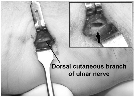
Preparation for the entrance site of bouquet pin intramedullary nail technique. The exposure was carried out through a small incision at the ulnar aspect of the 5th metacarpal. Protecting dorsal cutaneous branch of ulnar nerve, a cortical window at the base of metacarpal was created (arrow).
Fig. 2

Configuration of prebent K-wire. A K-wire was prebent to control pin direction (A) and to facilitate correction of rotational deformity (B).
Fig. 3
(B) Postoperative radiographs show 0° and 14° residual angulation, respectively.
(C) A 5-month postoperative radiographs show good union without any reduction loss or rotational deformity.

(A) A 28-year old man sustained a 5th metacarpal neck fracture with 34° angulation deformity in anteroposterior radiograph and 54° angulation deformity in oblique radiograph.


Figure & Data
REFERENCES
Citations
Citations to this article as recorded by 
- Percutaneous retrograde intramedullary single wire fixation for metacarpal shaft fracture of the little finger
Soo-Hong Han, Seung-Yong Rhee, Soon-Chul Lee, Seung-Chul Han, Yoon-Sik Cha
European Journal of Orthopaedic Surgery & Traumatology.2013; 23(8): 883. CrossRef - Treatment of 5th Metacarpal Neck Fracture Using Percutaneous Transverse Fixation with K-Wires
Jae-Hak Jung, Kwan-Hee Lee, Yong-Ju Kim, Woo-Jin Lee, Sung-Hyun Choi
Journal of the Korean Fracture Society.2012; 25(4): 317. CrossRef - Antegrade Intramedullary Prebent K-wire Fixation for the 5th Metacarpal Neck Fracture
Tae-Hyung Kim, Bo Hyeon Kim, In-Ho Jung, Dong-Hyun Kim
Journal of the Korean Fracture Society.2011; 24(1): 67. CrossRef - Percutaneous Retrograde Intramedullary Pin Fixation for Isolated Metacarpal Shaft Fracture of the Little Finger
Soo Hong Han, Hyung Ku Yoon, Dong Eun Shin, Seung Chul Han, Young Woong Kim
Journal of the Korean Fracture Society.2010; 23(4): 367. CrossRef
 Cite
CiteBouquet Pin Intramedullary Nail Technique of the 5th Metacarpal Neck Fractures


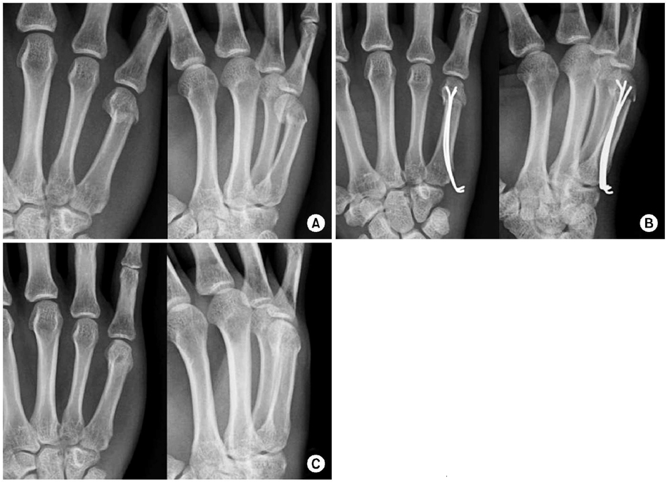
Fig. 1
Preparation for the entrance site of bouquet pin intramedullary nail technique. The exposure was carried out through a small incision at the ulnar aspect of the 5th metacarpal. Protecting dorsal cutaneous branch of ulnar nerve, a cortical window at the base of metacarpal was created (arrow).
Fig. 2
Configuration of prebent K-wire. A K-wire was prebent to control pin direction (A) and to facilitate correction of rotational deformity (B).
Fig. 3
(A) A 28-year old man sustained a 5th metacarpal neck fracture with 34° angulation deformity in anteroposterior radiograph and 54° angulation deformity in oblique radiograph.
(B) Postoperative radiographs show 0° and 14° residual angulation, respectively.
(C) A 5-month postoperative radiographs show good union without any reduction loss or rotational deformity.
Fig. 1
Fig. 2
Fig. 3
Bouquet Pin Intramedullary Nail Technique of the 5th Metacarpal Neck Fractures
Table 1
Grading the results of bouquet pin intramedullary nail technique of the 5th metacarpal neck fractures
From Kang et al.
Table 2
Radiographic angulations in patients with bouquet pin intramedullary nail technique of the 5th metacarpal neck fractures

