 E-submission
 TOTA
TOTA TOTS
TOTS
Search
- Page Path
- HOME > Search
Original Articles
- Clinical and radiographic outcomes of elastic stable intramedullary nailing for pediatric humeral shaft fractures: a retrospective case series
- Kang-San Lee, Dongju Shin, Sang Hee Kim, Il Seo, Tae-Hoon Kim, Sung Jung Kim
- J Musculoskelet Trauma 2026;39(2):156-161. Published online March 10, 2026
- DOI: https://doi.org/10.12671/jmt.2025.00381
-
 Abstract
 PDF
PDF - Background
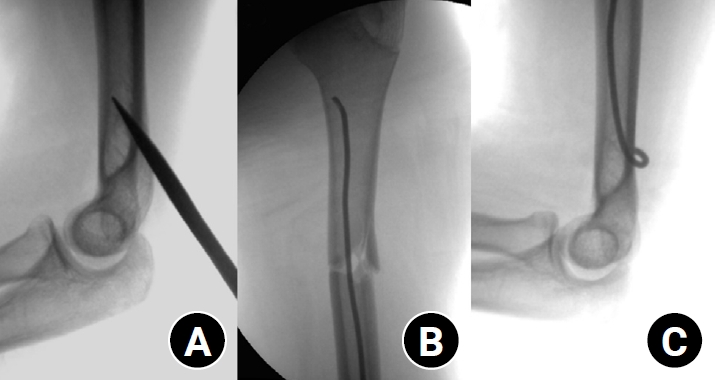
Pediatric humeral shaft fractures are uncommon and are generally treated conservatively, with satisfactory clinical outcomes reported in most cases. However, conservative management often necessitates prolonged immobilization and frequent outpatient follow-up visits, and it carries an inherent risk of residual angular or translational deformity. Elastic stable intramedullary nailing (ESIN) provides a simple and minimally invasive method of fracture fixation that offers adequate stability without disrupting the periosteal blood supply, thereby permitting early mobilization and promoting rapid bone union. The purpose of this study was to evaluate the clinical and radiological outcomes of ESIN fixation in pediatric patients with humeral shaft fractures.
Methods
The medical records of pediatric patients with humeral shaft fractures who underwent ESIN fixation between January 2015 and November 2025 were retrospectively reviewed. Data collected included patient demographics, mechanism of injury, fracture location, number of elastic nails used, time to union, degree of residual angulation, range of motion (ROM), and postoperative complications.
Results
The mean age of the patients was 10.0 years (range, 7 to 15 years). The mean time to radiographic union was 5.4 weeks (range, 2.4 to 10.4 weeks). The mean coronal angulation was 0.2° (range, −9.1° to 5.8°), while the mean sagittal angulation was −1.3° (range, −6.9° to 5.3°). No cases of infection, nerve injury, or nail migration were observed during the follow-up period. At the final follow-up assessment, all patients demonstrated full shoulder and elbow ROM, with no residual deformity or pain reported.
Conclusions
In this small retrospective case series, ESIN fixation resulted in favorable union rates and excellent functional outcomes in pediatric humeral shaft fractures. Level of evidence: IV.
- 567 View
- 19 Download

- Clinical Outcomes of Customized Staple Fixation Using K-wire in Metacarpal Base or Neck Fractures
- Hong-ki Jin, Hyoung Min Kim, Yong Seung Oh, Jihoon Kim
- J Korean Fract Soc 2021;34(1):23-29. Published online January 31, 2021
- DOI: https://doi.org/10.12671/jkfs.2021.34.1.23
-
Abstract
PDF
- Purpose
This study was designed to evaluate the radiological and clinical outcomes of a new surgical technique—customized staple fixation using K-wire—in displaced metacarpal neck or base fractures. Materials and Methods: From November 2016 to May 2017, 13 unstable metacarpal neck and base fractures (10 patients) were treated with II-shaped customized K-wire staples fixation, after performing open reductions through minimal dorsal incisions. The radiological and clinical outcomes were retrospectively evaluated. Results: A mean of 2.6 staples were used for each fracture fixation. Preoperative angulation of 36.3°was reduced to 3.1° postoperatively. A week after surgery, the volar short arm splint was replaced with a dorsal splint to initiate active range of motion exercise, and the splint was subsequently removed after 3 weeks. The radiologic union was achieved at a mean of 5.1 weeks, and total active motion was recovered at a mean of 7.4 weeks. On a mean, K-wire staples were removed at 16.5 weeks after the surgery, and the mean treatment took 18.6 weeks. At the final follow-up (at mean 27.3 weeks), no significant difference was observed for total active motion of the digits and grip strength, when compared to the contralateral hand. Complete union was achieved in all fractures without deformity, or complications such as infection or nerve injury. All patients were satisfied with the cosmetic and functional outcomes. Conclusion: K-wire stapling is an effective alternative modality in treating unstable displaced metacarpal neck or base fractures. It requires minimal incision to enable open reduction. In addition, early mobilization is ensured through the rigid fixations. Moreover, it prevents postoperative joint stiffness and reduces the time needed for treatment. -
Citations
Citations to this article as recorded by
- Individualized herbal prescriptions for delayed union: A case series
Jiyoon Won, Youngjin Choi, Lyang Sook Yoon, Jun-Hwan Lee, Keunsun Choi, Hyangsook Lee
EXPLORE.2023; 19(2): 260. CrossRef
- Individualized herbal prescriptions for delayed union: A case series
- 1,473 View
- 5 Download
- 1 Crossref
- Clinical Outcomes and Radiologic Characteristics of Insufficiency Femoral Neck Fracture in Elderly Patients
- Hee-Uk Ye, Kyung-Jae Lee, Byung-Woo Min, Kyung-Hwan Lim, Beom-Soo Kim, Young-Hoon Kim
- J Korean Fract Soc 2021;34(1):1-7. Published online January 31, 2021
- DOI: https://doi.org/10.12671/jkfs.2021.34.1.1
-
Abstract
PDF
- Purpose
In elderly patients, femoral neck insufficiency fractures that occur without a history of trauma are difficult to diagnose and treat, so it is emphasized that early suspicion of fractures and additional diagnostic tests are conducted. Materials and Methods: Between December 2010 to December 2019, 12 femoral neck insufficiency fractures (group 1) were evaluated by comparing them with 50 traumatic femoral neck fractures of a similar age. Along with demographic data, neck cortical thickness, shaft cortical thickness, head diameter, neck width, trochanter width, shaft width, neck-shaft angle, hip axis length, femoral neck index on the simple radiographic image were compared. Results: Seven of the 12 cases were non-displaced fractures, and it took an average of 19.2 days to diagnose the fracture after the symptoms occurred. The height was smaller than the control group at 149.1 cm in group 1 and 157.2 cm in group 2 (p<0.001). The cortical thickness of the medial femoral neck showed significant differences between the two groups: 3.16 mm in group 1 and 4.11 mm in group 2 (p=0.004). There was no statistical difference in the other measurements. Conclusion: Femoral neck insufficiency fracture often has a delayed diagnosis because of the characteristics of the fracture. The cortical thickness of the medial femoral neck in simple radiographic images can help suspect femoral insufficiency fractures in elderly patients when considered with detailed medical history taking and a physical examination.
- 1,186 View
- 12 Download
Case Report
- Atypical Fracture-Like Insufficiency Fracture of the Tibia with Prolonged Bisphosphonate Drug: A Case Report
- Min Jung Park, Su Jin Lee, Jin Hwa Kam, Yun Tae Lee, Ju Hyung Yoo, Hyun Cheol Oh, Joong Won Ha, Yung Park, Sang Hoon Park, Seong Hoon Kim, Han Kook Yoon
- J Korean Fract Soc 2017;30(3):137-141. Published online July 31, 2017
- DOI: https://doi.org/10.12671/jkfs.2017.30.3.137
-
Abstract
PDF
- Atypical femoral fracture related to a long-term bisphosphonate therapy has commonly been reported; however, a fracture at the site other than the femur has rarely been reported to date. Herein, we report a case of a patient on long-term bisphosphonate therapy who presented atypical tibial insufficiency fracture at the anterolateral aspect of diaphysis, without trauma. We, for the first time in Korea, present this case with a literature review.
-
Citations
Citations to this article as recorded by- Atypical Femoral Fracture Occurring at a Proximal Screw Insertion Site after Plate Removal in a Distal Femoral Fracture
Jin Woo Jin, Sung Jin Shin, Jong Min Jeon
Journal of the Korean Orthopaedic Association.2024; 59(4): 314. CrossRef
- Atypical Femoral Fracture Occurring at a Proximal Screw Insertion Site after Plate Removal in a Distal Femoral Fracture
- 1,117 View
- 3 Download
- 1 Crossref
Review Article
- Diagnosis and Management of Posttraumatic Chronic Osteomyelitis
- Jong Hoon Kim, Yong Cheol Yoon, Young Woo Kim, Sung Ho Jung, Jong Keon Oh
- J Korean Fract Soc 2014;27(1):88-104. Published online January 31, 2014
- DOI: https://doi.org/10.12671/jkfs.2014.27.1.88
-
Abstract
PDF
- No abstract available.
-
Citations
Citations to this article as recorded by- Treatment Strategy of Infected Nonunion
Hyoung-Keun Oh
Journal of the Korean Fracture Society.2017; 30(1): 52. CrossRef
- Treatment Strategy of Infected Nonunion
- 2,740 View
- 7 Download
- 1 Crossref
Case Report
- Acute Rupture of Subclavian Artery Pseudoaneurysm after Delayed Osteosynthesis of Clavicular Fracture: A Case Report
- Oog Jin Shon, Jee Hoon Kim, Kang Hyun Park
- J Korean Fract Soc 2014;27(1):82-87. Published online January 31, 2014
- DOI: https://doi.org/10.12671/jkfs.2014.27.1.82
-
Abstract
PDF
- Subclavian vessels are well protected by muscles, fascia and sheaths, so vascular complications associated with clavicular fractures are rare. Pseudoaneurysms after clavicular fractures have been reported, and the occurrence or rupture of pseudoaneurysm has been reported rarely as a late complication. However, cases of pseudoaneurysm after rupture of the clavicular fracture following delayed osteosynthesis of the clavicular fracture have not been reported. A 58-year-old female that presented with a right clavicular shaft fracture obtained conservative treatment. Surgery was performed after 4 months because of non-union in the local medical center. After operation, rupture of the subclavian pseudoaneurysm occurred following osteosynthesis of the clavicular shaft fracture. We report this case here with a review of the literature.
- 757 View
- 1 Download
Original Article
- Comparative Study of Proximal Femoral Nail Antirotation and Zimmer Natural Nail for the Treatment of Stable Intertrochanteric Fractures
- Jee Hoon Kim, Oog Jin Shon
- J Korean Fract Soc 2013;26(4):305-313. Published online October 31, 2013
- DOI: https://doi.org/10.12671/jkfs.2013.26.4.305
-
Abstract
PDF
- PURPOSE
To compare the results between Proximal femoral nail antirotation II (PFNA II) and Zimmer natural nail Asia type (ZNN) for the treatment of stable intertrochanteric fractures.
MATERIALS AND METHODS
Between September 2011 and September 2012, 40 consecutive patients with stable intertrochanteric femoral fractures were treated with PFNA II or ZNN. We reviewed 20 cases of PFNA II and 20 cases of ZNN prospectively. We evaluated the operation time, amount of bleeding, mean hospital day, and capability of mobility and function using 'mobility score of Parker and Palmer' and 'social score of Jensen'. We also evaluated the reduction state by the Fogagnolo, Cleveland index, change of tip and apex distance (TAD), sliding distance of cervical screw, change of neck shaft angle and bone union time.
RESULTS
There were no significant differences between the groups treated with PFNA and ZNN. Both groups showed good clinical results. PFNA showed less TAD change and ZNN showed a shorter sliding distance of cervical screw, but they were not statistically different. The bone union time was approximately 13 weeks in both groups.
CONCLUSION
PFNA and ZNN produced good clinical and radiologic results in the treatment of stable intertrochanteric fractures. There were no significant differences between the groups. Both implants provide good stability and union, so we can conclude that they are both suitable for the treatment of stable intertrochanteric fractures. -
Citations
Citations to this article as recorded by- Unable to Heel: The Clinical Journey of a Traumatic Near-Total Calcanectomy With 54 Years of Follow-up
Serge Andreou, Najeeb Baig, Rumyah Rafique, Ameen Suhrawardy, Pranav Khambete, Robert Meehan
Foot & Ankle Orthopaedics.2026;[Epub] CrossRef - Comparison of the Clinical and Radiological Outcomes of TFNA (Trochanteric Fixation Nail-Advanced) and PFNA-II (Proximal Femoral Nail Antirotation-II) Treatment in Elderly Patients with Intertrochanteric Fractures
Min Sung Kwon, Young Bok Kim, Gyu Min Kong
Journal of the Korean Fracture Society.2022; 35(4): 162. CrossRef - Clinical and Radiologic Outcome of Intertrochanteric Fracture Treatment Using TFNA (Trochanteric Fixation Nail-Advanced)
Hyeon Joon Lee, Hyun Bai Choi, Ba Rom Kim, Seung Hwan Jo, Sang Hong Lee
Journal of the Korean Fracture Society.2021; 34(3): 105. CrossRef - Comparison of osteoporotic intertrochanteric fracture fixation using a proximal femoral nail with a helical blade and lag screw type proximal femoral nail
Woong Chae Na, Chae Won Lim, Sang Hong Lee
Medical Biological Science and Engineering.2018; 1(2): 45. CrossRef - BONE HEALING PATTERNS OF INTERLOCKED INTRAMEDULLARY NAIL-FIXATED FEMORAL SHAFT FRACTURES: AGE-MATCHED RADIOGRAPHIC PRESENTATION OF UNION PATTERN
Myung-Sang Moon, Dong-Hyeon Kim, Bong-Keun Park, Min-Geun Yoon
Journal of Musculoskeletal Research.2017; 20(02): 1750010. CrossRef - The Curative Effect Comparison Between Prolonged Third Generation of Gamma Nail and Prolonged Dynamic Hip Screw Internal Fixation in Treating Femoral Intertrochanteric Fracture and the Effect on Infection
Wenye He, Wei Zhang
Cell Biochemistry and Biophysics.2015; 71(2): 695. CrossRef
- Unable to Heel: The Clinical Journey of a Traumatic Near-Total Calcanectomy With 54 Years of Follow-up
- 3,044 View
- 6 Download
- 6 Crossref
Case Report
- Treatment of Traumatic Posterior Dislocation of the Sternoclavicular Joint: A Case Report
- Dong Hee Kim, Do Hoon Kim, Seok Kwon Kang, Eui Chul Lee
- J Korean Fract Soc 2013;26(1):56-59. Published online January 31, 2013
- DOI: https://doi.org/10.12671/jkfs.2013.26.1.56
-
Abstract
PDF
- Compared with acromioclavicular dislocation, dislocation of the clavicle at its sternal end is uncommon and accounts for 3% of all injuries to the shoulder girdle. Furthermore, the posterior dislocation of the sternoclavicular joint is relatively a rare injury compared to the other types of sternoclavicular dislocation. We report this case since we have experience with similar cases of traumatic posterior dislocation at the sternoclavicular joint, which were successfully treated with x-ray guided reduction.
-
Citations
Citations to this article as recorded by- Posterior Sternoclavicular Dislocation: A Case Report
So Hwa Yoon, Sun Ki Kim, Ki Jun Kim
Journal of the Korean Society of Radiology.2015; 72(2): 128. CrossRef
- Posterior Sternoclavicular Dislocation: A Case Report
- 1,082 View
- 3 Download
- 1 Crossref
Original Articles
- Comparison of Surgical Outcomes in Thoracolumbar Fractures Having 6 or Less Scored by Load-Sharing Classification Based on Posterior Fusion Level
- Jung Hoon Kim, Sung Soo Kim, Jin Ho Cho, Bo Hoon Jang, Jin Hwan Kim
- J Korean Fract Soc 2013;26(1):21-26. Published online January 31, 2013
- DOI: https://doi.org/10.12671/jkfs.2013.26.1.21
-
Abstract
PDF
- PURPOSE
The aim of this study is to decide the optimal level of fusion with comparing the results between the short segment fusion and long segment fusion treated with pedicle screw instrumentation, including fractured vertebra in thoracolumbar junctional fractures.
MATERIALS AND METHODS
From February 2000 to November 2009, fifty three patients with junctional fracture of thoracolumbar spine were treated with pedicle screws and posterior fusion at our hospital. They were divided into two groups, the short segment group and long segment group. Preoperatively, immediate postoperative and last follow-up lateral radiological evaluation was done by measuring the correction and loss of segmental kyphosis, wedge angle, body compression rate and instrumented vertebra angle. In addition, operation time and amount of intraoperative bleeding were measured.
RESULTS
There were no significant differences of statistical analysis regarding the radiological variables between the two groups, especially the loss of corrected segmental kyphosis, wedge angle, body compression rate and instrumented vertebra angle (p>0.05). However, operative time in the short segment group (234 minutes) was shorter than the long segment group (284 minutes), and there was statistical significance (p=0.002).
CONCLUSION
We recommend the short segment transpediculr instrumentation one level above and one level below, including the fractured vertebra for thoracolumbar junctional fracture with 6 points or less of the load-sharing score.
- 677 View
- 0 Download
- Staged Minimally Invasive Plate Osteosynthesis of Distal Tibial Fractures
- Sung Ki Park, Chang Wug Oh, Jong Keon Oh, Kyung Hoon Kim, Woo Kie Min, Byung Chul Park, Won Ju Jeong, Joo Chul Ihn
- J Korean Fract Soc 2010;23(3):289-295. Published online July 31, 2010
- DOI: https://doi.org/10.12671/jkfs.2010.23.3.289
-
Abstract
PDF
- PURPOSE
To assess the result of staged minimally invasive plate osteosynthesis (MIPO) for distal tibial fracture with an open wound or injured soft tissue.
MATERIALS AND METHODS
In 20 patients (mean age, 47.8 year-old) with distal tibial fractures, there were 4 type A fractures and 16 type C fractures based on the AO classification system. Eight of the 20 patients had open fractures. MIPO was performed on average 23.9 days after bridging external fixation. At the final follow-up, we assessed the radiological results of bone union and alignment. Functional results were also evaluated by measuring the degrees of ankle motion and the American Orthopedic Foot & Ankle Society (AOFAS) scores.
RESULTS
Seventeen of 20 cases (85%) achieved primary union at an average of 21.3 weeks. There were 3 cases of nonunion requiring a bone graft. The mean AOFAS score was 88.5 (range, 67~92) and the average range of ankle motion was 49.2degrees (plantarflexion: 37.4degrees, dorsiflexion: 11.8degrees). Complication included 2 cases of minor mal-alignment, 1 case of claw toe and 1 case of peroneal neuropathy. Patients over the age of 60 had lower functional results. Additional factors did not affect the final results.
CONCLUSION
Staged MIPO may achieve satisfactory results in distal tibial fractures with soft tissue compromise, decreasing deep infections and soft tissue complications. -
Citations
Citations to this article as recorded by- Anterolateral Minimally Invasive Plate Osteosynthesis of Distal Tibial Fractures Using an Anterolateral Locking Plate
Dongwhan Suh, Hwan Hee Lee, Young Hoon Han, Jae Jung Jeong
Journal of Korean Foot and Ankle Society.2020; 24(1): 19. CrossRef - Minimally Invasive Osteosynthesis with Locking Compression Plate for Distal Tibia Fractures
Sung-Kyu Kim, Keun-Bae Lee, Keun-Young Lim, Eun-Sun Moon
Journal of the Korean Fracture Society.2011; 24(1): 33. CrossRef
- Anterolateral Minimally Invasive Plate Osteosynthesis of Distal Tibial Fractures Using an Anterolateral Locking Plate
- 1,419 View
- 6 Download
- 2 Crossref
- Staged Minimally Invasive Plate Osteosynthesis of Proximal Tibial Fracture
- Joon Woo Kim, Chang Wug Oh, Jong Keon Oh, Hee Soo Kyung, Woo Kie Min, Byung Chul Park, Kyung Hoon Kim, Hee Joon Kim
- J Korean Fract Soc 2009;22(1):6-12. Published online January 31, 2009
- DOI: https://doi.org/10.12671/jkfs.2009.22.1.6
-
Abstract
PDF
- PURPOSE
To assess the results of staged MIPO (Minimally Invasive Plate Osteosynthesis) for proximal tibial fractures with compromised soft tissue.
MATERIALS AND METHODS
Eighteen proximal tibial fractures (AO 41:9 cases, AO 42:9 cases) included this study. Ten were open fractures. After temporary external fixation until soft tissue healed (mean 27.3 days), MIPO was performed secondarily without bone graft. We assessed the bony union and knee function, and affecting factors of the results were investigated.
RESULTS
All fractures united at 20 weeks (range, 11~32) except 1 case. Mean range of knee flexion was 134.4degrees and mean IOWA knee score was 89.1. There were 2 superficial and 2 delayed deep infections from open fractures (grade II:1 case, grade III:3 cases), although they healed after implant removal. Open fractures seem to influence the infection rate. Otherwise, there was no related factor affecting the results.
CONCLUSION
MIPO after temporary external fixation can provide favorable results in proximal tibial fractures with soft tissue injuries, but attention of delayed infection should be paid in open fractures. -
Citations
Citations to this article as recorded by- MINIMALLY INVASIVE OSTEOSYNTHESIS WITH PLATE OR NAIL FOR META-DIAPHYSEAL TIBIAL FRACTURES - WHAT IS BETTER?
B. Makelov
Trakia Journal of Sciences.2023; 21(4): 357. CrossRef - Effect of Korean Medicine Treatments in Patients with Proximal Tibia Fracture: A Retrospective Observational Study
Jung Min Lee, Eun-Jung Lee
Journal of Korean Medicine Rehabilitation.2020; 30(3): 141. CrossRef - Comparison of Time to Operation and Efficacies of Ultrasound-Guided Nerve Block and General Anesthesia in Emergency External Fixation of Lower Leg Fractures (AO 42, 43, 44)
Chan Kang, Sang-Bum Kim, Youn-Moo Heo, You-Gun Won, Byung-Hak Oh, June-Bum Jun, Gi-Soo Lee
The Journal of Foot and Ankle Surgery.2017; 56(5): 1019. CrossRef - Minimally Invasive Plate Osteosynthesis for Proximal Tibial Shaft Fracture
Young-Soo Byun, Ki-Chul Park, Hyun-Jong Bong, Chang-Hoon Lee
Journal of the Korean Fracture Society.2011; 24(1): 23. CrossRef - The Use of Fresh Frozen Allogenic Bone Graft in the Impacted Tibial Plateau Fractures
Yeung Jin Kim, Soo Uk Chae, Jung Hwan Yang, Ji Wan Lee, Dae Han Wi, Duk Hwa Choi
Journal of the Korean Fracture Society.2010; 23(1): 26. CrossRef - Management of Open Fracture
Gu-Hee Jung
Journal of the Korean Fracture Society.2010; 23(2): 236. CrossRef - Staged Minimally Invasive Plate Osteosynthesis of Distal Tibial Fractures
Sung-Ki Park, Chang-Wug Oh, Jong-Keon Oh, Kyung-Hoon Kim, Woo-Kie Min, Byung-Chul Park, Won-Ju Jeong, Joo-Chul Ihn
Journal of the Korean Fracture Society.2010; 23(3): 289. CrossRef - Intramedullary Nailing of Proximal Tibial Fractures
Young-Soo Byun, Dong-Ju Shin
Journal of the Korean Fracture Society.2009; 22(3): 197. CrossRef - Proximal Tibia Fracture: Plating
Ki-Chul Park
Journal of the Korean Fracture Society.2009; 22(3): 206. CrossRef
- MINIMALLY INVASIVE OSTEOSYNTHESIS WITH PLATE OR NAIL FOR META-DIAPHYSEAL TIBIAL FRACTURES - WHAT IS BETTER?
- 1,119 View
- 0 Download
- 9 Crossref
- Hemiarthoplasty with Bone Block Graft and Low Profile Prosthesis for the Comminuted Proximal Humerus Fractures
- Chung Hee Oh, Joo Han Oh, Sae Hoon Kim, Ki Hyun Jo, Sung Woo Bin, Hyun Sik Gong
- J Korean Fract Soc 2008;21(3):213-219. Published online July 31, 2008
- DOI: https://doi.org/10.12671/jkfs.2008.21.3.213
-
Abstract
PDF
- PURPOSE
To evaluate the outcome of hemiarthroplasty with bone block graft and low profile prosthesis (Aequalis(R) fracture prosthesis) for the comminuted proximal humerus fractures.
MATERIALS AND METHODS
Sixteen low profile prostheses were used since July 2004, and 11 patients were followed-up for average 19.9 (12~30) months. Their mean age was 67.3 (52~78) years. Pain and satisfaction visual analog scale (VAS), range of motion, and modified UCLA score for hemiarthroplasty were evaluated at every visit. Radiography was also checked for stem position, loosening, and tuberosity union.
RESULTS
Mean pain VAS was 2.7 (0~5), and mean satisfaction VAS was 8.4 (5~10). Mean active forward flexion was 137 degrees (90~170), external rotation at side was 45.5 degrees (25~70), and internal rotation at back was T10 (T7~L1). Modified UCLA score was 19 (12~30) at final visit. All stems were stable, and there were no loosening at the final follow-up. All tuberosities were united except two tuberosity absorptions.
CONCLUSION
The outcome of hemiarthroplasty with bone block graft and low profile prosthesis was comparable to other implants for comminuted proximal humerus fractures. This system had unique advantages for tuberosity union. Further study with more patients and longer follow-up are necessary to clarify the effectiveness of this prosthesis. -
Citations
Citations to this article as recorded by- A Separate Approach and Cephalo-Diaphyseal Plate Fixation for the Comminuted Metadiaphyseal Fractures of the Proximal Humerus
Sung-Weon Jung
Journal of the Korean Fracture Society.2013; 26(1): 8. CrossRef - Comparison of Results between Internal Plate Fixation and Hemiarthroplasty in Comminuted Proximal Humerus Fracture
Doo-Sup Kim, Dong-Kyu Lee, Chang-Ho Yi, Jang-Hee Park, Jung-Ho Rah
Journal of the Korean Fracture Society.2011; 24(2): 144. CrossRef
- A Separate Approach and Cephalo-Diaphyseal Plate Fixation for the Comminuted Metadiaphyseal Fractures of the Proximal Humerus
- 1,031 View
- 0 Download
- 2 Crossref
- Posterior Plating in Distal Fibular Fracture
- Choong Hyeok Choi, Young A Cho, Jae Hoon Kim, Il Hoon Sung
- J Korean Fract Soc 2007;20(2):161-165. Published online April 30, 2007
- DOI: https://doi.org/10.12671/jkfs.2007.20.2.161
-
Abstract
PDF
- PURPOSE
To report the technical experience of posterior plating for the distal fibular fracture.
MATERIALS AND METHODS
20 Weber type-B fibular fractures were included in this study, which were treated with the posterior plating. 1/3 semitubular plate was used and orientation of all screws were intended to be perpendicular to the plate as possible. Fixation stability and maintenance of reduction after plating was assessed manually in the operating field. Clinical results were evaluated at least 1 year after operation, using American Orthopaedic Foot and Ankle Society (AFOAS) Ankle-Hindfoot score.
RESULTS
5 cases were firmly stabilized without using any lag screw or fixation of distal fragment. For improving stability or achieving proper reduction, a lag screw was placed posteroanteriorly through the plate in 14 cases. Anteroposterior interfragmentary fixation in 1 case before plating, and contouring of the plate in 3 cases were needed in cases of which the posterior plating impeded reduction of distal fibular fracture. In all cases, fracture was stabilized without fixation through the most distal hole. There were no major postoperative complications. AFOAS score was 95.5±5.2.
CONCLUSION
The posterior plating technique for distal fibular fracture is regarded as a recommendable option. Additional fixation with interfragmentary screw or contouring of the plate, however, would be needed in some cases to achieve anatomical reduction or sufficient stability. -
Citations
Citations to this article as recorded by- A Specialized Fibular Locking Plate for Lateral Malleolar Fractures
Eui Dong Yeo, Hak Jun Kim, Woo In Cho, Young Koo Lee
The Journal of Foot and Ankle Surgery.2015; 54(6): 1067. CrossRef
- A Specialized Fibular Locking Plate for Lateral Malleolar Fractures
- 1,215 View
- 20 Download
- 1 Crossref
- Correlation between Progression of Compression and Bone Densiometry Index in Osteoporotic Compression Fracture of Thoracolumbar Spine
- Jung Hoon Kim, Jeong Gook Seo, Jong Ho Ahn
- J Korean Fract Soc 2006;19(2):254-258. Published online April 30, 2006
- DOI: https://doi.org/10.12671/jkfs.2006.19.2.254
-
Abstract
- PURPOSE
To evaluate whether progression of compression correlates with bone densiometry index in patients who were treated conservatively for osteoporotic compression fracture of thoracolumbar spine.
MATERIALS AND METHODS
Using the results of bone densiometry, 30 patients who were treated conservatively for osteoporotic compression fracture of thoracolumbar spine between March 2002 to March 2005 were categorized into 4 groups; above 80%, 70 to 80%, 60 to 70%, and below 60%. We compared the measurements of sagittal index and anterior vertebral height from the plain radiographs taken at the time of injury and following three consecutive months after the injury.
RESULTS
Patients with lower bone densiometry index had greater amount of compression at the time of injury and more rapid progression of compression. We also found that progression of compression was lowest during the first month after injury in all groups.
CONCLUSION
Patients with low bone densiometry index in osteoporotic thoracolumbar compression fracture are susceptible to more rapid progression of compression and should have early brace application and longer duration of treatment for osteoporosis. -
Citations
Citations to this article as recorded by- Comparison of Outcomes of Conservative Treatment, Early Vertebroplasty, and Delayed Vertebroplasty in Patients with Osteoporotic Vertebral Compression Fractures
Se-Hyuk Im, Young-Joon Ahn, Bo-Kyu Yang, Seung-Rim Yi, Ye-Hyun Lee, Ji-Eun Kwon, Jong-Min Kim
Journal of Korean Society of Spine Surgery.2016; 23(3): 139. CrossRef - Comparison of Outcomes of Conservative Treatment, Early Vertebroplasty, and Delayed Vertebroplasty in Patients with Osteoporotic Vertebral Compression Fractures
Se-Hyuk Im, Young-Joon Ahn, Bo-Kyu Yang, Seung-Rim Yi, Ye-Hyun Lee, Ji-Eun Kwon, Jong-Min Kim
Journal of Korean Society of Spine Surgery.2016; 23(3): 139. CrossRef - Progression of Compression and Related Factors in Conservative Management of Osteoporotic Vertebral Compression Fractures
Young Do Koh, Jeong Soo Park
Journal of the Korean Fracture Society.2015; 28(2): 132. CrossRef
- Comparison of Outcomes of Conservative Treatment, Early Vertebroplasty, and Delayed Vertebroplasty in Patients with Osteoporotic Vertebral Compression Fractures
- 969 View
- 0 Download
- 3 Crossref
Case Report
- Spontaneous Intramuscular Hematoma associated with Acute Compartment Syndrome after Treatment of Low Molecular Weight Heparin: A Report of Two Cases
- Keun Woo Kim, Woo Dong Nam, Kee Hyung Rhyu, Byung Ryul Cho, Yong Hoon Kim, Soo Ik Awe
- J Korean Fract Soc 2006;19(1):89-92. Published online January 31, 2006
- DOI: https://doi.org/10.12671/jkfs.2006.19.1.89
-
Abstract
- Low-molecular-weight heparin (LMWH) has been considered superior to unfractionated heparin in several facets such as more effective anticoagulant, more predictable bioavailability, and less bleeding complications. We report two cases of LMWH, enoxaparin-induced spontaneous intramuscular hematoma with compartment syndrome of the lower extremity in patients with cardiac problems. The patients were treated with enoxaparin (LMWH) as bridging anticoagulation before use of warfarin due to cardiac problems. At the average 3 days of enoxaparin treatment, large and painful swelling was noticed in the lower extremities without intramuscular injection or trauma. The patients were diagnosed as having compartment syndrome with large intramuscular hematoma by CT. The patients underwent immediate fasciotomy and hematoma evacuation, and recovered without any complications.
- 1,095 View
- 0 Download
Original Articles
- Intramedullary K-wire Fixation for Displaced Fracture of Distal Radius
- Byung Woo Ahn, Chong Kwan Kim, Jong Youl Lee, Chae Ik Chung, Jong Ho Yoon, Young Min Kim, Jin Woo Jin, Kang Hoon Kim, Guk Sang Chung, Dong Wook Kim
- J Korean Fract Soc 2005;18(1):54-59. Published online January 31, 2005
- DOI: https://doi.org/10.12671/jkfs.2005.18.1.54
-
Abstract
PDF
- PURPOSE
To evaluate indications and effectiveness of intramedullary K-wire fixation for distal radial fractures.
MATERIALS AND METHODS
Twenty one fractures of distal radius treated with intramedullary K-wire fixation from April 2001 to September 2002 were evaluated. The mean age was 67.8(range 46~82). Severely comminuted intra-articular fractures and Barton's fractures were excluded. One or two K-wires were added percutaneously. To assess the functional result, we used Green and O'Brien score system. The radiographic assessment included a scoring system based on measurements of radial length, radial inclination, volar tilt and step-off of the radial articular surface.
RESULTS
Average follow up period was 13.5 months. In functional result, excellent and good result were obtained in 18 cases (86%). In radiologic result, mean loss of radial length, radial inclination and volar tilt were 0.9 mm, 1.4degrees, 0.9degrees, respectively.
CONCLUSION
The advantages of intramedullary K-wire fixation were relatively simple procedure, low occurrence of soft tissue complications and early wrist motion. Intramedullary K-wire fixation was good to maintaining reduction in osteoporotic bone.
- 640 View
- 3 Download
- Nailing in the Patients with Intertrochanteric Fractures of the Femur: Comparision Gamma Nail and Proximal Femoral Nail
- Sang Bong Ko, Myung Rae Cho, Tae Hoon Kim, Il Woong Chang
- J Korean Fract Soc 2004;17(4):295-300. Published online October 31, 2004
- DOI: https://doi.org/10.12671/jkfs.2004.17.4.295
-
Abstract
PDF
- PURPOSE
To evaluate the clinical and radiologic results of the fracture fixation using the Proximal Femoral Nail (PFN) or Gamma nail in patient with the intertrochanteric fractures.
MATERIALS AND METHODS
From January 1999 to February 2002, we reviewed 63 patients of intertrochanteric fractures including 31 patients managed with Gamma nail and 32 patients with PFN. In variate analysis of age, the pattern and stability of fracture, bone density (Singh index), preoperative patient activity, there was no difference between two groups. We evaluated the operation time, blood loss, stable reduction rate and varus reduction rate as operation-related factors, and radiologically investigated union time, the frequency of delayed union and nonunion, failure of fixation, status of the fracture line and complications. Functional results were evaluated by the possibility of early ambulation, ambulation ability at last follow up, and the pre and postoperative difference of ambulatory ability.
RESULTS
Mean operation time was 103+/-40 minutes in Gamma nail group and 101+/-28 minutes in PFN group. It was not statistically significant (p=0.801). Even though intraoperative blood loss was less in PFN group, it was not significant (p=0.601). Postoperative stable reduction rate was 31% in Gamma nail group and 30% in PFN group (p=0.934), suggesting not significant. Postoperative varus reduction and impacted amount of the fracture line was not significant. Delayed union developed in one case of Gamma nail group and 2 cases of PFN group. Mean loss of ambulation ability was 1.28 grade in Gamma nail group and 0.86 grade in PFN group (p=0.383). Statistical analysis was performed with the use of Student T test and Chi-square test. Analyses resulting in a p value of <0.05 were considered to show significance.
CONCLUSION
Generally, the radiologic and clinical results of intertrochanteric fractures were comparable for the PFN group and Gamma nail group. Even though it was not statistically significant, PFN group had superior results in operation time and blood loss than Gamma nail group for the fixation of the patients with intertrochanteric fracture. -
Citations
Citations to this article as recorded by- Treatment of Unstable Pertrochanteric Fractures with a Long Intramedullary Nail
Phil Hyun Chung, Suk Kang, Jong Pil Kim, Young Sung Kim, Ho Min Lee, Dae Jung Huh
Hip & Pelvis.2013; 25(1): 51. CrossRef - Hemiarthroplasty for Hip Fractures in Elderly Patients over 80 Years Old - Comparative Analysis between Femoral Neck Fracture and Intertrochanteric Fracture -
Chae-Hyun Lim, Young-Yool Chung, Jeong-Seok Kim, Chung-Young Kim
Hip & Pelvis.2013; 25(1): 44. CrossRef - Cementless Bipolar Hemiarthroplasty for Unstable Intertrochanteic Fractures in the Elderly
Byung-Hak Kim, Young-Yool Chung, Sung-Chang Ki, Dae-Hyun Yoon, Ji-Hoon Ryu
Journal of the Korean Orthopaedic Association.2011; 46(5): 399. CrossRef - Treatment of Intertrochanteric Fracture with Proximal Femoral Nail
Dae Joong Kim, Sung Chan Ki, Young Yool Chung
Journal of the Korean Fracture Society.2007; 20(1): 40. CrossRef
- Treatment of Unstable Pertrochanteric Fractures with a Long Intramedullary Nail
- 920 View
- 0 Download
- 4 Crossref
- Treatment of Proximal Shaft Fracture of Tibia with Intramedullary Nail: Analysis According to AO Classification and the Poller Screw
- Chang Wug Oh, Sung Jung Kim, In Ho Jeon, Hee Soo Kyung, Byung Chul Park, Joo Chul Ihn, Kyung Hoon Kim, Yun Kee Woo
- J Korean Fract Soc 2004;17(2):133-137. Published online April 30, 2004
- DOI: https://doi.org/10.12671/jkfs.2004.17.2.133
-
Abstract
PDF
- PURPOSE
This was a retrospective study to evaluate the results of intramedullary nailing in proximal shaft fracture of tibia. We analyzed those results according to AO classification and Poller screw.
MATERIALS AND METHODS
Thirty-three proximal tibial shaft fractures (32 patients) were followed for more than one year. In AO classification, there were 6 cases of type A, 14 cases of type B, and 13 cases of type C. We used Poller screws in 14 operations. We evaluated translation, angulation and nonunion after surgeries.
RESULTS
Twenty-eight cases (85%) were united primarily, but nonunions occurred in 5 cases. Malalignment (angulation>5degree or translation>5 mm) was found in 14 cases (42%). In Poller screw used and non-used groups, the malalignment was respectively showed in 2 cases (14%) and 12 cases (63%). According to AO classification, nonunion was found in only type B with 5 cases (36%).
CONCLUSION
Intramedullary nailing of proximal shaft fracture of tibia showed relatively lower rate of primary union. Especially, when initial fractures have a butterfly fragment, it showed the higher rate of nonunion. Moreover, the malalignment rate was relatively higher, yet it is possible to reduce the rate of malalignment by using Poller screw. -
Citations
Citations to this article as recorded by- Open Source-Based Surgical Navigation for Fracture Reduction of Lower Limb
Sanghyun Joung, Jaeyeong Park, Chul-Woo Park, Chang-Wug Oh, Il Hyung Park
Transactions of the Korean Society of Mechanical Engineers A.2014; 38(5): 497. CrossRef
- Open Source-Based Surgical Navigation for Fracture Reduction of Lower Limb
- 1,136 View
- 1 Download
- 1 Crossref
- Surgical and Conservative Treatment of Acetabular Fractures
- Chong Kwan Kim, Byung Woo Ahn, Saeng Guk Lee, Jae Ik Chung, Jin Woo Jin, Kwon Ho Kim, Kang Hoon Kim
- J Korean Soc Fract 2003;16(3):309-318. Published online July 31, 2003
- DOI: https://doi.org/10.12671/jksf.2003.16.3.309
-
Abstract
PDF
- PURPOSE
Fractures of the acetabulum remain a major challenge to the orthopedic surgeons. Although the operative treatment for the complex fractures is preferred, inaccurate reduction and then incongruity of the hip joint lead to serious complication such as premature osteoarthritis. We evaluated the results of surgical and conservative treatment for acetabular fractures.
MATERIALS AND METHODS
From January 1996 to March 2001, we reviewed 55 cases retrospectively. Posterior wall fracture (13 cases) was the most common by Letournel's classification and followed by both column fracture (10 cases). Causes of injuries included 41 cases of traffic accident and 8 cases of falling down. We divided the cases into an operation group (28 cases) and conservative group (27 cases) and evaluated the results as excellent, good, fair or poor according to Matta's clinical and radiological grade criteria.
RESULTS
Anatomical or satisfactory reduction was obtained in 22 cases of operative group and clinical results were excellent in 7 cases, good in 13 cases. Conservative group revealed excellent and good clinical results in 15 of 27 cases.
CONCLUSION
In cases of the displaced complex fractures, posterior wall fracture with instability and displaced fractures involving the weight bearing dome of the acetabulum, open reduction and internal fixation after accurate evaluation of the fracture pattern could allow earlier ROM exercise and have the result in better prognosis. -
Citations
Citations to this article as recorded by- Clinical Results of Surgical Treatment of Acetabular Fractures according to Quality of Reduction
Sang-Hong Lee, Min-Kyu Shin, Sueng-Hwan Jo
The Journal of the Korean Orthopaedic Association.2007; 42(2): 153. CrossRef
- Clinical Results of Surgical Treatment of Acetabular Fractures according to Quality of Reduction
- 1,177 View
- 19 Download
- 1 Crossref
- Retrograde Intramedullary Nail for Femoral Shaft Fracture with Limited Indications
- Sung Jung Kim, Chang Wug Oh, Joo Chul Ihn, Hee Soo Kim, In Ho Jeon, Hee Soo Kyung, Il Hyung Park, Kyung Hoon Kim
- J Korean Soc Fract 2003;16(1):45-51. Published online January 31, 2003
- DOI: https://doi.org/10.12671/jksf.2003.16.1.45
-
Abstract
PDF
- PURPOSE
This is a retrospective study to analyze the results after retrograde intramedullary(IM) nailing in femoral shaft fractures with limited indications.
MATERIALS AND METHODS
Twenty-four femoral shaft fractures(21 patients) were operated with unreamed IM nail(Unreamed femoral nail, SynthesR) in a retrograde method and were followed for more than 1 years. There were 16 men and 5 women, and the mean age at index operation was 41 years (range 18-76 years). In Winquist-Hansen classifications, there were 10 of type I, five of type II, three of type III, and six of type IV. All the patients had associated fractures or injuries, and there were eight ipsilateral tibia fractures, five ipsilateral proximal femoral fractures(including neck and trochanter), four ipsilateral pelvic or acetabular fracture, three bilateral femoral fractures, and one ipsilateral knee injury according to the used indications. In radiological study, we evaluated the time for union, non-unions and malunion, and clinical evaluation with Neer 's criteria was done.
RESULTS
Most fractures(87.5%) were primarily united cases, and the mean time for union was 15.8 weeks(range 12-20 weeks). Three cases of delayed union or nonunion were developed, but a shortening over 1cm or malunion over 10 degrees angular deformity were not found. Evaluating the knee functions, the Neer score was 86.9 in average and all the cases were above satisfactory grade. The average range of knee motion was 120.2 degrees, and the mild knee pain was developed in three cases.
CONCLUSION
The retrograde IM nailing can be a useful option for femoral shaft fractures with limited indications, including ipsilateral fractures of other areas or multiple fractures. -
Citations
Citations to this article as recorded by- Comparison of Operative Methods between Retrograde and Antegrade Nailing for Ipsilateral Femoral Shaft and Neck Fracture
Chang-Wug Oh, Jong-Keon Oh, Woo-Kie Min, Shin-Yoon Kim, Seung-Hoon Baek, Byung-Chul Park, Hyung-Soo Ahn, Tae-Gong Kim
Journal of the Korean Fracture Society.2007; 20(2): 135. CrossRef - Retrograde Intramedullary Nailing for the Treatment of Ipsilateral Femoral Shaft and Neck Fracture
Chang-Wug Oh, Jong-Keon Oh, Shin-Yoon Kim, Ki-Bong Cha, In-Ho Jeon, Byung-Chul Park, Woo-Kie Min, Tae-Gong Kim
The Journal of the Korean Orthopaedic Association.2007; 42(3): 380. CrossRef
- Comparison of Operative Methods between Retrograde and Antegrade Nailing for Ipsilateral Femoral Shaft and Neck Fracture
- 881 View
- 0 Download
- 2 Crossref
Case Report
- Fracture of the Os Peroneum with Rupture of the Peroneus Longus Tendon: A Case Report
- Yong Hoon Kim, Keun Woo Kim, Hak Jin Min, Eui Seong Yoon, Hee Oh Kim, Jae Seong Suh
- J Korean Soc Fract 2001;14(4):685-688. Published online October 31, 2001
- DOI: https://doi.org/10.12671/jksf.2001.14.4.685
-
Abstract
PDF
- Os peroneum is one of the normal sesamoids in the foot and it can be insertion of peroneus longus tendon. We report a case of as peroneum fracture with complete tear of peroneus longus tendon. This case was finally diagnosed in operation and treated by suture with peroneus brevis tendon and short leg cast.
-
Citations
Citations to this article as recorded by- Painful Os Peroneum Syndrome Presenting as Lateral Plantar Foot Pain
Seon Jeong Oh, Young Hoon Kim, Sun Ki Kim, Min-Wook Kim
Annals of Rehabilitation Medicine.2012; 36(1): 163. CrossRef
- Painful Os Peroneum Syndrome Presenting as Lateral Plantar Foot Pain
- 959 View
- 5 Download
- 1 Crossref
Original Articles
- Treatment of the Clavicular Shaft Fractures with K-wires
- Ho Choi, Hoon Kim, Neungcheol Shin, Dongsoo Han
- J Korean Soc Fract 2001;14(3):450-455. Published online July 31, 2001
- DOI: https://doi.org/10.12671/jksf.2001.14.3.450
-
Abstract
PDF
- PURPOSE
We report the results of treatment of the clavicular shaft fractures with Kwires.
MATERIALS AND METHODS
We treated 25 cases of clavicular shaft fractures with Kwires from June, 1998 to March, 2000 and analyzed the union period, functional results and complications.
RESULTS
Clinical union period was 3.2 weeks, radiologic union period was 8.3 weeks. The functional results were excellent or good in 21 cases(84 %) and there was no delayed or nonunion.
CONCLUSION
In the treatment of the clavicular shaft fractures, intramedullary fixation with K-wires is thought to be simple and effective method.
- 687 View
- 4 Download
- Treatment of Open Tibial Fracture with Repofix External Fixator
- Ho Choi, Hoon Kim, Neung cheol Shin, Sang Ho Shin, Dong Soo Han
- J Korean Soc Fract 2001;14(3):412-418. Published online July 31, 2001
- DOI: https://doi.org/10.12671/jksf.2001.14.3.412
-
Abstract
PDF
- PURPOSE
The purpose of this study is to evaluate the results of treatment of open tibial fracture with Repofix external fixator by clinical and radiological assessement. MATERIAL AND METHODS: 24 cases of open tibial fracture, who had been treated with Repofix external fixator from Oct. 1995 to June 1999, were evaluated about distribution of gender, age, cause of injury, fracture site and pattern, and assessed bony union time and complication.
RESULTS
The mean period of application of external fixator was 12.3 weeks and we kept PTB cast until bony union for average 11.5 weeks. According to Gustilo-Anderson's classification, the mean bony union time was 21.7 w e e k s ( type I ), 36 weeks(type II) and 39 weeks(type III), simple fracture was 22 weeks, comminuted fracture was 32 weeks(p<0.05). Complications were pin site infection(9 cases), delayed union(3 cases), nonunion(1 cases) and focal skin necrosis(1 case). There was no loss of motion about joint.
CONCLUSION
Repofix external fixator, which has the advantages of accurate reduction without extension of wound and decreases motion limitation of neighboring joint, is alternative useful appratus for open tibial fracture. -
Citations
Citations to this article as recorded by- Clinical Effectiveness of Korean Medical Rehabilitation Treatment after Patellar Fracture: A Report of 4 Cases
Ji-Hye Geum, Hyeon-Jun Woo, Jong-gyu Kim, Jung-Han Lee
Journal of Korean Medicine Rehabilitation.2020; 30(4): 203. CrossRef
- Clinical Effectiveness of Korean Medical Rehabilitation Treatment after Patellar Fracture: A Report of 4 Cases
- 870 View
- 0 Download
- 1 Crossref
- Treatment by Composite Fixation of Fractures in Osteoporotic Patients Aged over 60Years
- Keun Woo Kim, Yong Hoon Kim, Hak Jin Min, Ui Seoung Yoon, Hee Oh Kim, Young Joon Ahn, Yoon Jong Kim, Ki Chan Yoo, Sang Rim Kim
- J Korean Soc Fract 2001;14(1):121-127. Published online January 31, 2001
- DOI: https://doi.org/10.12671/jksf.2001.14.1.121
-
Abstract
PDF
- PURPOSE
This study summarizes the satisfactory results obtained using a composite fixation method for the surgical treatment of long bone fractures in elderly patients with osteoporosis.
MATERIALS AND METHODS
We reviewed 10 cases of long bone fractures, that were treated by composite fixation, involving patients over 60 years of age that presented with radiological osteoporosis. Composite fixation was applied incorporating, traditional plate and screw fixation in conjunction with bone graft, plate or intramedullary bone cement at four cases of humeral shaft fractures, three cases of femur supracondylar fractures, two cases of femur shaft fractures, and one case of tibia shaft fracture. Results were evaluated in methods of ambulation, range of motion, bony union and complications.
RESULTS
Satisfactory ambulation and range of motion was observed in all cases, which showed bony union without early implant failure. No re-operation were necessary due to nonunion. No medical complication was noted.
CONCLUSION
The results shows that the composite fixation method provided a stable reduction and a rigid fixation, which facilitated bony union, and allowed elderly patients with osteoporosis an early range of motion and mobility after the surgical treatment of long bone fractures.
- 655 View
- 0 Download
Case Report
- Radial Head Dislocation With Ipsilateral Radial Shaft Fracture: A Case Report
- Keun Woo Kim, Yong Hoon Kim, Hak Jin Min, Ui Seoung Yoon, Hee Oh Kim, Jae Hee Kim
- J Korean Soc Fract 2000;13(3):570-575. Published online July 31, 2000
- DOI: https://doi.org/10.12671/jksf.2000.13.3.570
-
Abstract
PDF
- Radial head dislocation with ipsilateral radial shaft fracture is one of the reportable trauma case with very low incidence. Only 4 cases of this type injury have been reported worldwide. In 3 cases of them, good results were achieved after early closed reduction of the radial head, followed by internal fixation of the radial shaft. In the a u t h o r s'case, the patient received the same method of treatment mentioned at the previous successful 3 cases. But after that, he suffered from repeated dislocations and finally nonunion of the fracture. At last, the authors could obtain bony union and stable elbow joint after radial head resection and osteosynthesis using autologous cancellous bone graft, but resulted in limitation of motion. We report the case and the experience of treatment.
-
Citations
Citations to this article as recorded by- Delayed radial head dislocation after radial shaft fracture fixation: a case report and review of the literature
Jiyong Yang, Jie Zhang, Zhengzhong Yang
BMC Surgery.2022;[Epub] CrossRef
- Delayed radial head dislocation after radial shaft fracture fixation: a case report and review of the literature
- 952 View
- 3 Download
- 1 Crossref
Original Articles
- Periprosthetic Femur Fracture due to Unrecognized Surgical Instrument Left in the Medullary Canal : A case report
- Ui Seoung Yoon, Keun Woo Kim, Yong Hoon Kim, Hak Jin Min, Kook Hyeung Cho, Min Sup Lee, Sang Rim Kim
- J Korean Soc Fract 2000;13(2):334-337. Published online April 30, 2000
- DOI: https://doi.org/10.12671/jksf.2000.13.2.334
-
Abstract
PDF
- Postoperative periprosthetic fracture of the femur after hip arthroplasty is a serious complication that can be difficult to treat. Moreover, it has become more common in recent years, as the cases of hip arthroplasty and revisonal hip arthroplasty increase. We have experienced a postoperative periprosthetic fracture of th femur probably caused by a surgical instrument left in the medullary canal during hip arthroplasty. We report this case with reference to other related articles.
- 549 View
- 0 Download
- Comparison between results of treatment of the Femoral Neck and Intertrochanteric Fractures : Focused on Mortality rate and Complications
- Chung Soo Hwang, Phil Hyun Chung, Suk Kang, Tae Hoon Kim, Han Chul Kim, Yong Soon Kim
- J Korean Soc Fract 1999;12(4):792-802. Published online October 31, 1999
- DOI: https://doi.org/10.12671/jksf.1999.12.4.792
-
Abstract
PDF
- The incidence of femoral neck and intertrochanteric fractures has steadily increased with lengthening of the life span. It is well known that anatomical characteristics of femoral neck may evoke complications such as nonunion and avascular necrosis. And there are many problems in the treatment of femoral intertrochanteric fractures due to osteoporosis, unstable pattern of fracture and poor general condition in elderly patients. The author analyzed 56 cases(56 patients) of femoral neck fractures and 63 cases(61 patients) of femoral intertrochanteric fractures which we have been able to follow up more than 1 year from March 1991 to March 1997. The purpose of this study is 1) to analyze results of treatment, predisposing factors, complications and mortality rates, and so 2) to reduce the mortality rate and complication in these fractures. The results were as follows , 1. The difference in union time between both type of fractures was not significant. 2. The mortality rate during admission was 1.8% in femoral neck fractures and 6.3% in intertrochanteric fractures. 3. The mortality rate during 1 years was 3.6% in femoral neck fractures and 9.5% in femoral intertrochanteric fractures. 4. The predisposing factors associated with postoperative mortality rate were malnutrition. chronic obstructive pulmonary disease, previous contralateral hip fracture, and operation within 3 days.
-
Citations
Citations to this article as recorded by- Anesthetic considerations for surgical treatment of geriatric hip fracture
Dong Kyu Lee, Seunguk Bang, Sangseok Lee
Anesthesia and Pain Medicine.2019; 14(1): 8. CrossRef - A Comparison of Clinical Results between Compression Hip Screw and Proximal Femoral Nail as the Treatment of AO/OTA 31-A2.2 Intertrochanteric Femoral Fractures
Phil Hyun Chung, Suk Kang, Jong Pil Kim, Young Sung Kim, Ho Min Lee, In Hwa Back, Kyeong Soo Eom
Journal of the Korean Orthopaedic Association.2016; 51(6): 493. CrossRef - Postoperative Mortality and the Associated Factors in Elderly Patients with Hip Fracture
You-Sung Suh, Yong-Beom Kim, Hyung-Suk Choi, Hong-Kee Yoon, Gi-Won Seo, Byung-Ill Lee
Journal of the Korean Orthopaedic Association.2012; 47(6): 445. CrossRef - One-Year Mortality Rate of Patients over 65 Years Old with a Hip Fracture
Phil Hyun Chung, Suk Kang, Jong Pil Kim, Young Sung Kim, Ho Min Lee, Young Hwa Choi
Hip & Pelvis.2011; 23(2): 137. CrossRef
- Anesthetic considerations for surgical treatment of geriatric hip fracture
- 1,801 View
- 4 Download
- 4 Crossref
- The Treatment of the Hip fracture in the Dementia Patients
- Ui Seoung Yoon, Keun Woo Kim, Yong Hoon Kim, Hak Jin Min, Kook Hyoung Cho, Sang Lim Kim, Hyong Tack Choi
- J Korean Soc Fract 1999;12(4):767-772. Published online October 31, 1999
- DOI: https://doi.org/10.12671/jksf.1999.12.4.767
-
Abstract
PDF
- PURPOSE
: The goal of treatment in elderly patients with hip fractures is restoration of function to preoperative ambulatory statuf as early as possible. The dementia patients who live in the asylum for the old need longer rehabilitation program for restoration of function, especially walking ability. The authors compare the modalities of the treatment for the hip fracture in the view point of walking ability.
MATERIALS and METHODS
: The twenty-eight dementia patients were operated due to hip fracture. Femur neck fractures were 7 cases, and femur intertrochanteric fractures were 21 cases. The authors analyze these patient on the recovery of walking ability. One patient who died immediately after operation was excluded in thil study. The patients were divided into two groups. Of 27 patients, 13 patients were treated with osteosynthesis(Group I), and remaining 14 patients were treated with hemidrthroplasy(Group II).
RESULTS
Fixation loss was treated with hemiarthroplasty in two case of osteosynthesis. The dislocation was treated with open reduction in one cafe of hemiarthioplasty. In the group I, the walking abilities were significantly different between the preoperative(3.85) and the postoperative at 2 weeks(2.46), at 2 months(2.73) and at the final follow-up(2.55)(P<0.05). In the group II, the walking abilities were not significantly different between the preoperative (2.57) and the postoperative at 2 weeks(2.14), at 2 months(2.36) and at the final follow- up(2.29)(P>0.05).
CONCLUSION
: Although there is no difference between two groups in final walking ability, The group treated with endoprosthesis showed earlier recovery of safting ability. -
Citations
Citations to this article as recorded by- CORR Insights®: What Are the Risk Factors for Dislocation of Hip Bipolar Hemiarthroplasty Through the Anterolateral Approach? A Nested Case-control Study
Eckart Mayr
Clinical Orthopaedics & Related Research.2016; 474(12): 2630. CrossRef - Risk Factors Associated with Dislocation after Bipolar Hemiarthroplasty in Elderly Patients with Femoral Neck Fracture
Yeesuk Kim, Joon-Kuk Kim, Il-Han Joo, Kyu-Tae Hwang, Young-Ho Kim
Hip & Pelvis.2016; 28(2): 104. CrossRef
- CORR Insights®: What Are the Risk Factors for Dislocation of Hip Bipolar Hemiarthroplasty Through the Anterolateral Approach? A Nested Case-control Study
- 878 View
- 0 Download
- 2 Crossref
- A Clinical Comparision of Multiple Pinning With Bipolar Endoprosthesis of the Femoral Neck Fractures in the Elderly Patients
- Jae Ik Lee, Myung Hwan Son, Jae Hong Park, Kang Hoon Kim
- J Korean Soc Fract 1998;11(2):321-327. Published online April 30, 1998
- DOI: https://doi.org/10.12671/jksf.1998.11.2.321
-
Abstract
PDF
- The goal of the treatment of femoral neck fractures is early ambulation to prevent the complications such as pneumoniae or atelectasis, deep vein thrombosis, pulmonary embolism, bed sore, general weakness. The authors have tried to asess the outcome and complication of multiple pinning (28 cases) and biploar endoprosthesis (35 cases) for femoral neck fractures in average 18 momths with the range of 12 months to 5 years from May 1990 to May 1996. The results were asessed by interval from injury to operation, post operative complication and mortality rate, weight bearing time, Lunceford hip grading method. The results were as folllows: 1. Post operative complications rate were 43% in multiple pinning, and 26% in bipolar group. 2. The mortality rate was 14.2% in multiple pinning group and 11.4% in bipolar endoprosthesis group. 3. The functional results evaluated by Lunceford's methods were satisfactory in 64% of multiple pinning group and 71% in bipolar endoprosthesis group. We concluded that displaced fractures, subcapital fracture, severe osteoporosis, old fracture over 3 weeks in elderly patients, bipolar endoprosthesis gave a more reliable results.
- 548 View
- 0 Download
- Treatment of Commiuted Fractures of the Proximal Tibia Using Ilizarov External Fixator
- Jae Ik Lee, Myung Hwan Son, Jae Hong Park, Kang Hoon Kim
- J Korean Soc Fract 1998;11(1):198-204. Published online January 31, 1998
- DOI: https://doi.org/10.12671/jksf.1998.11.1.198
-
Abstract
PDF
- Twenty one comminuted fractures of the proximal tibia in 21 patients were treated with Ilizarov external fixator from May 1990 to May 1996 in Pusan City Medical Center. The average duration of external fixation was 17weeks, and all of the fractures healed without bone graft. The mean time to union was 18weeks in closed comminuted fractures, and 21 weeks in Gustilo type I, II open fractures. The complications with this technique were pin tract infection 3 cases, pin loosening 6 cases, septic arthritis 1 case, post-traumatic osteoathritis 1 case, and joint ankylosis 1 case. The average duration of follow up was 18 months. The range of knee motion of 19 cases of the the 21 patients was at least 115-degrees arc. Post-operative immediate exercise was possible, and partial weight bearing with crutch was also possible after post-operative 4 weeks. We concluded that Ilizarov external fixation is a satisfactory technique for the treatment of selected comminuted fractures of the proximal tibia.
- 621 View
- 2 Download
- Treatment of Open Type-III Tibial Shaft Fractures -Comparison Between Ilizarov External Fixation and Secondary Intramedullary Nailing-
- Hoon Kim, Woo Dong Nam, Ki Chan Ahn, Seung Seok Seo, Young Chang Kim, Jang Seok Choi, Young Goo Lee
- J Korean Soc Fract 1998;11(1):191-197. Published online January 31, 1998
- DOI: https://doi.org/10.12671/jksf.1998.11.1.191
-
Abstract
PDF
- Open type III tibial shaft fractures have a high incidence of complication and a poor outcome. The most usual method of stabilization is by external fixation, but there are several complications as infection, delayed union and nonunion. We tried to compare the results of Ilizarov external fixation cases with the case of secondary intramedullary nailing after temporary Ilizarov fixation. There was no significant difference in the union time of Ilizarov external fixation and secondary intramedullary nailing after temporary Ilizarov fixation. But the patients were more comfortable in the secondary intramedullary nailing. In conclusions, secondary intramedullary nailing after temporary Ilizarov fixation is the useful method in the treatment of open type III tibial shaft fractures.
- 584 View
- 1 Download
Case Report
- Fatigue Fracture of the Distal femur Shaft: A Case Report
- Seung Gyun Cha, Kyoung Hoon Kim, Jong Ho Jang, Sang Jun Lee
- J Korean Soc Fract 1997;10(4):979-982. Published online October 31, 1997
- DOI: https://doi.org/10.12671/jksf.1997.10.4.979
-
Abstract
PDF
- A fatigue(stress) fracture begins as a gradual localized dissolution of bone as a result of repeated submaximal and generally unaccustomed stresses which may or may not result in a complete fracture. The most common sites of fatigue fracture are tibia, metatarsal area. We experienced peculiar faligue fracture in professional bowler. So we review the fatigue fracture article and reporte 1 case of fracture in distal part of femur with clinical and radiological findings.
- 673 View
- 0 Download
Original Articles
- Treatment of Supracondylar Fractures of the Humerus in Children
- Hyung Seok Kim, Ki Do Hong, Sung Sik Ha, Eu Suk Lew, Tae Hoon Kim
- J Korean Soc Fract 1997;10(4):964-971. Published online October 31, 1997
- DOI: https://doi.org/10.12671/jksf.1997.10.4.964
-
Abstract
PDF
- Supracondylar fracture of the humerus is the most common elbow fracture in children. It can be complicated by Volkmanns ischemic contracture, nerve injury, deformity of elbow, joint stiffness, and myositis ossificans, etc. even after the treatment. Various methods of treating displaced supracondylar rracture have been described, and the indications for open reduction and internal fixation are relatively established well. The surgeon, however, might be in trouble with making decision for closed reduction, whether to maintain by casting or by percutaneous pinning. From fan. 1990 to May 1990, 117 patients of supracondylar tracture had been treated in Seoul Adventist Hospital, and 82 patients of them were challenged initially by closed reduction and casting. Among them, 51 cases have been followed more than 1 year, and they are reviewed. 1. The average age was 7.2 years, and the patient population consisted of 33 boys and 18 girls. 2. 3 cases of nerve injuries, 2 cases of ipsilateral forearm bone fractures, 2 cases of impending Volkmanns ischemic contracture and 1 case of cerebral contusion were associated with supracondylar fracture. 3. Fracture pattern were classified as flexion type of 1 case and extension type of 50 cases. According to Gartland classification, type II was 16 cases(31.4%), type III-A was 27(52.9%) and III-B was 7(13.7%). Of all, 28 cases(54.9%) were comminuted and 23 were non-comminuted. On the other hand, 14 cases(27.5%) were oblique fracture and 37(72.5%) were transverse one, classified by pattern of fracture line. 4. Success rate of closed reduction and casting were 56.3% in Garland type II, 33.3% in type III-A, 42.9% in type III-B. In comminuted fracture, it was 21.4% and 65.2% in non-comminuted one. Success rate was 7.1% in oblique fracture, and 54.1% in transverse one. This suggests that comminuted fracture or oblique fracture should not be tried by closed reduction and casting but by percutaneous pinning. 87.5% of non-comminuted and transverse fractures were treated successfully by initial closed reduction and casting, and it also suggests that closed reduction and casting should be done only in this case selectively. 5. By Flynns criteria the results were excellent in 9 cases, good in 8, fair in 3 and poor in 1 with casting. Percutaneous pinning resulted in 15 excellent, 10 good, and 1 fair cases. Open reduction and internal fixation resulted in 2 excellent and 2 good cases.
- 653 View
- 0 Download
- Fracture of the Coracoid Process Associated with Acromioclavicular Dislocation
- Ui Seoung Yoon, Yong Hoon Kim, Keun Woo Kim, Hak Jin Min, Kook Hyeung Cho, Ki Seung Kum, Il Myung Kim
- J Korean Soc Fract 1997;10(3):640-644. Published online July 31, 1997
- DOI: https://doi.org/10.12671/jksf.1997.10.3.640
-
Abstract
PDF
- Acromioclavicular dislocation with a concomitant coracoid process fracture occurs infrequently, as only several cases have been reported in the literature. This injury usually occurs in the second or third decade of life. The mechanism of injury were sports, motor vehicle and direct trauma. An axillary lateral radiograph of the shoulder often is needed to detect this fracture. The authors present two cases of coracoid process fracture associated with acromioclavicular dislocation. Satisfactory function with minimal residual cosmetic deformity may be achieved by nonoperative measures. Also surgical treatment can produce good result in complete third acromioclavicular separation combined with the significantly displaced coracoid fracture.
- 639 View
- 5 Download
Case Report
- Fracture of Femur Neck Associated with Technical Errors in Closed Intramedullary Nailing of the Femur
- Yong Hoon Kim, Ki Chan Ahn, Sung Suk Seo, Young Chang Kim, Jang Suk Choi, Young Gu Lee
- J Korean Soc Fract 1997;10(1):73-78. Published online January 31, 1997
- DOI: https://doi.org/10.12671/jksf.1997.10.1.73
-
Abstract
PDF
- Closed intramedullary nailing has become increasingly popular in the management of fracture of the femur because of a high rate of union and a low rate of complications. Since the development, it has been widely used in more applicable level of femoral shaft fracture. Therefore, complications of intramedullary nailing was rarely seen, especially rare in case of fractures of the femoral neck associated with technical errors. The three cases of femoral neck fracture with technical errors during intramedullary nailing for treatment of femoral shaft fracture in Paik Hospital, Pusan from April 1994 to July 1995 are reported herein to document that this complication can occur. Three cases of the femoral neck fracture were treated by closed reduction and internal fixation with Knowles pin.
- 502 View
- 0 Download
Original Articles
- Femur Neck Fracture during Closed Nailing of Femur Shaft Fracture
- Keun Woe Kim, Ui Seong Yoon, Yong Hoon Kim, Hak Jin Min, Jin Sup Yeom, Young Ho Lee
- J Korean Soc Fract 1996;9(4):936-942. Published online October 31, 1996
- DOI: https://doi.org/10.12671/jksf.1996.9.4.936
-
Abstract
PDF
- Closed intramedullary nailing has become increasingly popular in the management of fractures of the femur because of a high rate of union, a low rate of infection, and excellent return of function. But it requires good availability of equipment and surgeons skill and experience. Iatrogenic femur neck fracture can occur as a complication of closed intramedullary nailing of femur shaft fracture. The authors report five cases of this complication and analyze these five cases in the viewpoint of causal technical errors and suggest some technical points to avoid this complication.
-
Citations
Citations to this article as recorded by- Some Series of Honey-Comb Spaces
Elena Barbieri, Alberto Cavicchioli, Fulvia Spaggiari
Rocky Mountain Journal of Mathematics.2009;[Epub] CrossRef
- Some Series of Honey-Comb Spaces
- 912 View
- 0 Download
- 1 Crossref
- Fracture-SeFaration Involving the Entire Distal Humeral Epiphysis in a Young Child(Salter-Harris type I injury): A Case Report
- Jong Ho Jang, Seung Gyun Cha, Kyoung Hoon Kim, Jeon Oh Kang
- J Korean Soc Fract 1996;9(3):674-677. Published online July 31, 1996
- DOI: https://doi.org/10.12671/jksf.1996.9.3.674
-
Abstract
PDF
- Fracture-separation of the distal humeral epiphysis is a rare injury, frequently misdiagnosed as a fracture of the lateral humeral condyle, a supracondylar fracture of the humerus or a dislocation of the elbow. Roentgenographic evaluation reveals posteromedial displacement of the distal epiphysis. Single contrast arthrography is performed in order to confirm diagnosis. Treatment is first directed toward prompt recognition of the injury. A manipulative closed reduction is usually recommended. We experienced a case of fracture-separation involving the entire distal humeral physis treated by closed reduction and percutaneous pinning. The result was excellent.
- 609 View
- 0 Download
- A Clinical Study of the Tibial Plafond Fractures
- Seung Gyun Cha, Won Suek Lee, Kyoung Hoon Kim, Jin Hak Kim, Jin Yong Jung, Jae Sung Lee
- J Korean Soc Fract 1992;5(2):268-274. Published online November 30, 1992
- DOI: https://doi.org/10.12671/jksf.1992.5.2.268
-
Abstract
PDF
- The tibial plafond fractures result from an axial compression and rotational forces causing variable degrees of metaphyseal disruption, articular damage, and malleolar displacement, it is relatively uncommon but a most difficult fracture to manage. It has been treated in a number of manners, but the best results have been occurred when stable internal fixation is accomplished using plates and screws, basic steps in reconstruction are restoration of anatomic length of the fibula with plate fixation, reconstruction of the tibial articular surface, cancellous bone grafting of the metaphyseal defect and buttress plating of the distal tibia. The author analysed the 22 cases of the tibial plafond fractures in 22 patients, which were treated at the department of orthopedic surgery in Dai Han hospital. From january 1989 to january 1991. The longest duration of follow up was 2 years and 6 months and shortest one was 7 months, and the average was 14 months The results were as follows ; 1. Among the 22 patients, male were 17 and female was five. 2. The major causes of injury were a fall from a height. 3. Regardless of the method of treatment the type I and II were In good and fair result, but the type III were fair in 4 cases and poor in 2 cases, other 5 cases were in good result. 4. It is important to remind the basic steps in reconstruction appllied in an indivisual case especially in type III. If anatomic reconstruction of the joint surface can be restored and stable fixation achieved, early motion of the ankle joint is possible. The early motion reduces stiffness of the ankle and will yeild the most satisfying rusult. But the anatomic reduction of ankle joint is difficult to be obtained in each case especlally in type III and may consider an ankle fusion.
- 591 View
- 0 Download
- A Clinical Study of the Childrens Anltle Fracture
- Seung Gyun Cha, Won Suck Lee, Kyoung Hoon Kim, Woo Tae Lee, Jin Hak Kim, Jae Sung Lee
- J Korean Soc Fract 1992;5(2):236-243. Published online November 30, 1992
- DOI: https://doi.org/10.12671/jksf.1992.5.2.236
-
Abstract
PDF
- The injuries of ankle in children occur mostly at the bony structure rather than ligamentous one, and some injuries of the growth plate may result in the arrest of entire growth an these may result to leg length discrepancy or deformity. Accurate treatment under the knowledge of injury mechanism is very important. We have reviewed 30 cases of childrens ankle fracture treated our clinics from Jan. 1987 to Dec. 1990. The results are as follows; 1. Traffic accident was most common cause of injury (50.0%). 2. According to Dias & Tachdjian classification, Supination-Invertion type was most common (11 cases). 3. The treatments performed were closed reduction & cast immobilization for 11 cases (36.7%), closed reduction & percutaneous pinning for 3 cases (10.0%), and open reduction & internal fixation for 16 cases(53.3%). 4. Among open reduction & internal fixation group, only 1 case(pronation-eversion-external rotation type) show angular deformaity. 5. For the prevention of complications, accurate reduction and adequate fixation was desired.
- 581 View
- 0 Download
- Clinical analysis of fixation failure in the treatment of intertro chanteric fracture of the femur
- Jong Deuk Rha, Young Hoon Kim, Sung Il Yoon, Jun Soon Kang, Duck Hyun Park
- J Korean Soc Fract 1991;4(2):298-305. Published online November 30, 1991
- DOI: https://doi.org/10.12671/jksf.1991.4.2.298
- 564 View
- 0 Download
- Analsis of the Distal Tibial Physeal and Epiphyseal Injury
- In Ho Choi, Choon Ki Lee, Duk Yong Lee, Se Il Suk, Song Choi, Yong Hoon Kim, Suk Kee Tae, Seong Il Kim
- J Korean Soc Fract 1989;2(2):164-173. Published online November 30, 1989
- DOI: https://doi.org/10.12671/jksf.1989.2.2.164
-
Abstract
PDF
- We have reviewed 21 cases of physeal and epiphyseal fracture of the distal tibia including one case of juvenile Tillaux fracture and four cases of triplane fracture. The patterns of fracture were correlated with the mechanism of injury using the modified Lauge-Hansen method and sysemic classification of the triplane frature was proposed. Treatment included closed reduction and cast(9 cases), closed reduction and percutaneous pin fixation(4 cases), and open reduction(8 cases). Two of the nine patients treated by means of closed reduction and cast had ankle joint incongruity or progressive varus deformity requiring corrective ost eotomy. These two patients had either Salter-Harris Type III ro Type IV fracture after supination-inversion injury. In one patient, who had 100 per cent displacement of the distal tibial epiphysis and degloving injury of the ankle, premature physeal arrest developed after open reduction and internal fixaion for Salter-Harris Type Tyre I fracture. If there are triagular metaphseal ledge along with the juvenile Tillaux fracture-like vertical epiphyseal fracture line on the antero-posterior view and Salter-Harris Type II or Type IV frature on the lateral view, a certain type of triplane fracture is strongly suggested. Plain radiographs, however, could not accurately demontrate the detailed configuration of the triplane fracture, instead computerized axial tomography was very helpful us to analyse the true dimensions of the triplane fracture. We agree that displace Salter-Harri Type III or Type IV and transitional fractures with a fracture gap of more than two millimeters in the weight-bearing portion of the epiphysis regure open reduction.
- 570 View
- 0 Download
 First
First Prev
Prev

