 E-submission
 TOTA
TOTA TOTS
TOTS
Articles
- Page Path
- HOME > J Musculoskelet Trauma > Volume 26(1); 2013 > Article
-
Case Report
- Treatment of Traumatic Posterior Dislocation of the Sternoclavicular Joint: A Case Report
- Dong-Hee Kim, M.D., Do-Hoon Kim, M.D., Seok-Kwon Kang, M.D., Eui-Chul Lee, M.D.
-
Journal of the Korean Fracture Society 2013;26(1):56-59.
DOI: https://doi.org/10.12671/jkfs.2013.26.1.56
Published online: January 17, 2013
Department of Orthopaedic Surgery, Samsung Changwon Hospital, Sungkyunkwan University School of Medicine, Changwon, Korea.
- Address reprint requests to: Do-Hoon Kim, M.D. Department of Orthopaedic Surgery, Samsung Changwon Hospital, 158, Paryong-ro, Masanhoewon-gu, Changwon 630-723, Korea. Tel: 82-55-290-6030, Fax: 82-55-290-6888, kdh7483@gmail.com
• Received: September 17, 2012 • Revised: October 17, 2012 • Accepted: October 23, 2012
Copyright © 2013 The Korean Fracture Society
- 1,081 Views
- 3 Download
- 1 Crossref
Abstract
- Compared with acromioclavicular dislocation, dislocation of the clavicle at its sternal end is uncommon and accounts for 3% of all injuries to the shoulder girdle. Furthermore, the posterior dislocation of the sternoclavicular joint is relatively a rare injury compared to the other types of sternoclavicular dislocation. We report this case since we have experience with similar cases of traumatic posterior dislocation at the sternoclavicular joint, which were successfully treated with x-ray guided reduction.
- 1. Aure A, Hetland KR, Rokkum M. Chronic posterior sternoclavicular dislocation. J Orthop Trauma, 2012;26:e33-e35.Article
- 2. Chaudhry FA, Killampalli VV, Chowdhry M, Holland P, Knebel RW. Posterior dislocation of the sternoclavicular joint in a young rugby player. Acta Orthop Traumatol Turc, 2011;45:376-378.
- 3. Friedrich L, Afifi FK, Skarvan J, Friederich NF, Hirschmann MT. Combined gracilis tendon autograft reconstruction and discus repair of a chronic anterior-superior sternoclavicular joint dislocation. Knee Surg Sports Traumatol Arthrosc, 2012;20:1978-1982.ArticlePDF
- 4. Glass ER, Thompson JD, Cole PA, Gause TM 2nd, Altman GT. Treatment of sternoclavicular joint dislocations: a systematic review of 251 dislocations in 24 case series. J Trauma, 2011;70:1294-1298.Article
- 5. Kumar P, Godbole R, Rees GM, Sarkar P. Intrathoracic migration of a Kirschner wire. J R Soc Med, 2002;95:198-199.ArticlePDF
- 6. Lee JS, Kim BG, Kim HJ, Jung SG, Son SJ. Traumatic posterior dislocation of the sternoclavicular joint: a case report. J Korean Orthop Assoc, 1990;25:579-583.ArticlePDF
- 7. Marcus MS, Tan V. Cerebrovascular accident in a 19-year-old patient: a case report of posterior sternoclavicular dislocation. J Shoulder Elbow Surg, 2011;20:e1-e4.Article
- 8. Sullivan JP, Warme BA, Wolf BR. Use of an O-arm intraoperative computed tomography scanner for closed reduction of posterior sternoclavicular dislocations. J Shoulder Elbow Surg, 2012;21:e17-e20.Article
REFERENCES

Fig. 2
(A) Initial chest radiograph. Antero-posterior radiograph of the left sternoclavicular dislocation. The left clavicle is slightly inferiorly displaced compared to the right side and has lost its normal articulation with the sternum.
(B) Initial lordotic view. Posteior dislocation of the left sternoclavicular joint. Note that the left medial clavicle is displaced inferiorly to the normal right medial clavicle.
(C) Computed tomography scan showing the posteriorly dislocated clavicle.
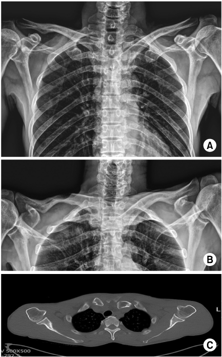


Figure & Data
REFERENCES
Citations
Citations to this article as recorded by 
- Posterior Sternoclavicular Dislocation: A Case Report
So Hwa Yoon, Sun Ki Kim, Ki Jun Kim
Journal of the Korean Society of Radiology.2015; 72(2): 128. CrossRef
 Cite
CiteTreatment of Traumatic Posterior Dislocation of the Sternoclavicular Joint: A Case Report
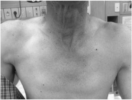

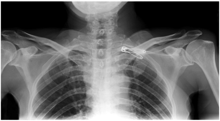
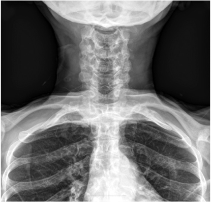
Fig. 1
Clinically evident posterior dislocation of the left sternoclavicular joint.
Fig. 2
(A) Initial chest radiograph. Antero-posterior radiograph of the left sternoclavicular dislocation. The left clavicle is slightly inferiorly displaced compared to the right side and has lost its normal articulation with the sternum.
(B) Initial lordotic view. Posteior dislocation of the left sternoclavicular joint. Note that the left medial clavicle is displaced inferiorly to the normal right medial clavicle.
(C) Computed tomography scan showing the posteriorly dislocated clavicle.
Fig. 3
Initial postoperative chest radiograph.
Fig. 4
Postoperative 2 months. Hobbs view.
Fig. 1
Fig. 2
Fig. 3
Fig. 4
Treatment of Traumatic Posterior Dislocation of the Sternoclavicular Joint: A Case Report

