 E-submission
 TOTA
TOTA TOTS
TOTS
Articles
- Page Path
- HOME > J Musculoskelet Trauma > Volume 26(1); 2013 > Article
-
Original Article
- Comparison of Surgical Outcomes in Thoracolumbar Fractures Having 6 or Less Scored by Load-Sharing Classification Based on Posterior Fusion Level
- Jung Hoon Kim, M.D., Sung Soo Kim, M.D., Jin Ho Cho, M.D., Bo Hoon Jang, M.D., Jin Hwan Kim, M.D.
-
Journal of the Korean Fracture Society 2013;26(1):21-26.
DOI: https://doi.org/10.12671/jkfs.2013.26.1.21
Published online: January 17, 2013
Department of Orthopaedic Surgery, Inje University Ilsan Paik Hospital, Goyang, Korea.
*Department of Orthopaedic Surgery, Inje University Sanggye Paik Hospital, Seoul, Korea.
- Address reprint requests to: Jin Hwan Kim, M.D. Department of Orthopaedic Surgery, Inje University Ilsan Paik Hospital, 170, Juhwa-ro, Ilsanseo-gu, Goyang 411-706, Korea. Tel: 82-31-910-7968, Fax: 82-31-910-7967, jhkim@paik.ac.kr
• Received: June 16, 2012 • Revised: July 23, 2012 • Accepted: October 26, 2012
Copyright © 2013 The Korean Fracture Society
- 602 Views
- 0 Download
Abstract
-
Purpose
- The aim of this study is to decide the optimal level of fusion with comparing the results between the short segment fusion and long segment fusion treated with pedicle screw instrumentation, including fractured vertebra in thoracolumbar junctional fractures.
-
Materials and Methods
- From February 2000 to November 2009, fifty three patients with junctional fracture of thoracolumbar spine were treated with pedicle screws and posterior fusion at our hospital. They were divided into two groups, the short segment group and long segment group. Preoperatively, immediate postoperative and last follow-up lateral radiological evaluation was done by measuring the correction and loss of segmental kyphosis, wedge angle, body compression rate and instrumented vertebra angle. In addition, operation time and amount of intraoperative bleeding were measured.
-
Results
- There were no significant differences of statistical analysis regarding the radiological variables between the two groups, especially the loss of corrected segmental kyphosis, wedge angle, body compression rate and instrumented vertebra angle (p>0.05). However, operative time in the short segment group (234 minutes) was shorter than the long segment group (284 minutes), and there was statistical significance (p=0.002).
-
Conclusion
- We recommend the short segment transpediculr instrumentation one level above and one level below, including the fractured vertebra for thoracolumbar junctional fracture with 6 points or less of the load-sharing score.
- 1. Been HD, Bouma GJ. Comparison of two types of surgery for thoraco-lumbar burst fractures: combined anterior and posterior stabilisation vs. posterior instrumentation only. Acta Neurochir (Wien), 1999;141:349-357.ArticlePDF
- 2. Carl AL, Tromanhauser SG, Roger DJ. Pedicle screw instrumentation for thoracolumbar burst fractures and fracture-dislocations. Spine (Phila Pa 1976), 1992;17:S317-S324.Article
- 3. Chung JY, Rhym IS. Short segment transpedicular cotrel-dubousset instrumentation including involved vertebra for fractures of thoracic and lumbar spine. J Korean Orthop Assoc, 1994;29:940-948.ArticlePDF
- 4. Cotrel Y, Dubousset J, Guillaumat M. New universal instrumentation in spinal surgery. Clin Orthop Relat Res, 1988;227:10-23.Article
- 5. Jeong ST, Cho SH, Song HR, Koo KH, Park HB, Chung UH. Comparison of short and long-segment fusion in thoracic and lumbar fractures. J Korean Soc Spine Surg, 1999;6:73-80.
- 6. Krag MH. Biomechanics of thoracolumbar spinal fixation. A review. Spine (Phila Pa 1976), 1991;16:S84-S99.
- 7. Lee CS, Chung SS, Jung HW, Kim ES. Decision of posterior fixation level by load-sharing classification in thoracolumbar and lumbar burst fracture. J Korean Soc Spine Surg, 2001;8:27-38.Article
- 8. Mann KA, McGowan DP, Fredrickson BE, Falahee M, Yuan HA. A biomechanical investigation of short segment spinal fixation for burst fractures with varying degrees of posterior disruption. Spine (Phila Pa 1976), 1990;15:470-478.Article
- 9. McCormack T, Karaikovic E, Gaines RW. The load sharing classification of spine fractures. Spine (Phila Pa 1976), 1994;19:1741-1744.Article
- 10. McLain RF, Sparling E, Benson DR. Early failure of short-segment pedicle instrumentation for thoracolumbar fractures. A preliminary report. J Bone Joint Surg Am, 1993;75:162-167.Article
- 11. Parker JW, Lane JR, Karaikovic EE, Gaines RW. Successful short-segment instrumentation and fusion for thoracolumbar spine fractures: a consecutive 41/2-year series. Spine (Phila Pa 1976), 2000;25:1157-1170.
- 12. Roy-Camille R, Saillant G, Mazel C. Internal fixation of the lumbar spine with pedicle screw plating. Clin Orthop Relat Res, 1986;(203):7-17.Article
- 13. Shono Y, McAfee PC, Cunningham BW. Experimental study of thoracolumbar burst fractures. A radiographic and biomechanical analysis of anterior and posterior instrumentation systems. Spine (Phila Pa 1976), 1994;19:1711-1722.
- 14. Sjostrom L, Karlstrom G, Pech P, Rauschning W. Indirect spinal canal decompression in burst fractures treated with pedicle screw instrumentation. Spine (Phila Pa 1976), 1996;21:113-123.Article
- 15. Steffee AD, Biscup RS, Sitkowski DJ. Segmental spine plates with pedicle screw fixation. A new internal fixation device for disorders of the lumbar and thoracolumbar spine. Clin Orthop Relat Res, 1986;(203):45-53.Article
REFERENCES

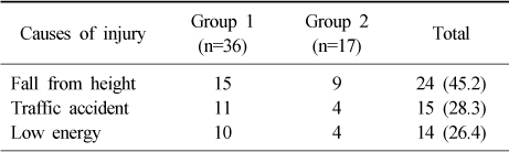


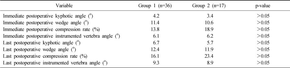
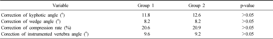
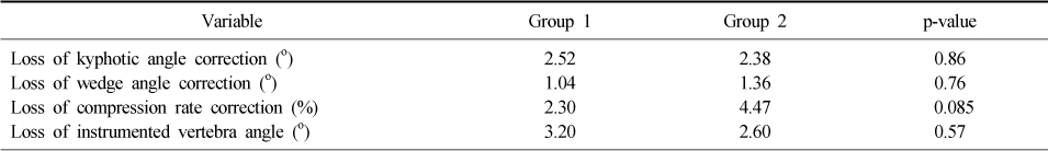

Figure & Data
REFERENCES
Citations
Citations to this article as recorded by 
 Cite
CiteComparison of Surgical Outcomes in Thoracolumbar Fractures Having 6 or Less Scored by Load-Sharing Classification Based on Posterior Fusion Level
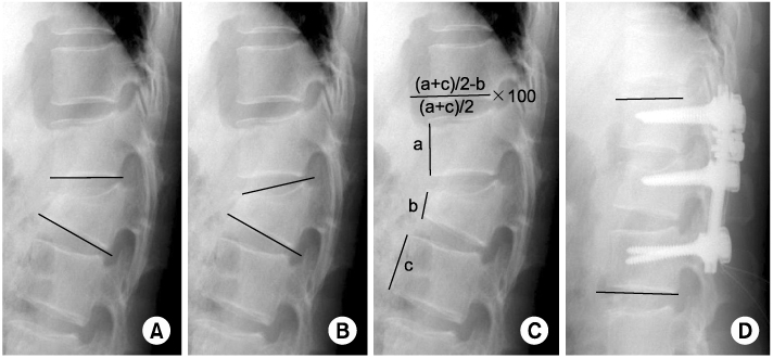
Fig. 1
Radiologic parameters.
(A) Kyphotic angle (°).
(B) Wedge angle (°).
(C) Compression rate (%).
(D) Instrumented vertebra angle (°).
Fig. 1
Comparison of Surgical Outcomes in Thoracolumbar Fractures Having 6 or Less Scored by Load-Sharing Classification Based on Posterior Fusion Level
Causes of Injury
Values are presented as number or number (%).
Injured Levels of Fractured Vertebra
Values are presented as number or number (%).
Patient Characteristics, Preoperative Variables
Immediate and Last Postoperative Variables
Correction of Postoperative Variables
Loss of Postoperative Variables
Loss of Postoperative Variables
*Significant differences (p-value<0.05).
Table 1
Causes of Injury
Values are presented as number or number (%).
Table 2
Injured Levels of Fractured Vertebra
Values are presented as number or number (%).
Table 3
Patient Characteristics, Preoperative Variables
Table 4
Immediate and Last Postoperative Variables
Table 5
Correction of Postoperative Variables
Table 6
Loss of Postoperative Variables
Table 7
Loss of Postoperative Variables
*Significant differences (p-value<0.05).

