 E-submission
 TOTA
TOTA TOTS
TOTS
Articles
- Page Path
- HOME > J Musculoskelet Trauma > Volume 30(3); 2017 > Article
-
Case Report
- Atypical Fracture-Like Insufficiency Fracture of the Tibia with Prolonged Bisphosphonate Drug: A Case Report
-
Min Jung Park, M.D.
 , Su Jin Lee, M.D., Jin Hwa Kam, M.D., Yun Tae Lee, M.D., Ju Hyung Yoo, M.D., Hyun Cheol Oh, M.D., Joong Won Ha, M.D., Yung Park, M.D., Sang Hoon Park, M.D., Seong Hoon Kim, M.D., Han Kook Yoon, M.D.
 -
Journal of the Korean Fracture Society 2017;30(3):137-141.
DOI: https://doi.org/10.12671/jkfs.2017.30.3.137
Published online: July 21, 2017
Department of Orthopaedic Surgery, National Health Insurance Service Ilsan Hospital, Goyang, Korea.
*Department of Internal Medicine, National Health Insurance Service Ilsan Hospital, Goyang, Korea.
†Department of Orthopaedic Surgery, Yonsei University College of Medicine, Seoul, Korea.
- Correspondence to: Han Kook Yoon, M.D. Department of Orthopaedic Surgery, National Health Insurance Service Ilsan Hospital, 100 Ilsan-ro, Ilsandong-gu, Goyang 10444, Korea. Tel: +82-31-900-0540, Fax: +82-31-900-0343, hangugi@gmail.com
• Received: February 20, 2017 • Revised: March 13, 2017 • Accepted: April 5, 2017
Copyright © 2017 The Korean Fracture Society. All rights reserved.
This is an Open Access article distributed under the terms of the Creative Commons Attribution Non-Commercial License (http://creativecommons.org/licenses/by-nc/4.0) which permits unrestricted non-commercial use, distribution, and reproduction in any medium, provided the original work is properly cited.
- 1,117 Views
- 3 Download
- 1 Crossref
Abstract
- Atypical femoral fracture related to a long-term bisphosphonate therapy has commonly been reported; however, a fracture at the site other than the femur has rarely been reported to date. Herein, we report a case of a patient on long-term bisphosphonate therapy who presented atypical tibial insufficiency fracture at the anterolateral aspect of diaphysis, without trauma. We, for the first time in Korea, present this case with a literature review.
- 1. Park JM, Sung KS. Stress fractures of the tibia. Arthrosc Orthop Sports Med, 2015;2:95-102.Article
- 2. Bone HG, Hosking D, Devogelaer JP, et al. Ten years' experience with alendronate for osteoporosis in postmenopausal women. N Engl J Med, 2004;350:1189-1199.Article
- 3. Shane E, Burr D, Abrahamsen B, et al. Atypical subtrochanteric and diaphyseal femoral fractures: second report of a task force of the American Society for Bone and Mineral Research. J Bone Miner Res, 2014;29:1-23.
- 4. Yang KH, Min BW, Ha YC. Atypical femoral fracture: 2015 position statement of the Korean Society for Bone and Mineral Research. J Bone Metab, 2015;22:87-91.Article
- 5. Rogers MJ, Crockett JC, Coxon FP, Mönkkönen J. Biochemical and molecular mechanisms of action of bisphosphonates. Bone, 2011;49:34-41.Article
- 6. Moon J, Bither N, Lee T. Atypical forearm fractures associated with long-term use of bisphosphonate. Arch Orthop Trauma Surg, 2013;133:889-892.ArticlePDF
- 7. Beals RK, Cook RD. Stress fractures of the anterior tibial diaphysis. Orthopedics, 1991;14:869-875.Article
- 8. Bissonnette L, April PM, Dumais R, Boire G, Roux S. Atypical fracture of the tibial diaphysis associated with bisphosphonate therapy: a case report. Bone, 2013;56:406-409.Article
- 9. Odvina CV, Levy S, Rao S, Zerwekh JE, Rao DS. Unusual mid-shaft fractures during long-term bisphosphonate therapy. Clin Endocrinol (Oxf), 2010;72:161-168.Article
- 10. Breglia MD, Carter JD. Atypical insufficiency fracture of the tibia associated with long-term bisphosphonate therapy. J Clin Rheumatol, 2010;16:76-78.Article
REFERENCES
Fig. 1

Preoperative radiograph shows atypical tibial diaphyseal fracture (white arrows), including periosteal reaction, endosteal thickening, and a radiolucent fracture line.
Fig. 2

Preoperative computed tomography shows periosteal reaction, cortical thickening, and transverse fracture line (white arrows).
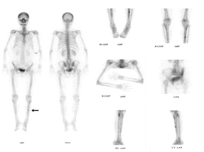
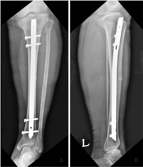
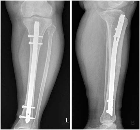
Figure & Data
REFERENCES
Citations
Citations to this article as recorded by 
- Atypical Femoral Fracture Occurring at a Proximal Screw Insertion Site after Plate Removal in a Distal Femoral Fracture
Jin Woo Jin, Sung Jin Shin, Jong Min Jeon
Journal of the Korean Orthopaedic Association.2024; 59(4): 314. CrossRef
 Cite
CiteAtypical Fracture-Like Insufficiency Fracture of the Tibia with Prolonged Bisphosphonate Drug: A Case Report
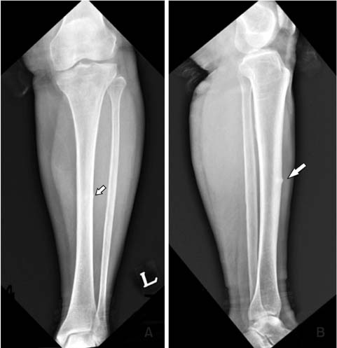
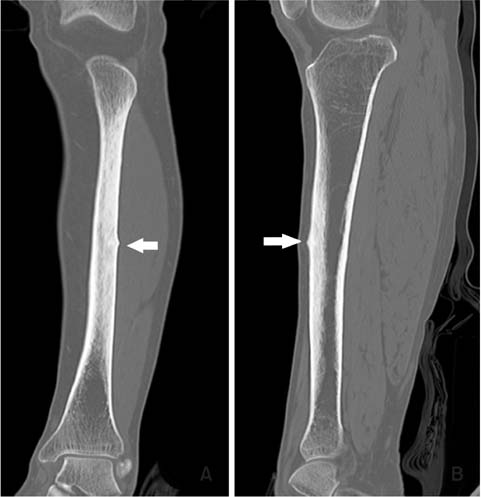


Fig. 1
Preoperative radiograph shows atypical tibial diaphyseal fracture (white arrows), including periosteal reaction, endosteal thickening, and a radiolucent fracture line.
Fig. 2
Preoperative computed tomography shows periosteal reaction, cortical thickening, and transverse fracture line (white arrows).
Fig. 3
Bone scan of this patient shows increased uptake (arrow) at the anterolateral area of tibial diaphysis.
Fig. 4
Using intramedullary nailing, operation was performed.
Fig. 5
The radiographs of tibia taken eight months after the operation show callus formation at the fracture site, with a diminished gap of fracture.
Fig. 1
Fig. 2
Fig. 3
Fig. 4
Fig. 5
Atypical Fracture-Like Insufficiency Fracture of the Tibia with Prolonged Bisphosphonate Drug: A Case Report

