 E-submission
 TOTA
TOTA TOTS
TOTS
Search
- Page Path
- HOME > Search
Original Articles
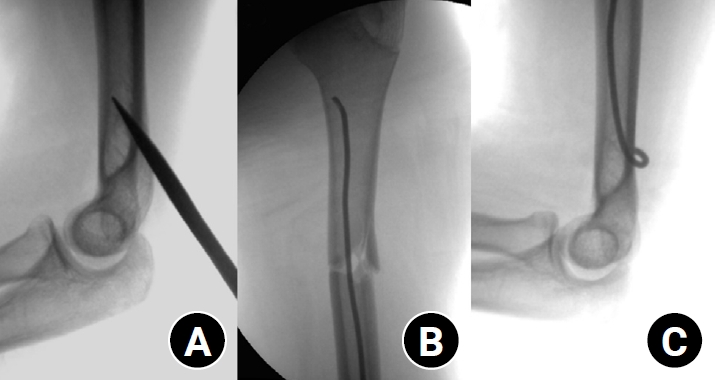
- Clinical and radiographic outcomes of elastic stable intramedullary nailing for pediatric humeral shaft fractures: a retrospective case series
- Kang-San Lee, Dongju Shin, Sang Hee Kim, Il Seo, Tae-Hoon Kim, Sung Jung Kim
- J Musculoskelet Trauma 2026;39(2):156-161. Published online March 10, 2026
- DOI: https://doi.org/10.12671/jmt.2025.00381
-
 Abstract
 PDF
PDF - Background
Pediatric humeral shaft fractures are uncommon and are generally treated conservatively, with satisfactory clinical outcomes reported in most cases. However, conservative management often necessitates prolonged immobilization and frequent outpatient follow-up visits, and it carries an inherent risk of residual angular or translational deformity. Elastic stable intramedullary nailing (ESIN) provides a simple and minimally invasive method of fracture fixation that offers adequate stability without disrupting the periosteal blood supply, thereby permitting early mobilization and promoting rapid bone union. The purpose of this study was to evaluate the clinical and radiological outcomes of ESIN fixation in pediatric patients with humeral shaft fractures.
Methods
The medical records of pediatric patients with humeral shaft fractures who underwent ESIN fixation between January 2015 and November 2025 were retrospectively reviewed. Data collected included patient demographics, mechanism of injury, fracture location, number of elastic nails used, time to union, degree of residual angulation, range of motion (ROM), and postoperative complications.
Results
The mean age of the patients was 10.0 years (range, 7 to 15 years). The mean time to radiographic union was 5.4 weeks (range, 2.4 to 10.4 weeks). The mean coronal angulation was 0.2° (range, −9.1° to 5.8°), while the mean sagittal angulation was −1.3° (range, −6.9° to 5.3°). No cases of infection, nerve injury, or nail migration were observed during the follow-up period. At the final follow-up assessment, all patients demonstrated full shoulder and elbow ROM, with no residual deformity or pain reported.
Conclusions
In this small retrospective case series, ESIN fixation resulted in favorable union rates and excellent functional outcomes in pediatric humeral shaft fractures. Level of evidence: IV.
- 634 View
- 21 Download

- Surgical Treatment of Pediatric Intra-Articular Proximal Phalangeal Head Fracture of the Big Toe
- Yeun Soo Kim, Geunwu Gimm, Il ung Hwang, Goo Hyun Baek, Jihyeung Kim
- J Korean Fract Soc 2020;33(1):9-15. Published online January 31, 2020
- DOI: https://doi.org/10.12671/jkfs.2020.33.1.9
-
Abstract
PDF
- PURPOSE
Pediatric intra-articularproximal phalangeal head fractures of the big toe are very rare and few studies on this have been published. The purpose of this study is to present the diagnostic approach and surgical management of these extremely rare fractures, which might be easily underestimated or misdiagnosed.
MATERIALS AND METHODS
The study retrospectively reviewed all the patients who were diagnosed as intra-articular proximal phalangeal head fracture of the big toe and who underwent surgical intervention in our institution. The size of the bony fragment and hallux valgus interphalangeus angle were measured on the preoperative X-rays. The size and rotation of the osteochondral fragment, the presence of avascular necrosis, ligamentous injury and soft tissue entrapment were assessed on the preoperative magnetic resonance images (MRIs). The radiologic and functional evaluation were performed at 1 year postoperatively.
RESULTS
The average size of the bony fragments measured on the X-rays was 4.1 mm in width and 2.3 mm in length. Two cases showed hallux valgus interphalangeus. Preoperative MRI was performed in four cases and the average size of any osteochondral lesion was 5.3 mm in width, 3.9 mm in length, and 4.7 mm in height. Rotation of the osteochondral fragment was observed in one patient, and soft tissue entrapment was noted in two patients. Postoperatively, successful bony union was achieved in all the patients and the average time to union was 74.4 days.
CONCLUSION
Intra-articular proximal phalangeal head fractures of the big toe are very rare and often neglected due to incomplete ossification in the pediatric population. It is important to suspect the presence of this intra-articular fracture and to appropriately implement further evaluation. Nonunion of chronic cases as well as acute fractures can be successfully treated through open reduction and internal fixation using multiple K-wires.
- 2,083 View
- 17 Download
Case Report
- Pediatric Cartilaginous Tibia Eminence Fracture Overlooked on Plain Radiograph: A Report of Two Cases
- Seong Eun Byun, Yunseong Choi, Wonchul Choi
- J Korean Fract Soc 2017;30(1):29-34. Published online January 31, 2017
- DOI: https://doi.org/10.12671/jkfs.2017.30.1.29
-
Abstract
PDF
- In children with open physis, avulsion fracture of the tibia eminence, as an anterior cruciate ligament (ACL) injury, is more commonly observed than an ACL rupture. Pure cartilaginous avulsions of the ACL tibia insertion seldom occurs. In such case, cartilaginous lesion is frequently overlooked or misdiagnosed on plain radiograph and may result in a less favorable treatment outcome. We report two cases of cartilaginous tibia eminence fractures of the children that were initially overlooked from plain radiographs, and then diagnosed by magnetic resonance imaging, which was ultimately treated by arthroscopyassisted headless compression screw fixation.
- 855 View
- 5 Download
Original Article
- The Pattern of Occurrence of Fractures in Children and Adolescents and Its Managements Based on the Database of the Health Insurance Review and Assessment Service
- Yong Wook Kwon, Soon Hyuck Lee, Hyun Woo Kim, Jin Ho Hwang
- J Korean Fract Soc 2014;27(4):308-314. Published online October 31, 2014
- DOI: https://doi.org/10.12671/jkfs.2014.27.4.308
-
Abstract
PDF
- PURPOSE
The purpose of this article is to report on the pattern of medical process and relative frequencies of fractures in children and adolescents.
MATERIALS AND METHODS
The authors retrospectively analyzed the database of the health insurance review and assessment service regarding children and adolescents under 20 years old treated from 2008 to 2010. Newly registered numbers of fractures in children and adolescents according to sex, month, institution, and anatomical location were also reviewed.
RESULTS
A total of 1,893,416 fractures occurred during three years; approximately 630,000 cases were treated during one year (approximately 562 cases among 10,000 people during one year). During one year, the most fractures occurred in June and the least in February. Senior general hospital consisted of 5.72%, 12.30% in general hospital, 19.28% in hospital, and 62.70% in clinics. Among the fracture sites, 0.05% were cervical fractures, 0.91% in sternum and thoracic vertebra, 1.35% in lumbar vertebra and pelvis, 12.79% in shoulder and upper extremities, 26.87% in lower extremities, 38.10% in wrist and hand, 1.01% in femur, 10.40% in lower extremities including ankle, and 8.52% in foot excluding ankle. The maximal incidence was age 14 years in male and 12 years in female.
CONCLUSION
The authors reviewed the pattern of medical process and relative frequencies of fractures in children and adolescents. -
Citations
Citations to this article as recorded by
- Temporal trend of pediatric fracture epidemiology in Thailand: a nationwide study 2018–2024
Nattakarn Numsriskulrat, Chatchai Suesirisawad, Ratikorn Chaisiwamongkol, Putthapong Ponpitak, Ouyporn Panamonta, Kaewjai Thepsuthammarat, Pattara Wiromrat
Osteoporosis International.2026;[Epub] CrossRef - Analysis of Computed Tomography Scans for Radiation Safety Management in the Republic of Korea
Min Young Lee, Ji Woo Kim, Ga Eun Oh, Geon Woo Son, Kwang Pyo Kim
Journal of Radiation Protection and Research.2024; 49(3): 141. CrossRef
- Temporal trend of pediatric fracture epidemiology in Thailand: a nationwide study 2018–2024
- 1,109 View
- 3 Download
- 2 Crossref
Case Report
- T-Condylar Fracture of Distal Humerus in a Child: A Case Report
- Young Ryeol Pae, Sang Soo Kang, Hyeong Min Kim, Min Jeong
- J Korean Fract Soc 2014;27(3):232-236. Published online July 31, 2014
- DOI: https://doi.org/10.12671/jkfs.2014.27.3.232
-
Abstract
PDF
- T-condylar fracture is a type of distal humerus fracture. T-condylar fracture in children is rare, with reported incidence of less than 1% of T-condylar fractures. The mean reported age of T-condylar fracture in children is 11. Cases in children under 5 years-old are extremely rare. Herein, we report on a T-condylar fracture of the distal humerus in a 5-year-old boy. This patient was treated with open reduction and K-wire fixation through the posterolateral approach. The result of treatment was satisfactory; therefore, we report this case.
- 1,241 View
- 14 Download
Original Articles
- Interposition of Periosteum in Distal Tibial Physeal Fractures of Children
- Phil Hyun Chung, Suk Kang, Jong Pil Kim, Young Sung Kim, Jae Woo Cho
- J Korean Fract Soc 2011;24(1):73-78. Published online January 31, 2011
- DOI: https://doi.org/10.12671/jkfs.2011.24.1.73
-
Abstract
PDF
- PURPOSE
To evaluate the factors influencing periosteal interposition in distal tibial physeal fractures of children.
MATERIALS AND METHODS
34 cases of distal tibial physeal fractures were analysed. We confirmed the presence of periosteal interposition with MRI in all cases and accessed the relationship between periosteal interposition and gender, age, cause of injury, type of fracture, degree of initial displacement and after closed reduction.
RESULTS
9 (26.5%) of 34 fractures had interposed periosteum. There was no statistically significant correlation between periosteal interposition and gender, age, cause of injury (p>0.05). 5 (83.3%) of 6 pronation-eversion-external rotation type of fractures according to Dias-Tachjian classification had interposed periosteum and that was a statistically significant correlation (p=0.006). As Salter-Harris type was toward to high degree, there were decreasing tendency of periosteal interposition (p=0.026). There was high rate of periosteal interposition in case of displacement more than 2 mm in each initial and after closed reduction (p<0.05).
CONCLUSION
There was high incidence of periosteal interposition in pronation-eversion-external rotation type with displacement more than 2 mm in distal tibial physeal fractures of children. But, periosteal interposition could occur in fractures with mild displacement less than 2 mm, if initial fracture displacement was more than 2 mm, the methods of treatment should be decided after confirm the presence of periosteal interposition with MRI after closed reduction.
- 1,124 View
- 8 Download
- Treatment of Femoral Shaft Fracture with Interlocking Humeral Nail in Older Children and Adolescent
- Kun Bo Park, Hoon Park, Hyun Woo Kim, Hui Wan Park, Jae Young Roh
- J Korean Fract Soc 2010;23(2):206-212. Published online April 30, 2010
- DOI: https://doi.org/10.12671/jkfs.2010.23.2.206
-
Abstract
PDF
- PURPOSE
To evaluate the results of interlocking humeral nail for femur shaft fractures through the greater trochanter in older children and adolescent.
MATERIALS AND METHODS
Eleven femoral shaft fractures in ten patients were selected. They were consisted of 9 boys and 1 girl. Two patients had osteogenesis imperfecta and one patient had a simple bone cyst as an underlying disease. 7 cases were right side and 4 cases were left side. The mean age at the time of operation was 12 years and 7 months (8 years 11 months~15 years 7 months). The mean follow-up period was 21 months and interlocking humeral nail was inserted at the greater trochanter in all patients.
RESULTS
All patients had a complete bony union without any complication such as infection, nonunion, leg length discrepancy and metal failure. Avascular necrosis of femoral head and coxa valga were not developed in all patients.
CONCLUSION
Intramedullary nailing through the greater trochanter using interlocking humeral nail is effective and safe treatment for the femoral shaft fracture in older children and adolescents.
- 996 View
- 2 Download
Case Report
- Bilateral Malunion and Distal Radioulnar Joint Dislocation after Operative Treatment of Bilateral Galeazzi Fractures in Child: A Case Report
- Sang Jin Cheon, Dong Joon Kang, Nam Hoon Moon, Seung Han Cha, He Myung Cho
- J Korean Fract Soc 2009;22(4):292-296. Published online October 31, 2009
- DOI: https://doi.org/10.12671/jkfs.2009.22.4.292
-
Abstract
PDF
- Galeazzi fractures in child is rare and seldom necessary of operative treatment because the result of conservative treatment is good. We present the patient who was a 11-year-old male and fell onto his both hands during a hundred-meter dash. His diagnosis was bilateral Galeazzi fractures and limited open reduction and internal fixation with Kirschner pins was initial treatment at local hospital. After 4 weeks postoperatively, Kirschner pins were removed and rehabilitating exercise was started. After 4 months postoperatively, he was transferred to our hospital due to malunion with severe angular deformities and distal radioulnar joint (DRUJ) dislocation. He was treated with corrective osteotomy. Thus, as in this case, we suggest more careful treatment and observation if conservative method of Galeazzi fracture in child is chosen and consider operative method as treatment according to age and pattern of fracture.
- 955 View
- 1 Download
Original Article
- Comparison of the Surgical Treatment Results of Avulsion Fracture of the Anterior Cruciate Ligament between Children and Adults
- Eun Kyoo Song, Sang Jin Park, Keun Bae Lee
- J Korean Fract Soc 2007;20(2):196-201. Published online April 30, 2007
- DOI: https://doi.org/10.12671/jkfs.2007.20.2.196
-
Abstract
PDF
- PURPOSE
To compare the clinical and radiological results after surgical treatments of the avulsion fractures of ACL between children and adults.
MATERIALS AND METHODS
40 cases (18 cases of children, 22 cases of adults), who underwent surgical treatments after avulsion fractures of the ACL and followed up more than one year, were enrolled. Fractures were classified by modified Meyers & McKeever criteria. Range of motion, LK score, Lachman test, Pivot-Shift test, quadriceps muscle atropy and Telos® stress arthrometer were compared.
RESULTS
The types of fracture in children were categorized into 8 cases of type II, 10 cases of type III, and 2, 15, 5 cases of type II, III, IV each in adult group. Mean LK score showed significant difference between 99.3 points in children and 89.5 points in adults (p<0.05). In addition, accompanied injuries and the high degree of fracture leaded low LK score. However, there was no significant difference in range of motion, Lachman test and Pivot-Shift test. Anterior laxity by Telos® device showed an average of 2.0 mm in children, 2.5 mm in adults (p>0.05).
CONCLUSION
Children group showed better treatment results of avulsion fracture of ACL. Higher incidence of type II fractures and less combined injuries considered to be factors for better results.
- 870 View
- 1 Download
Case Report
- Traumatic Pseudoaneurysm of Posterior Tibial Artery in a Child: A Case Report
- Tai Seung Kim, Kuhn Sung Whang, Woo Young Seo
- J Korean Fract Soc 2007;20(1):83-85. Published online January 31, 2007
- DOI: https://doi.org/10.12671/jkfs.2007.20.1.83
-
Abstract
PDF
- Pseudoaneurysm is one of the complications of arterial injuries by trauma. The case report in children is rare, although not in adult. A 7-year and 10-month girl was visited with the complaints of pain and a mass in her right leg. At first, the radiograph of right tibia showed a remarkable cortical erosion from without, suggesting mass effect by a soft tissue tumor. She had a history of fracture of right tibia, and then manipulative reduction and K-wire fixation at 11 months ago. Arteriography showed a formation of the pseudoaneurysm originated from the posterior tibial artery. The operation was done through the ligation of artery at proximal and distal to pseudoaneurysm, and then excision of mass. At 5 year follow-up, the configuration and function of right foot was normal. Eventually, the cause of the mass formation is thought by the trauma of fracture fragment at the time of accidents, but the possibility of penetrated injuries by K-wire should be ruled out, which is used frequently in children's fracture. We experienced a case of traumatic pseudoaneurysm of posterior tibal artery with tibial fracture, especially occurred in pediatric patient, and presented the result of long-term follow-up.
-
Citations
Citations to this article as recorded by- Coil Embolization of a Pseudoaneurysm of the Anterior Tibial Artery: A Case Report
Tae-Hyun Wang, Hyung-Lae Cho, Ki-Bong Park, Duc-Hee Kim
Journal of Korean Foot and Ankle Society.2016; 20(1): 43. CrossRef
- Coil Embolization of a Pseudoaneurysm of the Anterior Tibial Artery: A Case Report
- 1,220 View
- 3 Download
- 1 Crossref
Original Articles
- Lateral Condylar Fracture of the Humerus in Children: An Epidemiological Analysis of 158 Cases
- Chul Hyun Cho, Kwang Soon Song, Sung Won Sohn, Ki Chul Bae, Jung Hoon Lee
- J Korean Fract Soc 2006;19(4):466-470. Published online October 31, 2006
- DOI: https://doi.org/10.12671/jkfs.2006.19.4.466
-
Abstract
- PURPOSE
To analyze the correlation of various factors by examining the epidemiology of lateral condylar fracture of the humerus which is the second most fracture among elbow fractures in children.
MATERIALS AND METHODS
Of 158 cases treated for lateral condylar fracture of the humerus in children from April 1996 to March 2006, their age and sex distribution, the seasonal frequency, etiology, type of fracture, method of treatment, etc. were analyzed retrospectively.
RESULTS
Boys were 113 cases, girls were 45 cases, and the mean age was 5.4 years. Regarding the seasonal occurrence, spring 43 cases, summer 44 cases, autumn 48 cases, and winter 23 cases had occurred. It occurred preferentially during the season when outdoor activity was most active. As its etiology, the accident in a playground was 39 cases, sports activity was 32 cases, traffic accident was 17 cases, slipping accident at home was 15 cases, falling accident at home was 14 cases, slip while playing with friends was 6 cases, a missing step while walking on stairs was 6 cases, fall from a height more than 2 floors was 4 cases, and the cases with unknown cause were 25 cases. According to the Jakob stage, the stage I was 42 cases, the stage II 77 cases, and the stage III was 39 cases. As treatment, cast immobilization was performed in 34 cases, closed reduction and percutaneous K-wire fixation was performed in 68 cases, and open reduction and K-wire fixation was performed in 56 cases. The prevalent causalities were play devices, accident during sports activity, and traffic accident, and in such cases, the displacement of fracture was severe and thus surgical treatments were performed in many cases (94%).
CONCLUSION
It is thought that during the season when outdoor action is active, particularly, for kindergarten children or the lower grade primary school children, safety education is required to prevent the fracture by play devices, sports activity and traffic accident. -
Citations
Citations to this article as recorded by- The Pattern of Occurrence of Fractures in Children and Adolescents and Its Managements Based on the Database of the Health Insurance Review and Assessment Service
Yong-Wook Kwon, Soon-Hyuck Lee, Hyun-Woo Kim, Jin-Ho Hwang
Journal of the Korean Fracture Society.2014; 27(4): 308. CrossRef
- The Pattern of Occurrence of Fractures in Children and Adolescents and Its Managements Based on the Database of the Health Insurance Review and Assessment Service
- 991 View
- 0 Download
- 1 Crossref
- Separation of the Symphysis Pubis during Childbirth
- Dong Ju Shin, Young Soo Byun, Se Ang Chang, Ok Rang Park, Shin Yoon Kim, Dae Hee Hwang, Sung Rak Lee, Dong Young Kim
- J Korean Fract Soc 2006;19(4):412-417. Published online October 31, 2006
- DOI: https://doi.org/10.12671/jkfs.2006.19.4.412
-
Abstract
- PURPOSE
To evaluate the clinical features and incidence of separation of the symphysis pubis during childbirth, and to evaluate the risk factors of the lesion and the outcome of treatment.
MATERIALS AND METHODS
Seventy two cases of separation of symphysis pubis among 66,721 delivery between January 1992 and December 2004 was selected. The control group was composed of 498 cases without separation of symphysis pubis during childbirth. Several factors increasing the risk of this lesion were assessed using χ
- 686 View
- 0 Download
- External Fixation of Pediatric Femur Fractures
- Yeung Jin Kim, Tae Kyun Kim, Hwan Deok Yang, Hyung Joon Kim, Jin Young Park, Sang Jin Eun
- J Korean Fract Soc 2006;19(3):369-373. Published online July 31, 2006
- DOI: https://doi.org/10.12671/jkfs.2006.19.3.369
-
Abstract
- PURPOSE
To evaluate unilateral external fixation when applied as the standard treatment of displaced femoral shaft fractures in children.
MATERIALS AND METHODS
From 2000 through 2004, we used a unilateral external fixator (Any-fix(R)) to treat 24 femoral shaft fractures. The average age of the patients was 8.3 years (range, 5.6 to 14.8). 16 fractures were isolated, and 8 were associated with polytrauma. There were 4 open fractures. Patients were followed clinically and radiologically until healing and at 1 year.
RESULTS
Average time of external fixation was 97 days (range, 57 to 130 days). All patients regained the normal range of motion of knee joint without significant residual leg length discrepancy or growth disturbance. There were no nonunion, or rotationary deformities. There were 26 pin tract infection (total pin number: 108) (24%), all of which were resolved with antibiotics. No patient developed osteomyelitis. There were two refractures after fixator removal. There was one case of reduction loss and one of valgus deformity.
CONCLUSION
The external fixation is a useful alternative for operative management of femoral shaft fractures because of minimal invasive operation, and early mobilization in prepuberty.
- 776 View
- 0 Download
- Treatment of Pediatric Displaced Supracondylar Fractures of the Humerus by Pin Leverage Technique
- Han Yong Lee, Joo Hyoun Song
- J Korean Fract Soc 2006;19(1):83-88. Published online January 31, 2006
- DOI: https://doi.org/10.12671/jkfs.2006.19.1.83
-
Abstract
- PURPOSE
To evaluate a new treatment method by pin leverage technique in Gartland type III fractures to avoid forceful manipulation or open reduction.
MATERIALS AND METHODS
99 cases were included in this study and divided into 3 groups (I;open reduction, II; closed reduction and percutaneous pin fixation, III; pin leverage technique), and we analyzed timing to operation, length of operation, associated neurovascular injuries, complications, and clinical and radiological outcomes at final follow-up.
RESULTS
The average length of operation 119, 57, and 68 minutes respectively. The associated nerve injuries were 8, 2, and 2 cases respectively. There were a case of superficial pin tract infection in group I, three cases of superficial pin tract infection and a case of iatrogenic ulnar nerve injury in group II. At final follow-up, clinical results were excellent or good in all cases and there were 5 cases (8.3%) of fair results in group II radiologically. Closed reduction with pin leverage technique were failed in 5 cases.
CONCLUSION
In treatment of Gartland type III fractures, pin leverage reduction technique is considered to be a good alternative prior to open reduction, because it provides shortened length of operation, avoidance of forceful manipulation and open reduction. -
Citations
Citations to this article as recorded by- Recent Trends in Treatment of Supracondylar Fracture of Distal Humerus in Children
Soon Chul Lee, Jong Sup Shim
Journal of the Korean Fracture Society.2012; 25(1): 82. CrossRef
- Recent Trends in Treatment of Supracondylar Fracture of Distal Humerus in Children
- 905 View
- 0 Download
- 1 Crossref
- Treatment of Tibial Fractures In Children With Pin and Plaster Technique
- Byoung Ho Suh, Gyu Min Kong, Sang Ho Moon, Dong Joon Kim, Jin Woo Kwon, Se Won Park
- J Korean Fract Soc 2005;18(3):325-329. Published online July 31, 2005
- DOI: https://doi.org/10.12671/jkfs.2005.18.3.325
-
Abstract
PDF
- PURPOSE
To evaluate the result of tibial shaft fractures in children treated with pin and plaster method.
MATERIALS AND METHODS
From March 1998 to February 2003, Tibial shaft fractures in thirty six pediatric patients which were treated with pin and plaster method were clinically and radiologicaly evaluated retrospectively.
RESULTS
Mean bony union duration was 9.8 weeks. All fractures healed within acceptable angulations. There was neither delayed union nor nonunion. There were complications related to the pins, including superficial and deep infection, skin sloughing. There were 7 cases of tibial overgrowth but they had no functional disability.
CONCLUSION
Pin and plaster method can substitute other operative methods in tibial fractures in children which is difficult to reduce or maintain reduction by conservative treatment.
- 731 View
- 2 Download
- Refractures of Upper Extremity in Children
- Hui Wan Park, Dae Ya Kim, Hyun Woo Kim
- J Korean Fract Soc 2004;17(4):389-394. Published online October 31, 2004
- DOI: https://doi.org/10.12671/jkfs.2004.17.4.389
-
Abstract
PDF
- PURPOSE
To investigate the etiologic factors related to refractures of the upper extremity in children MATERIALS AND METHODS: 18 refractures of the upper extremity were divided into three groups according to the location of initial fractures: Supracondyle fractures of the humerus, lateral condyle fracture of the humerus, and the forearm bone fractures. They were analyzed in terms of the type of refractures (early refracture occurring at the immature callus and late refracture occurring at the remodeled bone), fracture patterns, and the existence of underlying deformity.
RESULTS
Nine supracondyle fractures had refractures at the supracondyle (2 cases) and the lateral condyle (7 cases), in which underlying cubitus varus were present in 6 cases. Three lateral condyle fractures had refractures at the supracondyle (1 case) and the lateral condyle (2 cases), in which one case had underlying cubitus varus. All but one case in the group of humerus fractures were late refractures and treated operatively except one. Of 6 refractures of forearm, 5 were early refractures and occurred within 9 weeks at the original site: 4 at the diaphysis of both bones of forearm and 1 at the diaphysis of ulna. All cases in the group of forearm fractures had volar angulation before the refracture, and treated conservatively except one CONCLUSION: In the humerus, underlying cubitus varus was the most important predisposing factor to refractures and the lateral condyle fractures were common. In the forearm, volar angulation of the diaphysis were related to refractures, and complete and circular consolidation of the primary fracture of forearm was thought to be important to prevent refracture. -
Citations
Citations to this article as recorded by- Characteristics and trends in heavy rainfall and storm damage to cultural heritage over the past 15 years (2007 ~ 2021)
Jisoo Kim
Journal of Climate Change Research.2023; 14(4): 425. CrossRef
- Characteristics and trends in heavy rainfall and storm damage to cultural heritage over the past 15 years (2007 ~ 2021)
- 933 View
- 1 Download
- 1 Crossref
- Treatment of Tibial Shaft Fractures in Children Using K-wires Fixation
- Phil Hyun Chung, Chung Soo Hwang, Suk Kang, Jong Pil Kim, Ho Jun Cheon
- J Korean Fract Soc 2004;17(4):384-388. Published online October 31, 2004
- DOI: https://doi.org/10.12671/jkfs.2004.17.4.384
-
Abstract
PDF
- PURPOSE
To report the effectiveness of Kirschner wire fixation for the treatment of unstable tibial shaft fractures in children.
MATERIALS AND METHODS
We analyzed 15 cases of pediatric tibial shaft fractures treated at our hospital with fixation using K-wire and followed up for more than 1 year from July 1998 to January 2002. The subjects included 11 boys and 4 girls. The ages ranged from 3 to 10 years at the time of injury, with the average age being 7.9 years. We examined the presence of angulation, leg length discrepancy, joint motion limitation, and complications.
RESULTS
Bony fusion was obtained in all patients by an average of postoperative 9.5 weeks. At the time of last follow-up (by an average of postoperative 1 year and 4 months), anterior and posterior radiographs showed an average of 4.2degree angulation, and lateral radiographs showed an average of 4.4degree angulation. The affected leg was extended by an average of 3.7 mm compared to the opposite leg according to Bell-Thompson's radiographs. As for complications, infection was developed around the pin in 3 cases but treated with the administration of oral antibiotics and sterilization around the site without progressing to deep infection. We could not observe joint motion limitation, pain and difficulties related with discrepancy in leg length.
CONCLUSION
We concluded that fixation using K-wire for children with tibial shaft fractures was a safe and effective method of surgery that could be performed easily, did not require secondary surgery to remove the wire, and showed sufficient stability after fixation.
- 891 View
- 6 Download
- Modified Step-cut Osteotomy of Distal Humerus for the Correction of Cubitus Varus Deformity in Children
- Yeo Hon Yun, Jun Gyu Moon, Duk Moon Chung
- J Korean Fract Soc 2004;17(3):287-294. Published online July 31, 2004
- DOI: https://doi.org/10.12671/jkfs.2004.17.3.287
-
Abstract
PDF
- PURPOSE
evaluate the radiologic and clinical results of modified step-cut osteotomy for correction of cubitus varus deformity in children.
MATERIALS AND METHODS
We analysed 16 children who had varus deformity preoperatively and received modified step-cut osteotomy. The results were evaluated by final follow-up radiographs and clinical results, which were humeral-elbow-wrist angle, lateral prominence, range of motion and complications.
RESULTS
The average preoperative humeral-elbow-wrist (HEW) angle was -15.8degrees and average last follow-up HEW angle was +6.7degrees Lateral prominence under 5 mm occurred in 3 cases and one children showed limited motion and transient ulna neuropathy.
CONCLUSION
The results demonstrate that modified step-cut osteotomy achieve good correction of cubitus varus without lateral bony prominence or complications.
- 724 View
- 3 Download
- Treatment of Fractures of the Hip in Children
- Do Hyun Moon, Jang Seok Choi, Jong Hun Lee
- J Korean Fract Soc 2004;17(3):283-286. Published online July 31, 2004
- DOI: https://doi.org/10.12671/jkfs.2004.17.3.283
-
Abstract
PDF
- PURPOSE
To evaluate the result of early anatomical reduction and internal fixation of hip fracture in children.
MATERIALS AND METHODS
From January 1996 to July 2002, 21 cases (mean, 9 years) of hip fracture were available for follow-up more than 1 year. We performed early anatomical reduction and internal fixation within 24 hours as possible. Fractures were classified according to the 4 types described by Delbet. The results were analyzed according to the functional results by Ratliff and the incidence of complication.
RESULTS
There were no type I, 7 type II, 10 type III and 4 type IV fractures. Avascular necrosis of femoral head in 2 cases (type II, III). Functional result was 18 Good, 1 Fair and 2 Poor.
CONCLUSION
Fractures of the hip in children have been associated with a very high rate of serious complications, but our treatment by early anatomical reduction and interal fixation reduced rates of complication and had good functional result.
- 689 View
- 3 Download
- Reduction of Pediatric Forearm Diaphyseal Fractures by Pin Leverage Technique
- Soo Hong Han, Duck Yun Cho, Hyung Ku Yoon, Byung Soon Kim, Sung Hoon Kang, Tae Hyung Kim
- J Korean Fract Soc 2004;17(1):59-63. Published online January 31, 2004
- DOI: https://doi.org/10.12671/jkfs.2004.17.1.59
-
Abstract
PDF
- PURPOSE
Although the majority of children's forearm diaphyseal fractures may be treated conservatively with closed reduction and cast immobilization, unstable or irreducible fractures are usually treated by surgical management. Authors performed percutaneous pin leverage reduction technique for irreducible displaced diaphyseal fractures. The aim of this study is to determine the efficacy of pin leverage technique in pediatric forearm diaphyseal fractures MATERIALS AND METHODS: In this retrospective study, we reviewed 22 cases of forearm diaphyseal fractures reduced by percutaneous pin leverage technique between 1997 and 2002. We analyzed radiographs, operation time, hospital stay and immobilization period, range of motion, postoperative complications and functional results by Thomas.
RESULTS
Average length of follow up was 28 months with mean age of 10.5 years. All fractures in this series healed less than 2 degrees of diaphyseal angulation. Average operation time including anesthesia was 42 minutes and hospital stay was 4.6 days. Time to union was 49.6 days in average and range of motion and functional results were satisfactory in all cases except one case of congenital radioulnar synostosis. There was one case of superficial pin track infection as complication.
CONCLUSION
In operative treatment of children's diaphyseal fractures of forearm bones, percutaneous pin leverage reduction technique is a good alternative method prior to open reduction in case of difficult closed reduction. -
Citations
Citations to this article as recorded by- Pediatric Forearm Bone Fractures Treated with Flexible Intramedullary Nail
Suk Kyu Choo, Jin Hwan Kim, Hyung Keun Oh, Dong Hyun Kim
Journal of the Korean Fracture Society.2007; 20(2): 190. CrossRef
- Pediatric Forearm Bone Fractures Treated with Flexible Intramedullary Nail
- 1,017 View
- 2 Download
- 1 Crossref
- Nancy Nail Fixation for Femur Shaft Fracture in Children
- Ki Do Hong, Sung Sik Ha, Nam Sik Chung, Jae Cheon Sim, Jae Young Kim
- J Korean Soc Fract 2003;16(4):592-599. Published online October 31, 2003
- DOI: https://doi.org/10.12671/jksf.2003.16.4.592
-
Abstract
PDF
- PURPOSE
To investigate, the radiologically, the duration of bone union, angular formation, leg length discrepancy, other complications and mean hospital stay after Nancy nail fixation has been performed in children with femoral shaft fracture, and also to inquire into the clinical validity of such procedure. MATERIAL AND METHOD: Included in this study were 12 patients who had been treated with the Nancy nail fixation for the femoral shaft fracture and then followed up for a year or more. The age distribution ranged from 4 to 12 years with mean age 7.2 years. After the fracture was reduced under an imaging intensifier, 2 or 3 Nancy nails were pinned onto the medial and lateral femur distally.
RESULTS
The average duration for complete union was 9.9 weeks. Any angular formation over 5 degrees was notfound. Leg length discrepancy ranged from 2 mm shortening to 12 mm overgrowth with a mean value of 2.8 cm. In one case, with overgrowth over 10 mm or more, there was no gait disturbance. In all cases, There was neither infection, delayed union, nor any motion disturbance. A nail was moved distally in one case and skin irritation was evident in another case. The mean hospital stay was 17.3 days.
CONCLUSION
Nancy nail fixation in pediatric femoral shaft fracture relatively has less complications and is a safe surgical procedure. In addition, it helps in reducing hospital stay. -
Citations
Citations to this article as recorded by- Comparison of Flexible Intramedullary Nailing with External Fixation in Pediatric Femoral Shaft Fractures
Do-Young Kim, Sung-Ryong Shin, Un-Seob Jeong, Yong-Wook Park, Sang-Soo Lee, Keun-Min Park
The Journal of the Korean Orthopaedic Association.2008; 43(1): 30. CrossRef
- Comparison of Flexible Intramedullary Nailing with External Fixation in Pediatric Femoral Shaft Fractures
- 2,073 View
- 5 Download
- 1 Crossref
- Treatment of Completely Displaced Supracondylar Fracture of Humerus in Children
- Bu Hwan Kim, Mu Jung Heo, Won Jun Hwang
- J Korean Soc Fract 2003;16(4):585-591. Published online October 31, 2003
- DOI: https://doi.org/10.12671/jksf.2003.16.4.585
-
Abstract
PDF
- PURPOSE
We performed a retrospective study of completely displaced supracondylar fracture of humerus in children to evaluate the result of primary open reduction and internal fixation for this injuries, and also to know whether it is recommendable or not for such injuries.
MATERIALS AND METHODS
For 5 years duration from March '96 to Feb. '01, we treated 58 cases of completely displaced supracondylar fractures of humerus in children. Among them we performed primary open reduction and internal fixation of this fracture in 14 cases. We followed up those cases more than 18 months and evaluated the results by Flynn et al. and Mark et al. criteria.
RESULTS
Eleven of them resulted in excellent grading by both Flynn and Mark criteria. Three cases not involved in excellent grading proved to be good result by Mark criteria. But by Flynn criteria, 2 cases showed good results and the other one proved to be fair. In all cases, the patient and parents were satisfied cosmetically and functionally. Only in one case, the recorded grading was different, fair by Flynn and good by Mark criteria.
CONCLUSION
Primary open reduction and internal fixation in the treatment of completely displaced and not easily reduced supracondylar fracture of humerus in children resulted excellent and good results with few complications, and patients were satisfied with the results functionally and cosmetically. So we recommend proceeding to primary open treatment and internal fixation for these difficult fractures with low threshold to open reduction. -
Citations
Citations to this article as recorded by- Treatment of displaced supracondylar fractures of the humerus in children by a pin leverage technique
H.-Y. Lee, S.-J. Kim
The Journal of Bone and Joint Surgery. British volume.2007; 89-B(5): 646. CrossRef
- Treatment of displaced supracondylar fractures of the humerus in children by a pin leverage technique
- 992 View
- 1 Download
- 1 Crossref
- Flexible Intramedullary Nailing in Children's Femoral Shaft Fractures
- Yeo Hon Yun, Chang Ho Choi, Jae Hak Jung
- J Korean Soc Fract 2003;16(3):385-391. Published online July 31, 2003
- DOI: https://doi.org/10.12671/jksf.2003.16.3.385
-
Abstract
PDF
- PURPOSE
We report a treatment result and the pros-cons of the flexible intramedullary nailing for femoral shaft fractures in children between the ages of 4 and 11 years.
MATERIALS AND METHODS
During the recent three years, 28 femoral shaft fractures in 27 consecutive pediatric patients were treated with flexible intramedullary nailing. We retrospectively reviewed their clinical and radiological records, followed-up for at least one year, in respects to the recovery of knee joint motion; time of weight bearing; time of fracture union; period of admission and rehabilitation; angular deformity and leg length discrepancy; and other complications.
RESULTS
In all children, the knee joint motion was rapidly recovered to near normal range within 2~4 weeks. Partial weight bearing with wearing functional brace was possible within 2~4 weeks, while full weight bearing without brace was started until 6~12 (average 8.4) weeks after the nailing. In the last follow-up radiographs, five cases (18%) showed an angular deformity in any direction of more than 5 degrees. Two children represented leg length discrepancy of more than 1 cm. Other complications were one fixation failure, and one deep soft tissue infection at the entry point of the nail.
CONCLUSION
We strongly recommend the flexible intramedullary nailing in this injury because the fixation is strong enough to permit early knee motion and weight bearing in orthosis, the fracture healing was so rapid without any case of delayed or nonunion, and the incidences of residual angular deformity and leg length discrepancy were significantly less than the nonoperative treatment. -
Citations
Citations to this article as recorded by- Comparison of Flexible Intramedullary Nailing with External Fixation in Pediatric Femoral Shaft Fractures
Do-Young Kim, Sung-Ryong Shin, Un-Seob Jeong, Yong-Wook Park, Sang-Soo Lee, Keun-Min Park
The Journal of the Korean Orthopaedic Association.2008; 43(1): 30. CrossRef
- Comparison of Flexible Intramedullary Nailing with External Fixation in Pediatric Femoral Shaft Fractures
- 1,017 View
- 0 Download
- 1 Crossref
- The Analysis of the Percutaneuos Pinning of Supracondylar Fractures in Children
- SangMok Lee, SoHak Jung, JaeHo Jang, JungHwan Son, JaeDo Kim
- J Korean Soc Fract 2003;16(2):284-291. Published online April 30, 2003
- DOI: https://doi.org/10.12671/jksf.2003.16.2.284
-
Abstract
PDF
- PURPOSE
Supracondylar fracture was known as supracondylar dilema because of complication and difficulty in treatment. Many methods were devised to prevent and minimize the complications. Among the treatment methods, closed reduction and percutaneous pinning was widely used for most of supracondylar fractures. But arguments was existed for the the pinning methods such as pinning site and number of pins.
MATERIALS AND METHODS
We reviewed 44 cases of displaced supracondylar fractures treated by closed reduction and percutaneous pining. The pins were removed about 4~5 weeks after operation and range of motion was begun. All cases was reviewed restropectively and results was analysed by Flynn grading system. The pinning method was analysed.
RESULTS
There were no significant clinical results among the methods of percutaneous pinnings. Even though two crossed pins from the lateral and medial condyles were preferable for most fractures. The results are that 1)At least 2 K-wires were needed. 2)Prognosis was not influenced by the pinning site. 3)The complication was prevented by accurate reduction using image intensifier during operation.
CONCLUSION
Two lateral parallel pins were alternative method for marked swelling and severely displaced fractures.
- 706 View
- 0 Download
- Analysis of Elbow Injuries 'pattern in Children
- Soon Hyuck Lee, Jong Woong Park, Sang Won Park, Kwang Suk Lee, Dang Jae Im, Tae Ha Kim, Sang Won Lee
- J Korean Soc Fract 2003;16(1):98-103. Published online January 31, 2003
- DOI: https://doi.org/10.12671/jksf.2003.16.1.98
-
Abstract
PDF
- PURPOSE
The purpose of this report is to describe the pattern of elbow injuries and the incidence of the different fracture types in children.
MATERIALS AND METHODS
The records of 445 in-patient children treated for the elbow injuries for 5 years were reviewed and analyzed statistically.
RESULTS
The average age of 445 children was 6.7 years. The average age of boys(6.9 years) was about 1 year older than girls(5.9 years). The age group of 4 - 7 years is the majority(50%), followed by the age group of 8 - 11 years(22%). The boy to girl ratio was approximately 2:1. The male predominance changed with the age and appeared dramatically in the age group of 12-16 years( 6.7:1 ). Left elbow was injured more frequently(60%). Left side predominance was accentuated in girls compared to boys(69% vs 55%), especially in the age group of 8 - 16 years (80% vs 50%). Thirty percent of the fractures occurred during the summer, followed by 27% the autumn, 26% the spring months and 17% the winter. The most common fracture was the supracondylar fractures of the humerus(52.3%), followed by lateral condylar fractures(25.4%), olecranon fractures(5.3%), radial head fractures(4.8%), medial epicondyle fractures(4.6%), transphyseal fractures(2.8%) and Monteggia fractures(2.2%). Medial condylar fractures(1.1%) and elbow dislocation(0.8%) were rare injuries. The average age was higher in radial head fracture(10.6 years) and medial epicondylar fracture(12.4years). Closed reduction and percutaneous pinning was the method of treatment in more than half(52%). Open reduction was performed in 32%. Sixteen percent was treated by closed reduction and cast immobilization.
CONCLUSION
The incidence and pattern of elbow injuries in children, which needed operative treatment in the majority, occured closely correlated with the amount of injury prone play and the pattern of behavior during the causative accidents.
- 588 View
- 0 Download
- Immediate hip spica cast application for femoral shaft fractures in children
- Han Yong Lee, Kee Won Rhyu, Jin Young Chung, Mun Ik Sohn, Chang Ki Kim, Yong Koo Kang
- J Korean Soc Fract 2003;16(1):91-97. Published online January 31, 2003
- DOI: https://doi.org/10.12671/jksf.2003.16.1.91
-
Abstract
PDF
- PURPOSE
To evaluate an efficacy of immediate closed reduction and hip spica casting in pediatric patients with femoral shaft fractures.
MATERIALS AND METHODS
27 cases of 27 pediatric patients who had been treated conservatively for the femoral shaft fractures under 10 years of age were retrospectively reviewed. The cases with serious associated injuries were excluded. The hospital stay, duration of traction and hip spica cast, frequencies of plain radiographs, clinical and radiological outcomes at the final follow-up, and financial aspects were evaluated comparatively between the groups of immediate hip spica casting(12 cases) and traction-casting(15 cases).
RESULTS
The patients treated with immediate hip spica casting had a mean hospital stay of 7.8 days, compared with a mean of 25.8 days for those treated with traction and casting. The mean duration of immobilization were 44.2 days in the group of immediate hip spica casting, and 65.1 days in the group of traction and casting. The radiologic examinations were performed 2 times and 6.9 times respectively. The clinical and radiological outcomes at the final follow-up were good in both groups. Insignificant leg length discrepancies were seen in 3 cases each group. The total charges of traction and casting was 2.4 times higher than that of immediate hip spica casting.
CONCLUSION
The immediate hip spica casting seems to be an effective method of treatment in femoral shaft fractures without serious associated injuries under 10 years of age because there are several advantages such as satisfactory result, shortened hospitalization and immobilization, reduction of total charges, and lowered the risk of radiologic exposures. -
Citations
Citations to this article as recorded by- Stiff Knee by Entrapment of Quadriceps Femoris Tendon at Fracture Site in Paediatric Distal Femur Shaft Fracture
Suk Kang, Jong Pil Kim, Chung Soo Hwang, Phil Hyun Chung, Young Sung Kim, Sang Ho Lee, Jin Wook Chung
Journal of the Korean Fracture Society.2007; 20(4): 339. CrossRef
- Stiff Knee by Entrapment of Quadriceps Femoris Tendon at Fracture Site in Paediatric Distal Femur Shaft Fracture
- 1,168 View
- 12 Download
- 1 Crossref
- Neurologic Complications of Elbow Fractures in Children
- Suk Kyu Choo, Gyu Won Park
- J Korean Soc Fract 2002;15(4):595-600. Published online October 31, 2002
- DOI: https://doi.org/10.12671/jksf.2002.15.4.595
-
Abstract
PDF
- PURPOSE
We analyzed neurologic complications of the elbow fractures in children and evaluated clinical results of type of fractures, frequency of nerve injuries and displacement of fracture fragments and spontaneous recovery of each nerve injuries.
MATERIALS AND METHODS
We analyzed 17 child-patients (20cases) with nerve injuries who were treated conservatively and follewed up for at least 1 year since December 1999. and we analyzed type of fractures, differences between fracture type and nerve injuries, frequency of each nerve injuries and periods of spontaneous recovery of each nerves.
RESULTS
There were all 148 elbow fractures in children. Children with neurologic complications were 17(20 nerves) and 14 in supracondylar and 3 in medial epicondylar fractures. There were 6 in radial nerve, 8 in ulnar nerve, 3 in median nerve and 3 in anterior interosseous nerve. Both ulnar and median nerve injuries were 3 patients. They were recovered spontaneously and mean periods of recovery was 7.3 weeks, 6.5 weeks in radial nerves, 7.0 in median nerves, 7.6 in anterior interosseous nerves, 7.8 in ulnar nerves and radial nerve recovery was most fast than any others. One patient with ulnar nerve injury who was diagnosed medial epicondylar fracture recovered 2 weeks after excision of nonuioned fragment. Among 14 supracondylar fractures, there was 11 posteromedial displacement, 1 posterolateral and 2 posterior. Most of them was displaced posteromedially.
CONCLUSION
All nerve injuries happened in supracondylar and medial condylar fractures and almost recovered. Nerve injuries in the supracondylar fractures was displaced fractures than nondisplaced simple fractures and displacement of fracture fragment and nerve injuries was not agreed with previous published books or papers. We recommand that observation is the appropriate way to manage these nerve injuries in most cases than immediate operation for excision.
- 767 View
- 1 Download
- Treatment of Femoral Shaft Fracture with External Fixator in Children
- Phil Hyun Chung, Suk Gang, Dong Ju Chae, Jong Pil Kim, Sung Pock Park
- J Korean Soc Fract 2002;15(3):421-426. Published online July 31, 2002
- DOI: https://doi.org/10.12671/jksf.2002.15.3.421
-
Abstract
PDF
- PURPOSE
Children with femoral shaft fractures in association with other injuries such as head injuries, abdominal injuries, open fractures, multiple fractures, or unstable displaced fractures require operative treatment rather than being treated in conservative methods. In this study, we compare the surgical result of femoral shaft fracture using external fixator in children, and evaluate the complications and the related factors as well as the advantage over the other management described in the literature.
MATERIALS AND METHODS
We reviewed 15 cases of femoral shaft fractures in children admitted between May, 1995 and May, 2000. The mean age was 8 years and 2months old (range: 6-12 year-old, 9 boys, 6 girls). All the evaluations were based on the postoperative radiologic studying and clinical findings. In the radiologic evaluations, bony union time, angular deformity, and leg length discrepancy in both sagittal and coronal plane were evaluated, and in the clinical evaluations, we analyzed the duration of external fixation, hospital day, range of motion in both hip and knee joint, and post-operative complications.
RESULTS
The average bony union time based on the radiologic studying was 10.9 weeks (ranging from 7 to 24 weeks). Angular deformity at the fracture site was less than 5 degrees and no rotational deformity was found in all 15 cases. The average length of overriding fracture fragment was 11.7 mm (ranging from 10 to 15 mm) and average leg length discrepancy was 2.1 mm (ranging from -3 to +10mm). Duration of external fixation averaged 12.1 weeks (ranging from 9 to 24 weeks) and average hospital day was 29.4 day (ranging from 21 to 48 day). None of the patient had limitation in hip or knee joint movement. There were 1 case of refracture (case of pathologic fracture) and 4 cases of pin tract infection(superficial infection).
CONCLUSION
At our institution, we observed average overgrowth of 2.1 mm and no severe complications excepts in 1 case of refracture due to pathologic fracture. Thus we concluded that closed reduction and external fixation is effective in treating open, or unstable displaced fracture of femoral shaft with other associated injuries in pediatric population, and it is also believed to be effective means in treating closed femoral fractures.
- 734 View
- 0 Download
- Treatment of femoral diaphyseal fractures in children: Comparison between conservative treatment and retrograde flexible intramedullary nailing
- Chang Wug Oh, Byung Chul Park, Joo Chul Ihn, Hyung Tae Soh, Seung Hoon Baek
- J Korean Soc Fract 2002;15(2):292-298. Published online April 30, 2002
- DOI: https://doi.org/10.12671/jksf.2002.15.2.292
-
Abstract
PDF
- PURPOSE
To compare clinical outcomes and complications between pediatric patients with femoral shaft fracture who had undergone conservative treatment and retrograde flexible intramedullary nailing.
MATERIALS AND METHODS
51 cases of 46 pediatric patients who had femoral shaft fracture were retrospectively studied. Hip spica cast was applied 3~6 weeks after traction in 24 cases of conservative treatment group and closed reduction and internal fixation with flexible nails were performed in 27 cases.
RESULT
Neither pain, limitation of joint motion, nor nonunion was reported in both groups. In radiologic evaluation, 4 cases of angulation more than 10 degrees were observed in conservative treatment group and none of surgical treatment group. In leg length discrepancy(LLD) over 10 mm, there was none in surgical treatment group, but 4 cases were seen in the conservative group. Two cases of limping were observed only in the conservative group. Mean time to weight bearing was earlier in surgical treatment group(7.5 weeks) than that in the conservative group(10.8 weeks).
CONCLUSION
As treatment of pediatric femoral shaft fracture, retrograde flexible intramedullary nailing had less complications such as LLD and angulation and enabled earlier rehabilitation than conservative treatment.
- 824 View
- 0 Download
- Treatment of Femoral Shaft Fractures with External Fixators in Children
- Ha Yong Kim, Jong Hyun Park, Seung Hun Lee, Kap Jung Kim, Kwang Won Lee, Byung Sung Kim, Won Sik Choy
- J Korean Soc Fract 2002;15(1):36-44. Published online January 31, 2002
- DOI: https://doi.org/10.12671/jksf.2002.15.1.36
-
Abstract
PDF
- PURPOSE
This study was to assess the amount of overgrowth and convenience after external fixation of pediatric femoral fracture.
MATERIALS AND METHODS
Followed-up more than 18 months were 20 childrens treated with external fixator for femoral fracture(mean follow-up periods: 25.5 months). Mean age was 7.15 years(range: 4-11 years). End to end apposition was done on the closed reduction. Evaluation of the result was done with five parameters; clinical results, radiological results, parents`satisfaction with questionnaire, hospital fee and complications.
RESULTS
Clincal results were not any disability in all cases. No angulation deformity was estimated in all cases, and overgrowth was estimated average 4.8mm (range: -1 ~ 13mm). Answer for questionnaire was revealed satisfactory result. Total hospital fee was average 831 thousand won in external fixator group, and average 289 thousand won in treated group with cast.
CONCLUSION
We propose that external fixation in closed femoral shaft fractures of children could be a rational alternative mode of therapy, because it has excellent clinical & radiological results and parents were satisfied with its convenience & final results. Total hospital fee was statistically higher in external fixator group. -
Citations
Citations to this article as recorded by- Comparison of Flexible Intramedullary Nailing with External Fixation in Pediatric Femoral Shaft Fractures
Do-Young Kim, Sung-Ryong Shin, Un-Seob Jeong, Yong-Wook Park, Sang-Soo Lee, Keun-Min Park
The Journal of the Korean Orthopaedic Association.2008; 43(1): 30. CrossRef - WDM-PON upstream transmission using Fabry–Perot laser diodes externally injected by polarization-insensitive spectrum-sliced supercontinuum pulses
Yang Jing Wen, Chang-Joon Chae
Optics Communications.2006; 260(2): 691. CrossRef
- Comparison of Flexible Intramedullary Nailing with External Fixation in Pediatric Femoral Shaft Fractures
- 1,074 View
- 5 Download
- 2 Crossref
- Operative treatment of fracture of medial epicondyle of humerus in children
- Ho Jung Kang, Yong Min Cheon, Kye Wook Song, Eung Shick Kang, Hui Wan Park
- J Korean Soc Fract 2001;14(4):762-768. Published online October 31, 2001
- DOI: https://doi.org/10.12671/jksf.2001.14.4.762
-
Abstract
PDF
- PURPOSE
We investigated injury mechanism, clinical feature, treatment, and prognosis in fracture of medial epicondyle of humerus in children. MATERIAL AND METHOD: From April 1997 to April 2000, 10 fractures of medial epicondyle of humerus treated by operative method and followed up for minimum 12 months were analyzed retrospectively.
RESULTS
The injury mechanism includes slip down with elbow outstretched in 8 case, throwing ball in one case, arm wrestling in other one case. Ulnar nerve symptom at the distal region of fracture site was noted in one case. 2 cases had elbow dislocation at the time of trauma. Fractured fragment displaced more than 5mm in 9 cases and fractured fragment incarcerated in elbow joint in one case. Open reduction and internal fixation was done with medial approach. The mean period of cast immobilization was 6 weeks postoperatively and after removal of cast, gentle exercise of range of motion was started. After operation and postoperative follow up, in all case except one, the full range of motion of elbow joint was recovered and there were no Unar nerve symptom and valgus instability in affected elbow joint.
CONCLUSION
The indication of operation for fracture of medial epicondyle of humerus is controversial, yet. We had done open reduction and internal fixation for medial epicondyle of humerus only in case of displacement of fractured fragment more than 5mm and incarceration in elbow joint after manual reduction, ulnar nerve symptom. In 90 percents of all case, the result was satisfactory.
- 1,251 View
- 0 Download
- Operative treatment of Radial neck fractures in Children
- Ho Jung Kang, Jae Hoon Jun, Kye Wook Song, Soo Bong Hahn, Eung Shick Kang
- J Korean Soc Fract 2001;14(4):745-752. Published online October 31, 2001
- DOI: https://doi.org/10.12671/jksf.2001.14.4.745
-
Abstract
PDF
- OBJECTS: Radial neck fractures are uncommon in children, and most cases were treated by conservative treatment or manual reduction. But if proximal fragment is angulated more than 30 degrees, and displaced more than 30%, operative treatment is needed. Operative treatment is also needed in cases of closed reduction failure or in type IV of Salter-Harris classification. If open reduction is not performed, limitation of motion, altered carrying angle and radiologic change occur. We retrospectively analyzed 12 patients who had operative treatment for radial neck fractures in children.
MATERIALS AND METHODS
From April 1996 to December 1998, 12 patients with radial head fracture, were admitted to our hospital and were treated by operation. The average age of 9 years and 6 months (range 5 years 11 months to 14 years). Falling down was most common cause of injury. Seven cases were treated by open reduction and 5 cases by closed reduction. On open reduction group, 3 cases were fixed by Kirschner wire and 4 cases fixed by mini-screw. On closed reduction group, 3 cases were reduced percutaneously using steinmann pin, 1 case reduced using curet, and I case was fixed with Kirschner wire.
RESULTS
Ten cases were evaluated as good or excellent by criteria for judging results of radial neck fracture by Tibone and Stortz. Three cases had complication of heterotopic ossification, two cases had complication of limitation of motion. and one case had complication of pin loosening.
CONCLUSION
The operative treatment for radial neck fracture in children, improved the results of physical examination and roentgenographic evaluation. So operative treatment is needed for radial neck fracture in children which are more than 30 degrees angulation, more than 30% displacement and with displaced epiphyseal plate injury.
- 593 View
- 0 Download
- Musculo-skeletal trauma of the children
- Ha Yong Kim, Kun Young Park, Kwang Won Lee, Jae Hoon Ahn, Jin Sup Yeom, Won Sik Choy
- J Korean Soc Fract 2001;14(1):128-134. Published online January 31, 2001
- DOI: https://doi.org/10.12671/jksf.2001.14.1.128
-
Abstract
PDF
- PURPOSE
The aim of study was to analyze the patterns of musculo-skeletal trauma of the children.
MATERIALS AND METHODS
From 1997 to 1999, the included for the study were 108 children, who had been admitted for the orthopedic treatment and followed-up. The analysis were done as for 1) children s biological characteristics, 2) the situations of trauma, 3) causes of trauma, 4) types of trauma and 5) locations of fractures.
RESULTS
The average age was 8.5 years at the time of trauma. Sixty cases (56%) were between 5 and 9 years old. Boys were 3 times more common than girls. Second children (61cases, 57%) were more prone to trauma. Half of trauma took place between July and October, and one third of trauma (36 cases, 33%) happened on the street. Ninety eight children (91%) were admitted due to fracture, and supracondyle fracture of humerus was the most common cause(48 cases).
CONCLUSION
Many of accidents could be attributed to children's mischievous play resulted from curiosity and freedom of thought. For the prevention of these accidents, therefore, environment should be restructured from the point of child's view, and not only the design of facility itself but also proper management and education on the facilities should be taken. -
Citations
Citations to this article as recorded by- The Pattern of Occurrence of Fractures in Children and Adolescents and Its Managements Based on the Database of the Health Insurance Review and Assessment Service
Yong-Wook Kwon, Soon-Hyuck Lee, Hyun-Woo Kim, Jin-Ho Hwang
Journal of the Korean Fracture Society.2014; 27(4): 308. CrossRef
- The Pattern of Occurrence of Fractures in Children and Adolescents and Its Managements Based on the Database of the Health Insurance Review and Assessment Service
- 909 View
- 3 Download
- 1 Crossref
- Treatment of Displaced Flexion Type Supracondylar Fractures of the Humerus in Children: Comparison with Extension Type
- Jong Sup Shim, Min Wook Jung
- J Korean Soc Fract 2000;13(4):1044-1052. Published online October 31, 2000
- DOI: https://doi.org/10.12671/jksf.2000.13.4.1044
-
Abstract
PDF
- PURPOSE
We tried to compare the clinical and the radiologic features between the extension and the flexion type of supracondylar fractures of the humerus in children. MATERIAL AND METHODS: Between May 1995 to September 1999, 68 extension type and 7 flexion type fractures were treated surgically. Baumann's angle, humeroulnar angle, carrying angle, clinical result and clinical features were analyzed and the flexion types were compared with the extension types. Changes in Baumann's angle, humeroulnar angle and carrying angle were evaluated as Excellent or Good, Fair, Poor with reference to the opposite elbow by Kallio method. The Clinical results were evaluated as Excellent or Good, Fair, Poor by the modifying Kallio method.
RESULTS
Mean changes in Baumann's angle were 4.6degrees , 9.3degrees and those in the humeroulnar angle were 3.8degrees , 8.0degrees ( respectively in extension and flexion type (P=0.047, P=0.021, respectively). Changes in carrying angle were Excellent or Good( <10degrees ), Fair(10 ~15degrees ), Poor(> or =15degrees ) in 65, 1, 2 children in extension type and 3, 4, 0 children in flexion type, respectively. Clinical results were Excellent or Good, Fair, Poor in 59, 6, 3 children in extension type and 3, 4, 0 children in flexion type, respectively (P=0.002).
CONCLUSION
Changes of Baumann's angle and carrying angle in flexion type of supracondylar fractures of the humerus in children were greater than those in extension type. The clinical results were poorer in flexion type. We suggested that treatment for flexion type supracondylar fractures should be careful and meticulous with frequent radiologic checkups and follow-ups. Also, if a reduction is not satisfactory with closed method, open reduction should not be hesitated.
- 696 View
- 3 Download
- Radiologic Pattern of Trochlear Ossification Center
- Jin Woo Kwon, Seung Ho Shin, Won Ho Cho, Woo Se Lee, Chun Ho Kim
- J Korean Soc Fract 2000;13(2):216-221. Published online April 30, 2000
- DOI: https://doi.org/10.12671/jksf.2000.13.2.216
-
Abstract
PDF
- PURPOSE
: The trochlear secondary ossification center may be variable in number, shape and size, thus it can be considered as fracture. The purpose of this study is to evaluate radiographic analysis of normal trochlear secondary ossification center.
METHOD
AND MATERIAL : We reviewed the 100 cases of normal elbow radiographs and classified the numbers and shapes of trochlear sencondary ossification center.
RESULT
: The number of trochear secondary ossification center was that one was 72 cases(72%), two 19 cases(19%), three 9 cases(9%). The shape of trochlear ossification center which has only one ossification center was classified as round was 11cases(15.3%), elliptical 24 cases(33.3%), irregular37(51.4%).
CONCLUSION
: The radiographic finding of trochlear secondary ossification center is variable in number, size and shape, thus authors concluded that fragmentation or bizzare shape of trochlea in painless elbow must be considered as secondary ossification center.
- 1,179 View
- 51 Download
- Treatment of Displaced Lateral Condylar Fracture of the Humerus in Children
- Jong Sup Shim, Sung Min Kim
- J Korean Soc Fract 2000;13(2):201-207. Published online April 30, 2000
- DOI: https://doi.org/10.12671/jksf.2000.13.2.201
-
Abstract
PDF
- PURPOSE
: We tried to define the complications related to the degree of injury and the treatment modalities in surgically treated lateral condylar fracture of the humerus in children. MATERIAL : We experienced 42 children under 15 years of age who were diagnosed lateral humeral condylar fracture and treated by operation. The follow up periods were from 6 months to 39 months, average 15.2 months. There were 4 cases of Milch type I fracture and 38 cases of Milch type II fractures. According to the displacement, there were 9 cases of stage I fracture, 20 cases of stage II fracture, and 13 cases of stage III fracture. The age distribution was from 1.5yrs. to 13yrs.(average ; 5.2years) and there were 26 male children and 16 female children.
METHOD
: 6 cases of stage I fracture and 2 cases of stage II fracture were treated by maunal reduction and percutaneous K-wire fixation. 3 cases of stage I fracture revealed incongruency of articular surface, 18 cases of stage II fractures and 13 cases of all stage III fracture were treated by open reduction and percutaneous K-wire fixation.
RESULT
: There 13 cases of lateral bony hypertrophic(30.9%), 4 cases of minimal limitation of elbow joint motion(9.5%), 3 cases of hypertrophic skin scar(7.1%), 2cases of pin tract infection(4.8%), one case of slight decrease of carrying angle(2.4%). Howerer there was no serious complication such as nonunio or cubitus valgus. Limitation of elbow joint motion occurred significantly higher in Milch type I fracture(2 cases, 50%) than Milch type II fracture(2 cases, 5.3%)(p<0.05). Lateral bony hypertroph occurred significantly higher in open reduction(12cases, 35.3%) than in manual reduction(one case, 12.5%)(p<0.05). Also lateral bony hypertrophy occurred higher in moderate and severe displaced fractures(stage II and III ; 12cases, 36.4%) than in minimal displaced fractures(stage I ; one cases, 11.1%)(p<0.05).
CONCLUSION
: In the displaced lateral humeral condylar fractures in children, the serious complications can be avoided through selecting adequate treatment modality. In the case of open reduction, the possibility, the possibility of lateral bony hypertrophy should be minded.
- 1,001 View
- 1 Download
- Leg Length Discrepancy after Ender Nail Fixation in Children Femoral Shaft Fracture
- Jin Woo Kwon, Seung Ho Shin, Won Ho Cho, Woo Se Lee, Jin Ho Park
- J Korean Soc Fract 2000;13(1):172-177. Published online January 31, 2000
- DOI: https://doi.org/10.12671/jksf.2000.13.1.172
-
Abstract
PDF
- PURPOSE
: The purpose of this study is to evaluate the seg length discrepancy after Ender nail fixation in children's femoral shaft fracture.
MATERIALS AND METHODS
: We reviewed 18 femoral shaft fracture that were treated with Ender nail and studied the relationship between the initial site of fracture, type of fracture and overgrowth. The age of children in this study ranged 6 to 13 years old and the average period of follow-up was 32 months.
RESULTS
: The range of leg length discrepancy was from 6 mm shortening to 16mm lengthening and average 3.4 mm lengthening. Only one patient had shortening, nine patients had limbs of equal length(less than 2 mm) and eight patients had lengthening. The average overgrowth was 1.3 mm in proximal 1/3, 7.0 mm in middle 1/3, 3.0 mm in distal 3/1 fractures. The average overgrowth was 7.3 mm in transverse, 1.3 mm in oblique and 2.3 mm in comminuted fractures.
Conclusions
: We consider closed Ender nailing in children femoral shaft fractures as a good treatment modality in the matter of leg length discrepancy.
- 733 View
- 1 Download
- Diagnosis and Treatment of the Lateral Condylar Fracture of Humerus Traversing the Capitulum in Children
- Kwang Soo Song, Dong Hwa Woo
- J Korean Soc Fract 2000;13(1):166-171. Published online January 31, 2000
- DOI: https://doi.org/10.12671/jksf.2000.13.1.166
-
Abstract
PDF
- PURPOSE
: To emphasize the importance of the oblique view of elbow in diagnosis of the lateral condylar fracture traversing the capitulum to propose an appropriate treatment reducing the complication.
MATERIALS AND METHODS
: We analyzed eight cases of lateral condylar fracture traversing the capitulum among the 192 cases of lateral condylar fracture with preoperative complete roentgenogram and medical record from April, 1992 to September, 1998.
RESULTS
: In seven cases, it was possible to diagnose as lateral condylar fracture of humerus in initial anteroposterior and lateral view, but it had a difficulty to decide whether fracture line traversing the capitulum. The oblique view provided accurate fracture line to diagnose. One case was diagnosed fracture line traversing the capitulum in lateral view. There was no considerable complications at 18 months follow up in average, except one case with malunion that was transferred form other hospital after operation.
CONCLUSION
: To make a diagnosis of the lateral condylar fracture traversing capitulum, the oblique view is helpful. We considered that internal fixation is require because the fragment can be displaced progressively.
- 755 View
- 0 Download
- Flexible Intramedullary Nailing for Femoral Shaft Fracture in Children
- Kwang Pyo Jeon, Kyung Hoon Kang, Jin Il Kim, Dong Soo Kim, Hyung Koo Yoon, Chul Weon Kang
- J Korean Soc Fract 1999;12(4):1051-1057. Published online October 31, 1999
- DOI: https://doi.org/10.12671/jksf.1999.12.4.1051
-
Abstract
PDF
- PURPOSE
: In children, fractures of the femoral shaft have been traditionally treated by immobilization in a spica cast, either immediately or after a period in traction except open fracture or for patient with head injuries. More recently, there has been a growing trend towards surgical treatment with widening of the indications including isolated femoral fractures. To evaluate the clinical, radiological results of surgical treatment for femoral shaft fractures in children, we reviewed 13 cases of femoral shaft fractures in children treated with flexible intramedullary nailing and followed for more than 12 months. MATERIALS & METHODS : The average age of the patients was 11 years and 4 months(range 10- 14 years). The average follow-up period was 1 years and 3 months(range 1 years-1 years and 8 months)after surgery. 10 cases and 3 cases were inserted through antegrade and retrograde entry, RESULTS: No major complications were found except 1 intraoperative iatrogenic fracture and 2 postoperative bursitis ;all fractures were united and radiologic union was obtained at 9.5 weeks after surgery.
CONCLUSION
: The clinical results obtained using flexible intramedullary nails for the stabilization of femoral shaft fracture in children are comparable to non-operative treatment, but with less disruption to family life and a shorter hospitalization while achieving near anatomic alingment, maintaining lengh, and allowing early active motion at the hip and knee.
- 663 View
- 0 Download
- The Result After Surgical Treatment on Lateral Condyle Fracture of Humenu in Children
- Dong Soo Kim, Kook Jin Chung, Jong Guk Ahn, Byung Hyun Jung, Yeol Bo Sung, Hyung Jin Chung, Chil Soo Kwon
- J Korean Soc Fract 1999;12(4):1034-1039. Published online October 31, 1999
- DOI: https://doi.org/10.12671/jksf.1999.12.4.1034
-
Abstract
PDF
- Fracture of the lateral condyle of humerus in children are common injury next to supracondylar fracture in children, account for 10 to 15% about the elbow and 18 to 20% about distal humerus fractures. This is the one fracture that can be overlooked clinically and that has a high potential for nonunion and cubitus valglls deformity. Sixty children treated by closed or open reduction with internal fixation on the lateral condyle fracture of humerus from January 1994 to August 1997, were reviewed. Almost fractures were treated within 12 hours after injury for the purpose of prevention of further displacement and occurrence of complication. According to Hardacre et als criteria, most patients showed excellent and good results. We report the result after surgical treatment on the laterdl condyle fracture of humerus in children.
- 721 View
- 2 Download
- Treatment of the Lateral Condyle Fractures of the Humerus in Children
- Duk Yong Lee, Jae Ik Shim, Taik Seon Kim, Sung Jong Lee, Suk Ha Lee, Dong Ki Lee, Yoen Sik Yu, Eun No Lee, Nak Hoon Seong
- J Korean Soc Fract 1999;12(3):667-673. Published online July 31, 1999
- DOI: https://doi.org/10.12671/jksf.1999.12.3.667
-
Abstract
PDF
- The authors analysed the 40 patients of the lateral condyle fracture of the humerus in children who were admitted in Korea Veterans Hospital in Seoul from Jan. 1990 to Dec. 1997. The fracture type and the displacement was classified according to Milch type and Jakob stage. Clinical analysis was performed on 40 patients with lateral condyle fracture of humerus, who could be followed up. The patients were followed up from 12 months to 63 months with an average of 23 months. Average duration of bone union was 6 weeks after operation. The significant differences in outcome were notified from open reduction and internal fixation in comparison to closed reduction and percutaneous pinning. According to the criteria of Hardacre, we obtained excellent result in 16 cases(40%), good result in 22 cases(55%) and poor results in 2 cases(5%).
- 834 View
- 1 Download
Case Report
- Traumatic dislocation of hip in children: A Case Report of 30 Months Followup
- Soo Jae Yim, Yeon Cheol Jeong, Seung Ryool Yoon, Joong Geun Choi, You Sung Suh, Yon Il Kim
- J Korean Soc Fract 1999;12(2):361-364. Published online April 30, 1999
- DOI: https://doi.org/10.12671/jksf.1999.12.2.361
-
Abstract
PDF
- Traumatic hip dislocation in childhood is rare. Factors predisposing to abnormal results are delayed reduction and severe trauma. We experienced 8 year-old girl with traumatic posterior hip dislocation and treated with immediate closed reduction. At 30 months follow-up, our patient had good functional and good roentgenographic result with no posttraumatic arthritis or posttraumatic avascular necrosis. So we report this case with review of literature.
- 593 View
- 0 Download
Original Articles
- Management of Displaced-Extension Type Supracondylar Fractures of Humerus in Children
- Suk Myun Ko, Myung Ku Kim, Kyoung Ho Moon, Ryuh Sup Kim, Kyu Jung Cho, Dong Hun Choi
- J Korean Soc Fract 1999;12(2):351-360. Published online April 30, 1999
- DOI: https://doi.org/10.12671/jksf.1999.12.2.351
-
Abstract
PDF
- From June 1996 to February 1998, we reviewed the cases of 27 patients who had a displaced - extension type supracondylar fracture of the humerus retrospectively. The mean length of follow-up was 1.3 years. According to Gartland classification, type II was 7 cases (26%), type III-A was 11 cases (41%), type III-B was 9 cases (33%). We treated with three different methods, including closed reduction and percutaneous Kirschner-wire fixation, skeletal traction, and cast application after closed reduction. In type II fractures, we used two parallel lateral pins. In type III fractures, we used 2 lateral parallel pins followed by 1 medial crossed pin predominantly. The results of treatment were assessed using the criteria of Flynn et al. We graded cosmetic and functional factors separately. Satisfactory results were achieved by percutaneous K-wire fixation(96%). There were seven neural lesions. These were the result of injury and not of the treatment, with exception of one case. Neural injuries spontaneously resolved at a mean of 2.5 months(range 1.5 to 5 months) after injury. Varus deformity was present in one patient. There was no Volkman's ischemic contracture. Percutaneous Kirschnerwire fixation is advocated as the method of choice for the majority of displaced-extension type supracondylar humerus fractures in children.
- 767 View
- 0 Download
- Treatment of Supracondylar Fractures of the Humerus in Children
- Duk Yong Lee, Jae Ik Shim, Taek Seon Kim, Sung Jong Lee, Suk Ha Lee, Dong Ki Lee, Yoen Sik Yu, Young Jin Park
- J Korean Soc Fract 1999;12(1):179-186. Published online January 31, 1999
- DOI: https://doi.org/10.12671/jksf.1999.12.1.179
-
Abstract
PDF
- The supracondylar fracture of the humerus is the most common fracture of the elbow in children and is occasionally associated with significant residual complications such as cubitus varus deformity. Clincal analysis was performed on 32 patients with displaced supracondylar fractures of humerus, who were treated either by closed reduction and percutaneous pinning or by open reduction and internal fixation. The results were as follows : 1. The average age of the children was 6.8 years old in both sexes, and male to female was 21 : 11. 2. The extension type was 26 cases(82%), flexion type was 6 cases(18%) and the left side was more frequently involved. 3. According to Pirone classification, type II was 14 cases, type III 18 cases. 4. There was no significant difference in the mean duration of bony union between the percutaneous group and the open reduction group. 5. Cubitus varus deformity and limitation of elbow motion were more common complications in open reduction group.
-
Citations
Citations to this article as recorded by- The Effect of Rotational Deformities on Cubitus Varus for Supracondylar Humerus Fractures in Children
Hyun Dae Shin, Kyung Cheon Kim, Dong Kyu Kim, Woo Yong Lee
Journal of the Korean Orthopaedic Association.2010; 45(5): 373. CrossRef
- The Effect of Rotational Deformities on Cubitus Varus for Supracondylar Humerus Fractures in Children
- 1,033 View
- 2 Download
- 1 Crossref
- AClinical study of the Lateral Humeral Condyle Fractures in children
- Hyung Seok Kim, Ki Do Hong, Sung Sik Ha, Beom soo Kim
- J Korean Soc Fract 1998;11(4):994-1000. Published online October 31, 1998
- DOI: https://doi.org/10.12671/jksf.1998.11.4.994
-
Abstract
PDF
- Fracture of the lateral humeral condyle is a relatively common injury in children. It is an itraarticular fracture involving the growth plate so, the treatment should be done very carefully to reduce aditional damage and complication. The authors analyzed 34 fractures of the lateral humeral condyle in children who were treated from Mar. 1993 to Feb. 1998, to know the releationship between the factors affecting the development of complications and the results. The results were summerized as follows ; 1. There were 1 case (2.9 %) of Milch type I and 33 cases (97.1 %) of Milch ype II, and according to Jakob' stage, 9 case (26.5 %) of stage I, 19 cases (55.9 %) of stage II, and 6 cases (17.6 %) of stage III. 2. They were treated with cast immobilization in 5 cases (14.7 %), with closed rduction and percutaneous K-wire pinning in 8 cases (23.5%), and with open reduction and K-wire fixation in 21 cases (61.8%). 3. According to the criteria of Hardacre, the result of 5 cases of cast immobilization were excellent, 8 cases of closed reduction and percutaneous K-wire pinning were 3 excellent, 4 good, 1 poor, and 22 cases of open reduction and K-wire fixation were 19 excellent, 2 good, so there were 22 cases (64.7%) excellent, 11 cases (32.4%) good, and 1 case (2.9%)poor. 4. The complications were 11 cases (32.4%) of bony spur, 6 cases (17.6%) of overgrowth of the lateral condylar or capitellum, 1 case (2.9%) of premature epiphyseal fusion, and 2 cases (5.9%) of pinning site infection.
-
Citations
Citations to this article as recorded by- Synthesis and Characterization of Large NaX Zeolite Crystals by Continuous Crystallization
Hae Jin Lee, Hyung Mi Lim, Ik Jin Kim
Materials Science Forum.2005; 492-493: 731. CrossRef
- Synthesis and Characterization of Large NaX Zeolite Crystals by Continuous Crystallization
- 1,036 View
- 0 Download
- 1 Crossref
- The Comparative Sturdy of Treatment between the Skeletal Traction and the Operation of the Supracondylar Fracture in Children
- Eu Sub Chung, Young Kee Lee, Byoung Ho Lee
- J Korean Soc Fract 1998;11(4):970-976. Published online October 31, 1998
- DOI: https://doi.org/10.12671/jksf.1998.11.4.970
-
Abstract
PDF
- The most common fracture about the elbow joint in children is supracondylar fracture. That treatment has many fitsfalls and had writtened about its method and the prevention of serious complication such as Volkmann ischemic contractre and cubitus varus deformity. the authors analyzed the follow-up results of 58 cases of supracondylar fractures treated by skeletal traction, percutaneous pinning and open reductionin Persbyterian Medical Center from March 1992 to November 1996. The average follow-up was 20 months(12-54 months). The results were as follows. 1. The mean age was 7.8 years, ranged from 1 year to 15 years, and the male and the female ratio was 1.9:1. 2. Seventeen cases were treated with skeletal traction group, thirty cases with closed reduction and percutaneous pinning group, eleven cases with open reduction and internal fixation group. 3. The average duration of admission in skeletal traction group was 23.5 days, 5.2 days in percutaneous pinning group and 14.7 days in open reduction and internal fixation group. 4. The ration of hospital cost to that in the skeletal group were 0.85+/-0.18 in the percutneous pinning group, and 1.43+/-0.21 in the open reduction and internal fixation group. 5. According to the Mitchell and Adams' criteria, the results of skeletal tracion group were excellent in 58.8%, good in 35.3% and poor in 5.9%; the results of percutaneous pinning group were excellent in 60%, good in 36.7% and poor in 3.3% ; and those of open reduction group were excellent in 54.5%, good in 27.3% and poor in 18.2%, respectively, on mean follow-up of 1 year and 8 month. So, our result indicated that the closed reducion and percutaneous pinning is a simple and cost-effective method for treatment of supracondylar fractures of the humerus in children, but some instances, the skeletal traction might be another treatment method in supracondylar fractures.
- 637 View
- 0 Download
- Spontaneous Correction of Angular Deformity after femoral Shaft Fracture in children
- Eu Sub Chung, Jong Bone Lee, Boyung Ho Lee
- J Korean Soc Fract 1998;11(3):644-649. Published online July 31, 1998
- DOI: https://doi.org/10.12671/jksf.1998.11.3.644
-
Abstract
PDF
- Remodeling of long bones in children after posttraumatic deformity was accomplished by two distinctive mechanisms that axially oriented compression forces will slow physeal growth and simillarly oriented tensile forces tend to enhance physeal growth, which occur at the fracture site and physis. We reviewed 17 childrens with unilateral fractures of femoral shaft who had an angular deformity after union of 10degreewere selected and measured by C-T scans. The average correction rate was 84.9% of the initial deformity and was no relation between the remodeling rate and degree of malunion. The correction of angulation, only 25% had occurred at the fracture site and 75% at physis. Under the 10 years old, malunion as much as 23degreein any plane will remodel enough to give normal alignment of the joints surfaces.
-
Citations
Citations to this article as recorded by- Stiff Knee by Entrapment of Quadriceps Femoris Tendon at Fracture Site in Paediatric Distal Femur Shaft Fracture
Suk Kang, Jong Pil Kim, Chung Soo Hwang, Phil Hyun Chung, Young Sung Kim, Sang Ho Lee, Jin Wook Chung
Journal of the Korean Fracture Society.2007; 20(4): 339. CrossRef
- Stiff Knee by Entrapment of Quadriceps Femoris Tendon at Fracture Site in Paediatric Distal Femur Shaft Fracture
- 1,044 View
- 2 Download
- 1 Crossref
- The Results and Complications After Treatment of Open Tibia Fractures in Children
- Chung Nam Kang, Jong Ho Kim, Dong Wook Kim, Young Do Gho, Jae Doo You, Jin Chang
- J Korean Soc Fract 1998;11(2):464-470. Published online April 30, 1998
- DOI: https://doi.org/10.12671/jksf.1998.11.2.464
-
Abstract
PDF
- We describe the results of treatment and complication of open tibial fractures in 44 children. There were 30 males and 14 females with an average age of the 6.7 years(range 3~2 years). The mean follow up period was 15 months(range 1.4~28month). According to the classification of Gustilo et al, Type I were 17 cases, Type II were 13 cases, Type IIIA were 9 cases and Type IIIB were 5 cases. All patient received tetanus prophylaxis, and systemic thirty-four with minimal soft tissue injury were closed primarily. The other 10 were initially left open; of these, 7 wounds were allowed to heal secondarily and 3 larger wounds required split skin grafts. The average time to healing of the fracture was 12.9weeks(range 6.9~22.4weeks). The complication included superficial infection(7%), malunion(5%), delayed union(2%), synostosis(2%), and leg length discrepancy(5%): these incidences are similar to those reported in adult. The osteomyelitis, compartment syndrome, and vascular injury didn't developed at any case. The K-wire fixation of open tibia fractures of the childrens is very useful to prevent the displacement and to care for the openwound without the risk of deep infection.
-
Citations
Citations to this article as recorded by- Analysis of Treatment Outcomes for Open Fractures of the Tibia in Children
Jong-Hyuk Park, Jung Ryul Kim, Dong Hun Ham, Hyung Suk Lee, Sung Jin Shin
Journal of the Korean Orthopaedic Association.2010; 45(6): 440. CrossRef
- Analysis of Treatment Outcomes for Open Fractures of the Tibia in Children
- 1,193 View
- 2 Download
- 1 Crossref
- Correction Angular Deformity & Evaluation of Overgrowth for Femoral Shaft Fractures in Children
- Chung Nam Kang, Jong Oh Kim, Dong Wook Kim, Young Do Koh, Jae Doo You, Seong Man Kim
- J Korean Soc Fract 1998;11(1):123-128. Published online January 31, 1998
- DOI: https://doi.org/10.12671/jksf.1998.11.1.123
-
Abstract
PDF
- We reviewed 40 cases of femoral shaft fractures in children treated with Russel traction. The treatment of femoral shaft fractures in children is various according to age. Satisfactory result have been reported with russel traction. At an average follow-up of 30 months, We obtained following results. 1. The average time of traction was 18 days, and hip spica cast was applied for 28 days. 2. Malunion exceeding 25degree in flexion & valgus & varus was not developed. 3. Remodeling of angulation deformities occurred slowly over the duration of the follow-up period in our patients. At the last evaluation no patient was aware that he had any residual angulation but many did have X-ray evidence of an abnormal contour of the femur, that is average rate of spontaneous correction is 83% in anterior angulation, 87% in varus deformities, 88% in valgus deformities. 4. Fracture occurred most commonly in the middle third of the femoral shaft, but overgrowth was the greatest in proximal one third fracture. The average overgrowth was 1.2 cm in length and, growth acceleration was the greatest in oblique fracture and, among the children 4 to 9 years of age, significant overgrth(average 1.2 cm) was seen. 5. Skeletal Russel traction is easy, safe and convenient to treat all femoral shaft fractures of the children between four and ten years old.
- 702 View
- 2 Download
- Remodelling of Angular DeforMity after Femoral Shaft Fractures in Children
- Young Bae Pyo, Sang Ho Ha, Hong Moon Sohn
- J Korean Soc Fract 1998;11(1):116-122. Published online January 31, 1998
- DOI: https://doi.org/10.12671/jksf.1998.11.1.116
-
Abstract
PDF
- The common sequelae after femoral shaft fractures in children are leg-length discrepancy, angular deformity and rotational deformity. Overgrowth after the conservative management of fractures has been clearly defined, and it has been reported that rotational deformities can remodel. The importance of angular deformity after fracture is less clear although it has been reported as many as 40% of cases. So, authors reviewed 15 children (16 cases) with unilaterral femoral shaft fractures who had more than 10 degrees angular deformity after conservative treatment and observed the remodelling of deformity both at the fracture site and the physes. after average follow up of 34 months, the results were as follows 1. The average correction was 84% of the initial angular deformity; the physes contributed more than the fracture site. 2. The anterior angulation remodelled better than the varus angulation. 3. Younger children remodelled better and the magnitude of the angulation influenced the degree of remodelling.
- 737 View
- 6 Download
 First
First Prev
Prev

