 E-submission
 TOTA
TOTA TOTS
TOTS
Articles
- Page Path
- HOME > J Musculoskelet Trauma > Volume 27(3); 2014 > Article
-
Case Report
- T-Condylar Fracture of Distal Humerus in a Child: A Case Report
- Young-Ryeol Pae, M.D., Sang-Soo Kang, M.D., Hyeong-Min Kim, M.D., Min Jeong, M.D.
-
Journal of the Korean Fracture Society 2014;27(3):232-236.
DOI: https://doi.org/10.12671/jkfs.2014.27.3.232
Published online: July 16, 2014
Department of Orthopaedic Surgery, Samsung Changwon Hospital, Sungkyunkwan University School of Medicine, Changwon, Korea.
- Address reprint requests to: Min Jeong, M.D. Department of Orthopaedic Surgery, Samsung Changwon Hospital, Sungkyunkwan University School of Medicine, 158 Paryong-ro, Masanhoewon-gu, Changwon 630-723, Korea. Tel: 82-55-290-6030, Fax: 82-55-290-6117, jeongmin3744@daum.com
• Received: May 9, 2013 • Revised: June 6, 2014 • Accepted: June 9, 2014
Copyright © 2014 The Korean Fracture Society. All rights reserved.
This is an Open Access article distributed under the terms of the Creative Commons Attribution Non-Commercial License (http://creativecommons.org/licenses/by-nc/3.0/) which permits unrestricted non-commercial use, distribution, and reproduction in any medium, provided the original work is properly cited.
- 1,085 Views
- 13 Download
Abstract
- T-condylar fracture is a type of distal humerus fracture. T-condylar fracture in children is rare, with reported incidence of less than 1% of T-condylar fractures. The mean reported age of T-condylar fracture in children is 11. Cases in children under 5 years-old are extremely rare. Herein, we report on a T-condylar fracture of the distal humerus in a 5-year-old boy. This patient was treated with open reduction and K-wire fixation through the posterolateral approach. The result of treatment was satisfactory; therefore, we report this case.
- 1. Hasankhani E. T-condylar fracture of distal humerus in 5-year-old child. A report of one case. Iran J Med Sci, 2004;29:195-197.
- 2. Beghin JL, Bucholz RW, Wenger DR. Intercondylar fractures of the humerus in young children. A report of two cases. J Bone Joint Surg Am, 1982;64:1083-1087.ArticlePubMed
- 3. Charles A, Rockwood JR. Fracture in children. 5th ed. Philadelphia: Lippincott-Raven Co; 2001. p. 687.
- 4. Mihran O. Pediatric orthopedics. 3rd ed. Philadelphia: WB Saunders Co; 2002. p. 2202.
- 5. Papavasiliou VA, Beslikas TA. T-condylar fractures of the distal humeral condyles during childhood: an analysis of six cases. J Pediatr Orthop, 1986;6:302-305.ArticlePubMed
- 6. Kanellopoulos AD, Yiannakopoulos CK. Closed reduction and percutaneous stabilization of pediatric T-condylar fractures of the humerus. J Pediatr Orthop, 2004;24:13-16.ArticlePubMed
- 7. Re PR, Waters PM, Hresko T. T-condylar fractures of the distal humerus in children and adolescents. J Pediatr Orthop, 1999;19:313-318.ArticlePubMed
- 8. Kasser JR, Beaty JH. Rockwood and Wilkins' fractures in children. 5th ed. Philadelphia: Lippincott Williams and Wilkins; 2001. p. 577.
- 9. Kantharajanna SB, Goni V, Sudesh P, Gopinathan NR. T-condylar fracture delayed for 10 days in a 5-year-old boy: a case report and review of the literature. Chin J Traumatol, 2013;16:58-60.
REFERENCES
Fig. 1Initial plain antero-posterior (AP) and lateral view of x-ray. (A) AP view of plain x-ray shows the intercondylar fracture line. (B) Lateral view of plain x-ray shows posterior displacement at the lateral condyle of the left distal humerus.
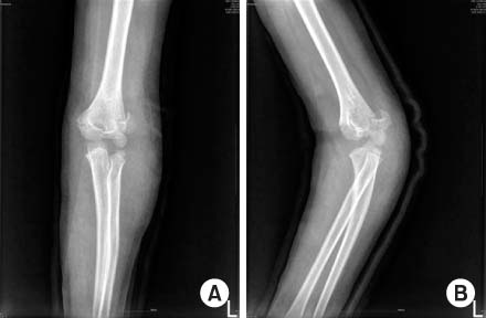
Fig. 2Pre-operative computed tomography (CT) image of the left elbow. (A) Axial view shows the supracondylar fracture line. (B) Lateral condylar fracture line is definite in the coronal view. (C) T-condylar fracture and posterior displacement of the lateral condyle is remarkable in 3-dimensional CT image.
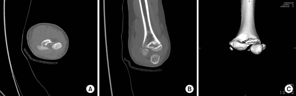
Fig. 3Plain x-ray after open reduction and Kirschner wire fixation. (A) Antero-posterior view of plain x-ray shows reduced, anatomically. (B) Shaft-condylar angle was recovered on lateral view x-ray.
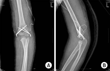
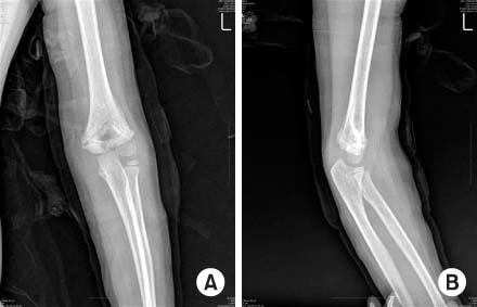

Figure & Data
REFERENCES
Citations
Citations to this article as recorded by 
 Cite
CiteT-Condylar Fracture of Distal Humerus in a Child: A Case Report


Fig. 1
Initial plain antero-posterior (AP) and lateral view of x-ray. (A) AP view of plain x-ray shows the intercondylar fracture line. (B) Lateral view of plain x-ray shows posterior displacement at the lateral condyle of the left distal humerus.
Fig. 2
Pre-operative computed tomography (CT) image of the left elbow. (A) Axial view shows the supracondylar fracture line. (B) Lateral condylar fracture line is definite in the coronal view. (C) T-condylar fracture and posterior displacement of the lateral condyle is remarkable in 3-dimensional CT image.
Fig. 3
Plain x-ray after open reduction and Kirschner wire fixation. (A) Antero-posterior view of plain x-ray shows reduced, anatomically. (B) Shaft-condylar angle was recovered on lateral view x-ray.
Fig. 4
Five weeks after the operation, Kirschner-wires are removed. (A) Well-bone union is checked in the anteroposterior view of plain x-ray. (B) Posterior displacement is reduced successfully in lateral view of plain x-ray.
Fig. 5
Two years and seven months after surgery. (A) Valgus or varus deformity is not found in long bone view of plain x-ray. (B) 125 degrees of active elbow flexion was possible. (C) There was no flexion contracture at the elbow.
Fig. 1
Fig. 2
Fig. 3
Fig. 4
Fig. 5
T-Condylar Fracture of Distal Humerus in a Child: A Case Report

