 E-submission
 TOTA
TOTA TOTS
TOTS
Articles
- Page Path
- HOME > J Musculoskelet Trauma > Volume 30(1); 2017 > Article
-
Case Report
- Pediatric Cartilaginous Tibia Eminence Fracture Overlooked on Plain Radiograph: A Report of Two Cases
- Seong-Eun Byun, M.D., Yunseong Choi, M.D., Wonchul Choi, M.D., Ph.D.
-
Journal of the Korean Fracture Society 2017;30(1):29-34.
DOI: https://doi.org/10.12671/jkfs.2017.30.1.29
Published online: January 20, 2017
Department of Orthopaedic Surgery, Bundang CHA Hospital, CHA University, Seongnam, Korea.
- Correspondence to: Wonchul Choi, M.D., Ph.D. Department of Orthopaedic Surgery, Bundang CHA Hospital, CHA University, 59 Yatap-ro, Bundang-gu, Seongnam 13496, Korea. Tel: +82-31-780-5289, Fax: +82-31-708-3578, wcchoios@chamc.co.kr
• Received: November 15, 2016 • Revised: December 11, 2016 • Accepted: December 27, 2016
Copyright © 2017 The Korean Fracture Society. All rights reserved.
This is an Open Access article distributed under the terms of the Creative Commons Attribution Non-Commercial License (http://creativecommons.org/licenses/by-nc/4.0) which permits unrestricted non-commercial use, distribution, and reproduction in any medium, provided the original work is properly cited.
- 808 Views
- 5 Download
Abstract
- In children with open physis, avulsion fracture of the tibia eminence, as an anterior cruciate ligament (ACL) injury, is more commonly observed than an ACL rupture. Pure cartilaginous avulsions of the ACL tibia insertion seldom occurs. In such case, cartilaginous lesion is frequently overlooked or misdiagnosed on plain radiograph and may result in a less favorable treatment outcome. We report two cases of cartilaginous tibia eminence fractures of the children that were initially overlooked from plain radiographs, and then diagnosed by magnetic resonance imaging, which was ultimately treated by arthroscopyassisted headless compression screw fixation.
- 1. Anderson CN, Anderson AF. Tibial eminence fractures. Clin Sports Med, 2011;30:727-742.Article
- 2. Accousti WK, Willis RB. Tibial eminence fractures. Orthop Clin North Am, 2003;34:365-375.Article
- 3. Chotel F, Seil R, Greiner P, Chaker MM, Berard J, Raux S. The difficult diagnosis of cartilaginous tibial eminence fractures in young children. Knee Surg Sports Traumatol Arthrosc, 2014;22:1511-1516.ArticlePDF
- 4. Meyers MH, McKeever FM. Fracture of the intercondylar eminence of the tibia. J Bone Joint Surg Am, 1970;52:1677-1684.Article
- 5. Zaricznyj B. Avulsion fracture of the tibial eminence: treatment by open reduction and pinning. J Bone Joint Surg Am, 1977;59:1111-1114.
- 6. Bogunovic L, Tarabichi M, Harris D, Wright R. Treatment of tibial eminence fractures: a systematic review. J Knee Surg, 2015;28:255-262.Article
- 7. Chotel F, Raux S, Accadbled F, et al. Cartilaginous tibial eminence fractures in children: which recommendations for management of this new entity. Knee Surg Sports Traumatol Arthrosc, 2016;24:688-696.ArticlePDF
- 8. Feucht MJ, Brucker PU, Camathias C, et al. Meniscal injuries in children and adolescents undergoing surgical treatment for tibial eminence fractures. Knee Surg Sports Traumatol Arthrosc, 2016;[epub]. ArticlePDF
- 9. Ahn JH, Yoo JC. Clinical outcome of arthroscopic reduction and suture for displaced acute and chronic tibial spine fractures. Knee Surg Sports Traumatol Arthrosc, 2005;13:116-121.ArticlePDF
- 10. Johnson DL, Durbin TC. Physeal-sparing tibial eminence fracture fixation with a headless compression screw. Orthopedics, 2012;35:604-608.Article
REFERENCES
Fig. 1
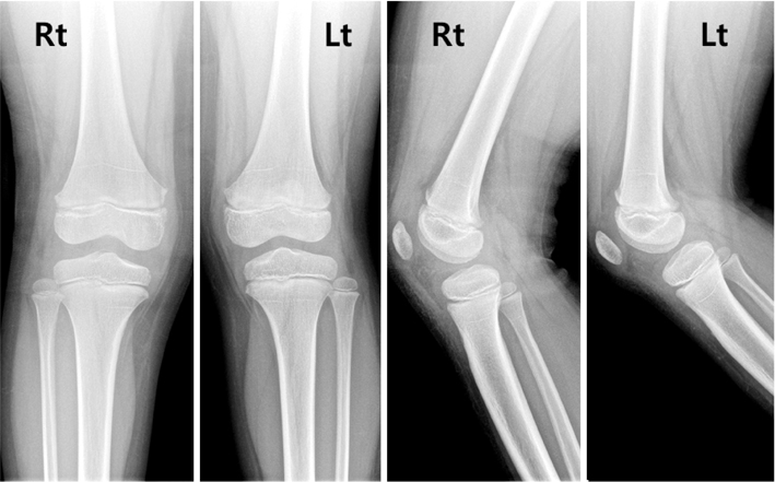
Initial both knee anteroposterior and lateral plain radiograph images show no definite sign of bony fracture. Rt: right, Lt: left.
Fig. 2
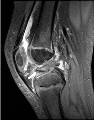
Preoperative right knee magnetic resonance imaging image shows fractured cartilaginous fragment attached to the anterior cruciate ligament (arrow).
Fig. 3

(A) Arthroscopic finding via anterolateral portal shows displaced fracture fragment. (B) Fragment is raised by a probe and fracture between cartilage and subchondral bone was identified.
Fig. 4

Follow-up plain radiographs taken at 3 years after the operation shows healed fracture without any sign of screw loosening or leg length discrepancy.
Fig. 5

The existence of thin ossification (arrows) inside the right knee joint is suspected from the initial both knee anteroposterior and lateral plain radiograph images. Rt: right, Lt: left.
Fig. 6
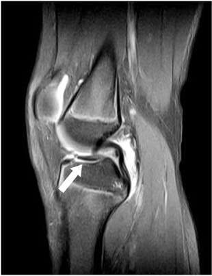
Preoperative right knee magnetic resonance imaging shows disrupted proximal tibia articular cartilage continuity with fluid signal between cartilage and subchondral bone (arrow).
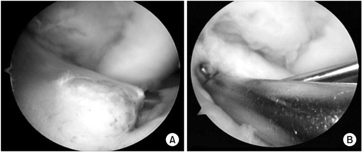

Figure & Data
REFERENCES
Citations
Citations to this article as recorded by 
 Cite
CitePediatric Cartilaginous Tibia Eminence Fracture Overlooked on Plain Radiograph: A Report of Two Cases


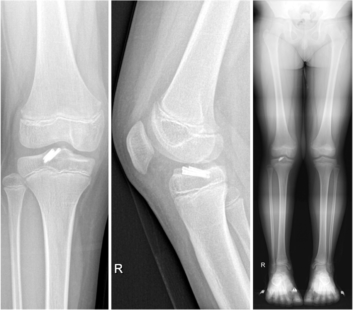
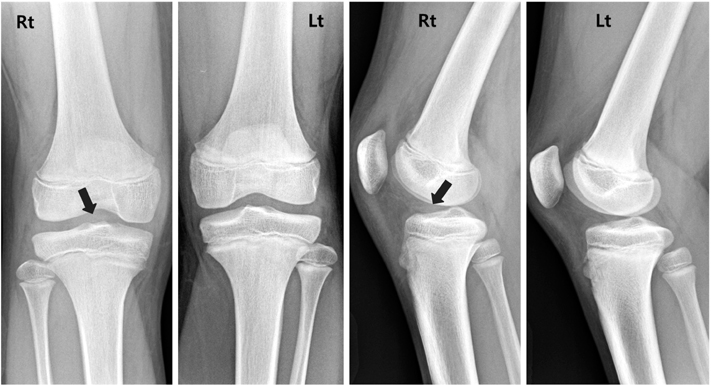


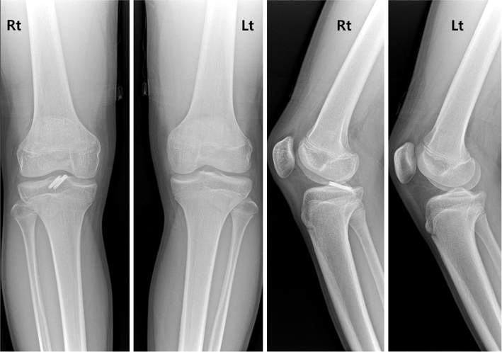
Fig. 1
Initial both knee anteroposterior and lateral plain radiograph images show no definite sign of bony fracture. Rt: right, Lt: left.
Fig. 2
Preoperative right knee magnetic resonance imaging image shows fractured cartilaginous fragment attached to the anterior cruciate ligament (arrow).
Fig. 3
(A) Arthroscopic finding via anterolateral portal shows displaced fracture fragment. (B) Fragment is raised by a probe and fracture between cartilage and subchondral bone was identified.
Fig. 4
Follow-up plain radiographs taken at 3 years after the operation shows healed fracture without any sign of screw loosening or leg length discrepancy.
Fig. 5
The existence of thin ossification (arrows) inside the right knee joint is suspected from the initial both knee anteroposterior and lateral plain radiograph images. Rt: right, Lt: left.
Fig. 6
Preoperative right knee magnetic resonance imaging shows disrupted proximal tibia articular cartilage continuity with fluid signal between cartilage and subchondral bone (arrow).
Fig. 7
(A) Arthroscopic finding via the anterolateral portal shows avulsed fracture of anterior cruciate tibia insertion. (B) Fracture reduction is maintained with an aid from the anterior cruciate ligament tibial guide and was held with a Kirschner wire.
Fig. 8
Follow-up plain radiographs taken at 1 year and 6 months after the operation show a healed fracture without any signs of screw loosening or growth disturbance. Rt: right, Lt: left.
Fig. 1
Fig. 2
Fig. 3
Fig. 4
Fig. 5
Fig. 6
Fig. 7
Fig. 8
Pediatric Cartilaginous Tibia Eminence Fracture Overlooked on Plain Radiograph: A Report of Two Cases

