Articles
- Page Path
- HOME > J Musculoskelet Trauma > Volume 33(1); 2020 > Article
- Original Article Surgical Treatment of Pediatric Intra-Articular Proximal Phalangeal Head Fracture of the Big Toe
- Yeun Soo Kim, Geunwu Gimm, Il ung Hwang, Goo Hyun Baek, Jihyeung Kim
-
Journal of Musculoskeletal Trauma 2020;33(1):9-15.
DOI: https://doi.org/10.12671/jkfs.2020.33.1.9
Published online: January 31, 2020

- 825 Views
- 3 Download
- 0 Crossref
- 0 Scopus
Abstract
PURPOSE
Pediatric intra-articularproximal phalangeal head fractures of the big toe are very rare and few studies on this have been published. The purpose of this study is to present the diagnostic approach and surgical management of these extremely rare fractures, which might be easily underestimated or misdiagnosed.
MATERIALS AND METHODS
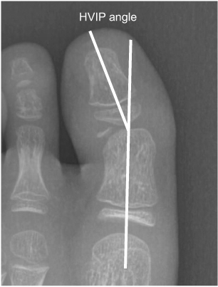
The study retrospectively reviewed all the patients who were diagnosed as intra-articular proximal phalangeal head fracture of the big toe and who underwent surgical intervention in our institution. The size of the bony fragment and hallux valgus interphalangeus angle were measured on the preoperative X-rays. The size and rotation of the osteochondral fragment, the presence of avascular necrosis, ligamentous injury and soft tissue entrapment were assessed on the preoperative magnetic resonance images (MRIs). The radiologic and functional evaluation were performed at 1 year postoperatively.
RESULTS
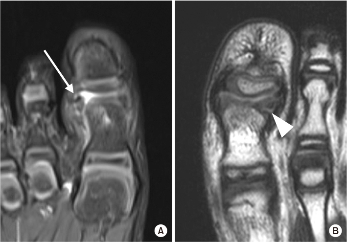
The average size of the bony fragments measured on the X-rays was 4.1 mm in width and 2.3 mm in length. Two cases showed hallux valgus interphalangeus. Preoperative MRI was performed in four cases and the average size of any osteochondral lesion was 5.3 mm in width, 3.9 mm in length, and 4.7 mm in height. Rotation of the osteochondral fragment was observed in one patient, and soft tissue entrapment was noted in two patients. Postoperatively, successful bony union was achieved in all the patients and the average time to union was 74.4 days.
CONCLUSION
Intra-articular proximal phalangeal head fractures of the big toe are very rare and often neglected due to incomplete ossification in the pediatric population. It is important to suspect the presence of this intra-articular fracture and to appropriately implement further evaluation. Nonunion of chronic cases as well as acute fractures can be successfully treated through open reduction and internal fixation using multiple K-wires.
Published online Jan 23, 2020.
https://doi.org/10.12671/jkfs.2020.33.1.9
Surgical Treatment of Pediatric Intra-Articular Proximal Phalangeal Head Fracture of the Big Toe
 , M.D.,
Geunwu Gimm
 , M.D.,
Il-ung Hwang
 , M.D., Ph.D.,
Goo Hyun Baek
 , M.D., Ph.D.
and Jihyeung Kim
 , M.D., Ph.D.
Abstract
Purpose
Pediatric intra-articularproximal phalangeal head fractures of the big toe are very rare and few studies on this have been published. The purpose of this study is to present the diagnostic approach and surgical management of these extremely rare fractures, which might be easily underestimated or misdiagnosed.
Materials and Methods
The study retrospectively reviewed all the patients who were diagnosed as intra-articular proximal phalangeal head fracture of the big toe and who underwent surgical intervention in our institution. The size of the bony fragment and hallux valgus interphalangeus angle were measured on the preoperative X-rays. The size and rotation of the osteochondral fragment, the presence of avascular necrosis, ligamentous injury and soft tissue entrapment were assessed on the preoperative magnetic resonance images (MRIs). The radiologic and functional evaluation were performed at 1 year postoperatively.
Results
The average size of the bony fragments measured on the X-rays was 4.1 mm in width and 2.3 mm in length. Two cases showed hallux valgus interphalangeus. Preoperative MRI was performed in four cases and the average size of any osteochondral lesion was 5.3 mm in width, 3.9 mm in length, and 4.7 mm in height. Rotation of the osteochondral fragment was observed in one patient, and soft tissue entrapment was noted in two patients. Postoperatively, successful bony union was achieved in all the patients and the average time to union was 74.4 days.
Conclusion
Intra-articular proximal phalangeal head fractures of the big toe are very rare and often neglected due to incomplete ossification in the pediatric population. It is important to suspect the presence of this intra-articular fracture and to appropriately implement further evaluation. Nonunion of chronic cases as well as acute fractures can be successfully treated through open reduction and internal fixation using multiple K-wires.
Fig. 1
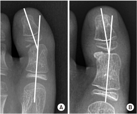
Measurement of the hallux valgus interphalangeus (HVIP) angle.
Fig. 2
(A) Rotation of the osteochondral fragment (arrow). (B) Entrapment of the periosteum in the fracture site (arrowhead).
Fig. 3
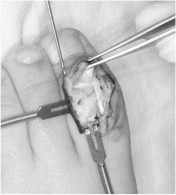
The joint surface was exposed through the dorsal approach. Sclerotic margin of the fracture site was observed.
Fig. 4
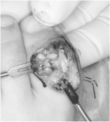
A K-wire was inserted into the fragment and retracted from the opposite side to avoid unwanted traction when removing the wire.
Fig. 5
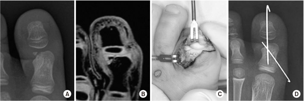
(A, B) The size of the osteochondral fragment might be underestimated on the plain X-ray. (C, D) Minimally displaced osteochondral fracture was fixed with two K-wires.
Fig. 6
Preoperative X-ray (A) and X-ray at 2 months postoperatively (B). The hallux valgus interphalangeus angle was reduced after the surgery.
Fig. 7
(A, B) A case of acute fracture with forefoot laceration. (C) Stable fixation was achieved using two K-wires. (D) Follow-up X-ray at 2 years postoperatively.
Table 1
Demographics and Preoperative Radiologic Evaluations
Financial support:None.
Conflict of interests:None.

 E-submission
 KOTA
KOTA TOTA
TOTA TOTS
TOTS


 PubReader
 Cite
Cite

