 E-submission
 TOTA
TOTA TOTS
TOTS
Articles
- Page Path
- HOME > J Musculoskelet Trauma > Volume 20(2); 2007 > Article
-
Original Article
- Comparison of the Surgical Treatment Results of Avulsion Fracture of the Anterior Cruciate Ligament between Children and Adults
- Eun Kyoo Song, M.D., Sang Jin Park, M.D., Keun Bae Lee, M.D.
-
Journal of the Korean Fracture Society 2007;20(2):196-201.
DOI: https://doi.org/10.12671/jkfs.2007.20.2.196
Published online: June 14, 2016
Center for Joint Disease, Chonnam National University Hwasun Hospital, Hwasun, Korea.
- Address reprint requests to: Sang Jin Park, M.D. Department of Orthopaedic Surgery, Chonnam National University Hwasun Hospital, 160, Ilsim-ri, Hwasun-eup, Hwasun 519-809, Korea. Tel: 061-379-7676, Fax: 82-61-379-7681, park5962@paran.com
Copyright © The Korean Fracture Society. All rights reserved
- 686 Views
- 1 Download
Abstract
-
Purpose
- To compare the clinical and radiological results after surgical treatments of the avulsion fractures of ACL between children and adults.
-
Materials and Methods
- 40 cases (18 cases of children, 22 cases of adults), who underwent surgical treatments after avulsion fractures of the ACL and followed up more than one year, were enrolled. Fractures were classified by modified Meyers & McKeever criteria. Range of motion, LK score, Lachman test, Pivot-Shift test, quadriceps muscle atropy and Telos® stress arthrometer were compared.
-
Results
- The types of fracture in children were categorized into 8 cases of type II, 10 cases of type III, and 2, 15, 5 cases of type II, III, IV each in adult group. Mean LK score showed significant difference between 99.3 points in children and 89.5 points in adults (p<0.05). In addition, accompanied injuries and the high degree of fracture leaded low LK score. However, there was no significant difference in range of motion, Lachman test and Pivot-Shift test. Anterior laxity by Telos® device showed an average of 2.0 mm in children, 2.5 mm in adults (p>0.05).
-
Conclusion
- Children group showed better treatment results of avulsion fracture of ACL. Higher incidence of type II fractures and less combined injuries considered to be factors for better results.
- 1. Baxter MP, Wiley JJ. Fracture of the tibial spine in children. An evaluation of knee stability. J Bone Joint Surg Br, 1988;70:228-230.
- 2. Burstein DB, Viola A, Fulkerson JP. Entrapment of the medial meniscus in a fracture of the tibial eminence. Arthroscopy, 1988;4:47-50.Article
- 3. Grönkvist H, Hirsch G, Johansson L. Fracture of the anterior tibial spine in children. J Pediatr Orthop, 1984;4:465-468.Article
- 4. Hess T, Rupp S, Hopf T, Gleitz M, Liebler J. Lateral tibial avulsion fractures and disruptions to the anterior cruciate ligament. A clinical study of their incidence and correlation. Clin Orthop Relat Res, 1994;303:193-197.
- 5. Iobst CA, Stanitski CL. Acute knee injuries. Clin Sports Med, 2000;19:621-635.Article
- 6. Kendall NS, Hsu SY, Chan KM. Fracture of the tibial spine in adults and children. A review of 31 cases. J Bone Joint Surg Br, 1992;74:848-852.ArticlePDF
- 7. Keys GW, Walters J. Nonunion of intercondylar eminence fracture of the tibia. J Trauma, 1988;28:870-871.Article
- 8. Lubowitz JH, Grauer JD. Arthroscopic treatment of anterior cruciate ligament avulsion. Clin Orthop Relat Res, 1993;(294):242-246.Article
- 9. McLennan JG. The role of arthroscopic surgery in the treatment of fractures of the intercondylar eminence of the tibia. J Bone Joint Surg Br, 1982;64:477-480.ArticlePDF
- 10. McNair PJ, Marshall RN, Matheson JA. Important features associated with acute anterior cruciate ligament injury. N Z Med J, 1990;103:537-539.
- 11. Meyers MH, Mckeever FM. Fracture of the intercondylar eminence of the tibia. J Bone Joint Surg Am, 1970;52:1677-1684.Article
- 12. Song EK, Seol JY, Choi J. The surgical treatment of chronic avulsion fracture of the anterior curciate ligament. J Korean Arthrosc Soc, 2002;6:31-36.
- 13. Wiley JJ, Baxter MP. Tibial spine fractures in children. Clin Orthop Relat Res, 1990;255:54-60.Article
REFERENCES

Fig. 2
(B) Radiographs 2 years after pull-out suture operation shows the complete healing state.
(C) Type II ACL avulsion fracture of 24-year old female patient.
(D) Radiographs 8 years after screw fixation shows the complete healing state of the ACL fracture.
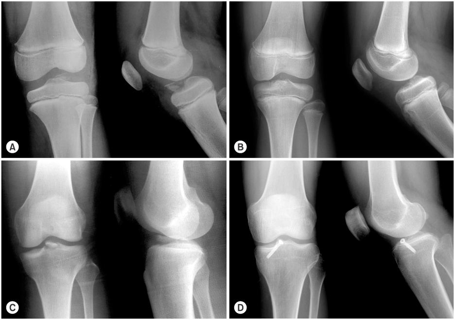
(A) Type III ACL avulsion fracture of 7-year old male patient.
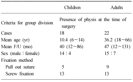
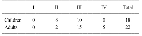
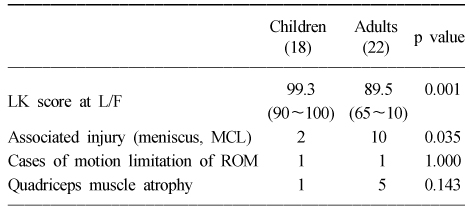
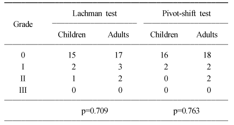
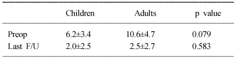
Figure & Data
REFERENCES
Citations
Citations to this article as recorded by 
 Cite
CiteComparison of the Surgical Treatment Results of Avulsion Fracture of the Anterior Cruciate Ligament between Children and Adults
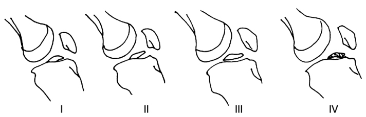

Fig. 1
Classification of the ACL avulsion fractures by modified Meyers & McKeever classification.
Fig. 2
(A) Type III ACL avulsion fracture of 7-year old male patient.
(B) Radiographs 2 years after pull-out suture operation shows the complete healing state.
(C) Type II ACL avulsion fracture of 24-year old female patient.
(D) Radiographs 8 years after screw fixation shows the complete healing state of the ACL fracture.
Fig. 1
Fig. 2
Comparison of the Surgical Treatment Results of Avulsion Fracture of the Anterior Cruciate Ligament between Children and Adults
Dermographic data of the 40 patients of avulsion fractures of the anterior cruciate ligament
Classification of the avulsion fractures of the anterior cruciate ligament according to the Modified Meyers and McKeever Classification
The difference of clinical results between children and adults groups
The Lachman test and Pivot shift test at last follow up
The difference of anterior knee laxity with Telos® from opposite side (mm)
Table 1
Dermographic data of the 40 patients of avulsion fractures of the anterior cruciate ligament
Table 2
Classification of the avulsion fractures of the anterior cruciate ligament according to the Modified Meyers and McKeever Classification
Table 3
The difference of clinical results between children and adults groups
Table 4
The Lachman test and Pivot shift test at last follow up
Table 5
The difference of anterior knee laxity with Telos® from opposite side (mm)

