 E-submission
 TOTA
TOTA TOTS
TOTS
Articles
- Page Path
- HOME > J Musculoskelet Trauma > Volume 20(1); 2007 > Article
-
Case Report
- Traumatic Pseudoaneurysm of Posterior Tibial Artery in a Child: A Case Report
- Tai-Seung Kim, M.D., Kuhn-Sung Whang, M.D., Woo-Young Seo, M.D.
-
Journal of the Korean Fracture Society 2007;20(1):83-85.
DOI: https://doi.org/10.12671/jkfs.2007.20.1.83
Published online: June 14, 2016
Department of Orthopaedic Surgery, Hanyang University College of Medicine, Seoul, Korea.
- Address reprint requests to: Kuhn-Sung Whang, M.D. Department of Orthopaedic Surgery, College of Medicine, Hanyang University, 17, Haengdang-dong, Seongdong-gu, Seoul 133-792, Korea. Tel: 2-2-2290-8479, Fax: 2-2-2299-3774, whangks@hanyang.ac.kr
Copyright © The Korean Fracture Society. All rights reserved
- 1,152 Views
- 3 Download
- 1 Crossref
Abstract
- Pseudoaneurysm is one of the complications of arterial injuries by trauma. The case report in children is rare, although not in adult. A 7-year and 10-month girl was visited with the complaints of pain and a mass in her right leg. At first, the radiograph of right tibia showed a remarkable cortical erosion from without, suggesting mass effect by a soft tissue tumor. She had a history of fracture of right tibia, and then manipulative reduction and K-wire fixation at 11 months ago. Arteriography showed a formation of the pseudoaneurysm originated from the posterior tibial artery. The operation was done through the ligation of artery at proximal and distal to pseudoaneurysm, and then excision of mass. At 5 year follow-up, the configuration and function of right foot was normal. Eventually, the cause of the mass formation is thought by the trauma of fracture fragment at the time of accidents, but the possibility of penetrated injuries by K-wire should be ruled out, which is used frequently in children's fracture. We experienced a case of traumatic pseudoaneurysm of posterior tibal artery with tibial fracture, especially occurred in pediatric patient, and presented the result of long-term follow-up.
- 1. Gantz ED, Sweet MB, Jakim I. False aneurysm mimicking an aggressive soft-tissue tumor. A case report. J Bone Joint Surg Am, 1988;70:1090-1092.Article
- 2. Georgiadis GS, Deftereos SP, Eleftheriadou E, Zacharouli D, Lazarides MK. Delayed presentation of a posterior tibial false aneurysm. Surgery, 2006;139:446-447.Article
- 3. Ha KI, Hahn SH, Chung MY, Yang BK, Shin KH. Traumatic false aneurysm at fracture site. J Korean Orthop Assoc, 1992;27:408-411.ArticlePDF
- 4. Kang KH, Kang ST, Kwon DJ, Suh DH. Traumatic false aneurysm: report of two cases. J Korean Orthop Assoc, 2002;37:678-681.ArticlePDF
- 5. Lee JY, Yoo CI, Kim BH, Shon BH. Traumatic false aneurysm of ulnar artery. A case report. J Korean Orthop Assoc, 1975;10:302-305.ArticlePDF
- 6. Sung YB, Kim DY. Traumatic false aneurysm of posterior tibial artery: a case report. J Korean Orthop Assoc, 1997;32:202-207.ArticlePDF
REFERENCES
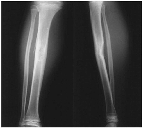
Fig. 2

Peripheral portion of mass is high signal intensity at T1, T2 and central portion is heterogenous signal intensity. The size of mass in the calf deep portion is 5.5 cm in diameter, and oval shaped.
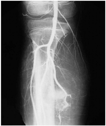
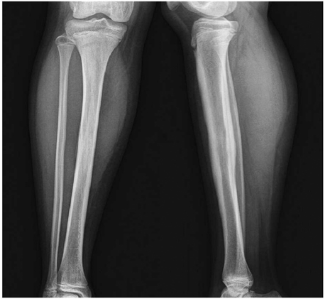
Figure & Data
REFERENCES
Citations
Citations to this article as recorded by 
- Coil Embolization of a Pseudoaneurysm of the Anterior Tibial Artery: A Case Report
Tae-Hyun Wang, Hyung-Lae Cho, Ki-Bong Park, Duc-Hee Kim
Journal of Korean Foot and Ankle Society.2016; 20(1): 43. CrossRef
 Cite
CiteTraumatic Pseudoaneurysm of Posterior Tibial Artery in a Child: A Case Report

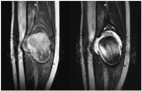


Fig. 1
Preoperative plain radiography shows posterior cortical erosion by a mass in diaphysis.
Fig. 2
Peripheral portion of mass is high signal intensity at T1, T2 and central portion is heterogenous signal intensity. The size of mass in the calf deep portion is 5.5 cm in diameter, and oval shaped.
Fig. 3
An angiography of the right lower extremity shows a 4 cm by 3 cm size of a pseudoaneurysm from the posterior tibial artery.
Fig. 4
Follow-up radiography in 5 years after excision of aneurysm shows complete restoration of posterior cortex in diaphys.
Fig. 1
Fig. 2
Fig. 3
Fig. 4
Traumatic Pseudoaneurysm of Posterior Tibial Artery in a Child: A Case Report

