-
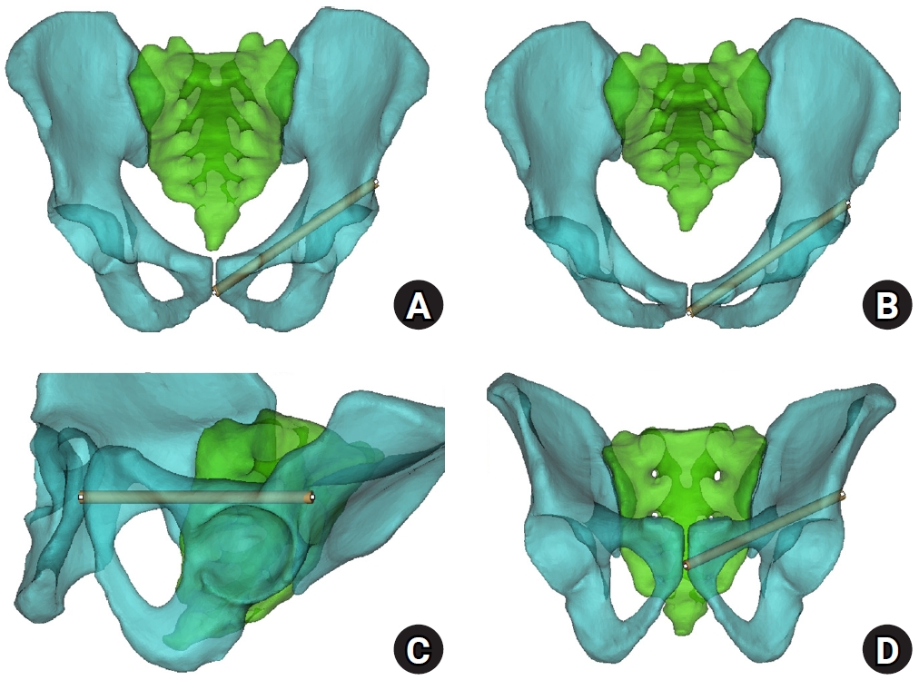
Sex-specific bottlenecks and risk zones in the retrograde superior pubic ramus screw corridor: a 3D CT-based morphometric cadaver study
-
Ji Won Jeong, Jung Tae Ahn, Gu Hee Jung, Kun Tae Kim
-
J Musculoskelet Trauma 2026;39(2):103-116. Published online March 26, 2026
-
DOI: https://doi.org/10.12671/jmt.2026.00066
-
-
 Abstract Abstract
 PDF PDF Supplementary Material Supplementary Material
- Background
Superior ramus screw fixation is commonly used to stabilize anterior pelvic ring injuries but is constrained by a narrow, irregular, and curved intraosseous corridor. Trajectory-based morphometric analysis may assist in screw diameter selection and enable identification of reproducible anatomic constriction zones.
Methods
We conducted a cross-sectional computed tomography (CT)-based morphometric study of 82 cadaveric pelvises (42 males, 40 females). Bottleneck diameter was defined as the diameter of the largest fully contained virtual cylinder along the planned trajectory, and cylinder length was recorded. Orthogonal cross-sections at 9.5-mm intervals (up to 12 segments) were generated to measure segment-wise effective diameter (defined as twice the minimum centerline-to-cortex distance) and cortical clearance, which was used as a diameter-based safety margin. Segments were realigned to the acetabular start segment to define relative segment positions (Δ seg). Feasibility was assessed for prespecified screw diameters ranging from 3.5 to 7.3 mm.
Results
Mean bottleneck diameter was larger in males than in females (7.34±1.10 vs. 5.93±0.98 mm), whereas trajectory length was similar between sexes (127.85±8.54 vs. 128.85±8.20 mm). Δ seg realignment localized corridor constriction to two discrete zones: a preacetabular zone (Δ seg −6 to −4) and a periacetabular zone (Δ seg 1 to 2), where effective diameter and cortical clearance were most limited. Feasibility rates were 100% at 3.5–4.5 mm, 95.2% vs. 82.5% at 5.0 mm, 81.0% vs. 27.5% at 6.5 mm, and 59.5% vs. 10.0% at 7.3 mm in males and females, respectively.
Conclusions
Female models demonstrated smaller trajectory-wide bottleneck diameters and segment-wise effective diameters than male models. Acetabular-referenced Δ seg realignment identified two reproducible anatomic risk zones: a preacetabular zone adjacent to the obturator neurovascular bundle and a periacetabular zone near the external iliac vessels. At diameters ≥6.5 mm, cortical proximity increased more prominently in females than in males.
Level of evidence: III.
-
Fixation Options of Unstable Posterior Pelvic Ring Disruption: Ilio-Sacral Screw Fixation, S2AI Fixation, Posterior Tension Band Plate Fixation, and Spino-Pelvic Fixation
-
Dong Hee Kim, Jae Hoon Jang, Myungji Shin, Gu Hee Jung
-
J Korean Fract Soc 2019;32(4):240-247. Published online October 31, 2019
-
DOI: https://doi.org/10.12671/jkfs.2019.32.4.240
-
-
Abstract
PDF
- The fixation methods that can be used for unstable posterior pelvic ring injuries have undergone many innovative changes due to the recent development of surgical and imaging techniques. After understanding the appropriate indications of first and second sacroiliac screw fixation and spinopelvic fixation, innovative methods, including the trans-sacral screw fixation, posterior tension-band plate fixation, and the S2AI screw, would be chosen and applied. Considering the anatomical complexity and proximity to the surrounding vessels and nerves in the posterior fixation, the safe zone according to the fixation options should be well understood in preoperative planning. Moreover, the functional reduction of the posterior pelvic ring through the reduction and fixation of the anterior lesion should be achieved before placing the implant to reduce the number of malposition-related complications.
-
Citations
Citations to this article as recorded by  - Clinical Research through Computational Anatomy and Virtual Fixation
Ju Yeong Kim, Dong-Geun Kang, Gu-Hee Jung
Journal of the Korean Orthopaedic Association.2023; 58(4): 299. CrossRef
-
1,580
View
-
19
Download
-
1
Crossref
-
Computational Simulation of Multiple Cannulated Screw Fixation for Femoral Neck Fractures and the Anatomic Features for Clinical Applications
-
Jin Hoon Jeong, Gu Hee Jung
-
J Korean Fract Soc 2018;31(2):37-44. Published online April 30, 2018
-
DOI: https://doi.org/10.12671/jkfs.2018.31.2.37
-
-
Abstract
PDF
- PURPOSE
To identify the anatomic features for clinical applications through a computational simulation of the fixation of three cannulated screws for a femoral neck fracture.
MATERIALS AND METHODS
Thirty cadaveric femurs underwent computed tomography and the images were transferred to the Mimics® program, resulting in three-dimensional proximal femur models. A three-dimensional scan of the 7.0 mm cannulated screw was performed to enable computerized virtual fixation of multiple cannulated screws for femoral neck fractures. After positioning the screws definitively for cortical support, the intraosseous position of the cannulated screws was evaluated in the anteroposterior image and axial image direction.
RESULTS
Three cannulated screws located at the each ideal site showed an array of tilted triangles with anterior screw attachment and the shortest spacing between posterior and central screws. The central screw located at the lower side was placed in the mid-height of the lesser trochanter and slightly posterior, and directed toward the junction of femoral head and neck to achieve medial cortical support. All the posterior screws were limited in height by the trochanteric fossa and were located below the vastus ridge, but the anterior screws were located higher than the vastus ridge in 10 cases. To obtain the maximum spacing of the anterior and posterior screws on the axial plane, they should be positioned parallel to the cervical region nearest the cortical bone at a height not exceeding the vastus ridge.
CONCLUSION
The position of cannulated screws for cortical support were irregular triangular arrangements with the anterosuperior apex. The position of the ideal central screw in the anteroposterior view was at the mid-height of the lesser trochanter toward the junction of the femoral head and neck, and the anterior and posterior screws were parallel to the neck with a maximal spread just inferior to the vastus ridge.
-
Citations
Citations to this article as recorded by - Computational Simulation of Femoral Neck System and Additional Cannulated Screws Fixation for Unstable Femoral Neck Fractures and the Biomechanical Features for Clinical Applications
Ju-Yeong Kim
Journal of the Korean Fracture Society.2023; 36(1): 1. CrossRef
-
988
View
-
0
Download
-
1
Crossref
-
The Determination of Optimal Entry Point for Proximal Femoral Nail Antirotation-II by Fluoroscopic Simulation: A Cadaveric Study
-
Jin Hoon Jeong, Gu Hee Jung
-
J Korean Fract Soc 2017;30(4):173-179. Published online October 31, 2017
-
DOI: https://doi.org/10.12671/jkfs.2017.30.4.173
-
-
Abstract
PDF
- PURPOSE
This study seeks to determine the anatomically optimal entry point of proximal femoral nail antirotation-II (PFNA-II®) according to geographic features of Korean cadaveric femoral trochanters for successful reduction of osteoporotic proximal femoral fractures.
MATERIALS AND METHODS
Forty-three adult cadaveric femurs without previous fractures or surgeries were included. Anteroposterior (AP) and lateral images of all femurs and PFNA-II® were taken with an image intensifier. Using the image synthesis process via the image editing program (Adobe Photoshop CS6), the optimal entry point was verified and compared with the tip of the greater trochanter (GT) and the cervicotro-chanteric junction on AP images, as well as the width of the trochanter and the neck on lateral images.
RESULTS
The optimal entry point of PFNA-II® was an average distance of 9.1 mm (range, 7–15 mm) medially from the tip of GT on AP images. The center of the nail was located at an average of 30% (range, 21%–44%) area from the posterior margin of the middle neck, which is an average area of 38% (range, 26%–48%) from the posterior cortex of the trochanter on lateral images. Furthermore, the ideal entry point was at the extended line of the cervico-trochanteric junction.
CONCLUSION
The optimal entry point, which was found to be medial to the tip of the GT and posterior to the center of the middle femoral neck and the trochanter, was at on the extended line of the cervicotrochanteric junction.
-
Citations
Citations to this article as recorded by - Clinical Research through Computational Anatomy and Virtual Fixation
Ju Yeong Kim, Dong-Geun Kang, Gu-Hee Jung
Journal of the Korean Orthopaedic Association.2023; 58(4): 299. CrossRef - Does the Entry Point of Proximal Femoral Nail Antirotation Affect the Malalignment of Intertrochanteric Fracture? A Cadaveric Study
Chittawee Jiamton, Nonpawit Nimmankiatkul, Pongsakorn Rungchamrassopa, Wichan Kanchanatawan, Pariyut Chiarapatanakom, Wirat Kongcharoensombat
Journal of Southeast Asian Orthopaedics.2022;[Epub] CrossRef
-
2,623
View
-
69
Download
-
2
Crossref
-
Analysis of Low-Energy Trochanter Fracture Using the Multiplanar Computed Tomography Image: Application for Intramedullary Nail Fixation
-
Gu Hee Jung, Sung Keun Heo, Hyun Je Seo
-
J Korean Fract Soc 2015;28(3):155-162. Published online July 31, 2015
-
DOI: https://doi.org/10.12671/jkfs.2015.28.3.155
-
-
Abstract
PDF
- PURPOSE
The purpose of this radiologic study was to evaluate the geographic patterns of low-energy trochanteric fractures using multiplanar computed tomography (CT) images for application of intramedullary nailing.
MATERIALS AND METHODS
In this study, 117 trochanteric fractures (stable fracture, 39 cases, unstable fractures, 78 cases) sustained from simple slip-down were assessed. The mean age was 78.4 years (range, 60-96 years). Multiplanar CT images were assessed for evaluation of geographic features of trochanteric fracture, and the fracture exit and geographic patterns were analyzed.
RESULTS
The medial and lateral exit of the trochanteric fracture showed no statistical difference by age, bone density, and comorbid disease. The exit was located at an average distance of 10.2 mm (range, 1.0-22.2 mm) from the tip of the greater trochanter (GT), and the medial exit, average distance of 8.1 mm (range, 0.0-18.3 mm) from the tip of the lesser trochanter. It was also found that there was no comminution around the anteromedial cortex of the fracture, and its contact loss was from fracture deformity.
CONCLUSION
Because of no comminution, the contact restoration of the anteromedial cortex resulted in correction of fracture deformity and reduction. Trochanteric nailing by GT tip could be fixed through the proximal fragment of the fracture because the lateral exit is placed at an average distance of 10.2 mm from the GT tip.
-
Surgical Fitness for Trochanteric Fracture in Elderly: Prospective Study
-
Gu Hee Jung, Jong Seo Lee, Sung Gun Heo, Jae Do Kim, Hyun Ik Cho
-
J Korean Fract Soc 2014;27(4):261-266. Published online October 31, 2014
-
DOI: https://doi.org/10.12671/jkfs.2014.27.4.261
-
-
Abstract
PDF
- PURPOSE
The purpose of this study was to evaluate the risks of undergoing intramedullary nailing with minimum surgical optimization (fast-track) for geriatric trochanter fracture due to fall from a standing height.
MATERIALS AND METHODS
From May 2006 to August 2013, 48 fractures were enrolled in fast-track, and were an average age of patients was 77.6 years (range, 62-97 years). They underwent primary testing for anesthesia, including basic body fluid test, arterial blood, electrocardiography, and chest radiographs. The time from visit to surgery was 28.9 hours (range, 1-96 hours).
RESULTS
During hospitalization, there was one case of stress-induced cardiac arrest; however, other complications, infection, and 30-day mortality did not occur. According to preoperative classic test, the average albumin was 3.45 g/dl, blood sugar, 169 mg/dl, blood urea nitrogen, 20.5 mg/dl, Cr, 1.5 mg/dl, Na, 135.3 mEq/L, and K, 4.21 mEq/L. The average PaCO2 of arterial blood was 37.6 mmHg.
CONCLUSION
We found that the fast-track for trochanteric fracture due to slip-down was relatively safe, and could be considered as a therapeutic approach.
-
Granulation Tissue Formed by Stimulating K-Wire Mimicking Tuberculous Cervical Lymphadenopathy: A Case Report
-
Gu Hee Jung, Tae Hun Kim, Hyun Ik Cho
-
J Korean Fract Soc 2014;27(3):227-231. Published online July 31, 2014
-
DOI: https://doi.org/10.12671/jkfs.2014.27.3.227
-
-
Abstract
PDF
- Pins and wires are still used frequently in surgeries of the shoulder; however, these can cause breakage or migration to surrounding tissues, leading to complications. We report on case of a patient with a neck mass who had a past history of pulmonary tuberculosis and distal clavicle fracture with internally fixated state. She was misdiagnosed as tuberculous cervical lymphadenopathy and treated for approximately one year, but was finally revealed as granulation tissue around the internally fixated distal clavicle fracture site, thus, mass excision and metal removal was performed. This case shows the importance of a proper selection device, internal fixation technique, duration, and close follow-up after the operation.
-
Citations
Citations to this article as recorded by - Kirschner Wire Migration into SpinaL Canal after Acromioclavicular Joint Fixation (Literature Review and Clinical Case)
D. A. Gulyaev, D. S. Godanyuk, T. A. Kaurova, P. V. Krasnoshlyk, S. V. Maikov
Traumatology and Orthopedics of Russia.2018; 24(4): 121. CrossRef
-
836
View
-
0
Download
-
1
Crossref
-
Radiation Exposure Over the Course of a Year from an Image Intensifier in the Orthopaedic Operating Room
-
Gu Hee Jung, Jae Ho Jang, Jae Do Kim, Chung Kyu Kim
-
J Korean Fract Soc 2012;25(1):58-63. Published online January 31, 2012
-
DOI: https://doi.org/10.12671/jkfs.2012.25.1.58
-
-
Abstract
PDF
- PURPOSE
To measure the annual radiation exposure of staff in the orthopaedic surgical room.
MATERIALS AND METHODS
From January 2010 to December 2010, we measured the radiation exposure of a tumor surgeon, spine surgeon, trauma surgeon, six residents, and six scrub nurses. Radiation was monitored with the use of thermoluminescent dosimeters placed on the chest under the lead apron. The annual dose of radiation exposure was compared to the maximum yearly permissible dose (20 mSv). During the study period, the trauma surgeon made a deliberate effort to minimize the radiation time and maintain a distance of 1 m from the image intensifier.
RESULTS
The annual exposure levels were 0.04 mSv (radiation time, 34 min 50 s), 0.08 mSv (151 min 46 s), and 0.12 mSv (135 min 27 s) for the tumor surgeon, trauma surgeon, and spine surgeon, respectively. The mean exposure was 0.0146 mSv (range, 0.4~0.39 mSv) for the residents and 0.06 mSv (range, 0.04~0.13 mSv) for the scrub nurses. Overall, the annual radiation exposure was 0.2~1.95% of the maximal yearly permissible dose. Despite the longer period of radiation exposure, the trauma surgeon was exposed to a lower dose of radiation than the spine surgeon.
CONCLUSION
The annual radiation exposure of a trauma surgeon can be reduced by a deliberate effort to decrease exposure time and maintain a distance of at least 1 m from the image intensifier.
-
Citations
Citations to this article as recorded by - How to obtain the desired results from distal tibial nailing based on anatomy, biomechanics, and reduction techniques
Jungtae Ahn, Se-Lin Jeong, Gu-Hee Jung
Journal of Musculoskeletal Trauma.2025; 38(2): 74. CrossRef - Current status of occupational radiation exposure and protection among medical interns and residents
Seungwon Cho, Hangyeol Lee, Minku Kang, Won Jin Lee, Seulki Ko
Journal of the Korean Medical Association.2024; 67(2): 134. CrossRef - Radiation exposure and fluoroscopically-guided interventional procedures among orthopedic surgeons in South Korea
Seonghoon Kang, Eun Shil Cha, Ye Jin Bang, Teresa W. Na, Dalnim Lee, Sang Youn Song, Won Jin Lee
Journal of Occupational Medicine and Toxicology.2020;[Epub] CrossRef
-
968
View
-
2
Download
-
3
Crossref
-
Treatment of the Intertrochanteric Femoral Fracture with Proximal Femoral Nail: Nailing Using the Provisional K-wire Fixation
-
Gu Hee Jung
-
J Korean Fract Soc 2011;24(3):223-229. Published online July 31, 2011
-
DOI: https://doi.org/10.12671/jkfs.2011.24.3.223
-
-
Abstract
PDF
- PURPOSE
To evaluate the efficiency of provisional K-wire fixation in intertrochanteric fractures treated with proximal femoral nail (PFN).
MATERIALS AND METHODS
Twenty seven patients (by AO/OTA classification, A1 8 cases, A2 19 cases) were treated with PFN with percutaneous reduction and provisional K-wire fixation, and followed a mean 24.5 months. The adequacy of fracture reduction was assessed by Fogagnolo's classification and reestablishment of bone-to-bone contact with the medial anatomy. Functional results were evaluated by postoperative complications, Jensen's method and Harris hip score (HHS).
RESULTS
In all cases, the bone-to-bone contact with the medial anatomy was reestablished by percutaneous reduction and examination of Fogagnolo's classification showed a good reduction. The technical complications and error of starting point were not occurred. The mean HHS was 76.5 and means Jensen's grade was 2.1 grades. Complications included excessive sliding in 1 case and early cutting-out of screw in 1 case.
CONCLUSION
The provisional K-wire fixation in trochanteric fracture treated with PFN had an advantage in preventing technical complications because it facilitates a nail insertion in ideal position.
-
Citations
Citations to this article as recorded by - Analysis of Low-Energy Trochanter Fracture Using the Multiplanar Computed Tomography Image: Application for Intramedullary Nail Fixation
Gu-Hee Jung, Sung-Keun Heo, Hyun-Je Seo
Journal of the Korean Fracture Society.2015; 28(3): 155. CrossRef - Morbidity and Mortality of the Elderly after Early Operation for Trochanteric Fractures
Se-Ang Jang, Young-Ho Cho, Young-Soo Byun, Ki-Hong Park, Hyun-Seong Yoo, Chul Jung
Journal of the Korean Fracture Society.2013; 26(3): 199. CrossRef
-
1,225
View
-
6
Download
-
2
Crossref
-
Intramedullary Nailing for Complex Fractures of the Proximal and Midshaft of the Humerus
-
Chul Hyun Cho, Gu Hee Jung, Kyo Wook Kim
-
J Korean Fract Soc 2011;24(3):237-242. Published online July 31, 2011
-
DOI: https://doi.org/10.12671/jkfs.2011.24.3.237
-
-
Abstract
PDF
- PURPOSE
To evaluate the results of antegrade interlocking intramedullary nailing for complex fractures of the proximal and midshaft of the humerus.
MATERIALS AND METHODS
We retrospectively analyzed the clinical and radiologic results in 11 cases, which were treated by antegrade interlocking intramedullary nail. We assessed clinical outcomes according to ASES scoring system and radiological result.
RESULTS
All cases had bony union and the mean union period was 14.7 weeks. Postoperative complications were 1 loss of fixation, 2 proximal protrusion of nail and 2 temporary shoulder pain. A case with loss of fixation was treated open reduction and refixation and had union at 14 weeks postoperatively. The mean ASES score was 85.9 and the clinical outcomes were 4 excellent, 5 good, 1 fair and 1 poor.
CONCLUSION
Intramedullary nailing for complex fractures of the proximal and midshaft of the humerus can offer a reliable treatment option.
-
Minimally Invasive Plate Osteosynthesis for the Upper Extremity Fracture Using a Lumbar Spreader: Surgical Technique
-
Gu Hee Jung, Chyul Hyun Cho, Jae Do Kim
-
J Korean Fract Soc 2011;24(1):83-86. Published online January 31, 2011
-
DOI: https://doi.org/10.12671/jkfs.2011.24.1.83
-
-
Abstract
PDF
- The minimally invasive plate osteosynthesis (MIPO) which is extensively performed, is very dependent on the indirect reduction technique to prevent the exposure of fracture sites. Indirect reduction with the use of the femoral distractor is a much more efficient technique to restore the length in the fracture of lower limbs. However, the femoral distractor cannot be used for fracture of upper limbs, and other instruments for indirect reduction have not yet been reported. Therefore, we introduce the novel indirect reduction technique with the use of the lumbar spreader for the MIPO of upper limbs.
-
Management of Open Fracture
-
Gu Hee Jung
-
J Korean Fract Soc 2010;23(2):236-250. Published online April 30, 2010
-
DOI: https://doi.org/10.12671/jkfs.2010.23.2.236
-
-
Abstract
PDF
- No abstract available.
|
 E-submission
 TOTA
TOTA TOTS
TOTS