 E-submission
 TOTA
TOTA TOTS
TOTS
Articles
- Page Path
- HOME > J Musculoskelet Trauma > Volume 28(3); 2015 > Article
-
Original Article
- Analysis of Low-Energy Trochanter Fracture Using the Multiplanar Computed Tomography Image: Application for Intramedullary Nail Fixation
- Gu-Hee Jung, M.D., Ph.D., Sung-Keun Heo, M.D., Ph.D., Hyun-Je Seo, M.D.
-
Journal of the Korean Fracture Society 2015;28(3):155-162.
DOI: https://doi.org/10.12671/jkfs.2015.28.3.155
Published online: July 22, 2015
Department of Orthopaedic Surgery, Kosin University College of Medicine, Busan, Korea.
*Department of Orthopaedic Surgery, Gijang Hospital, Busan, Korea.
- Address reprint requests to: Gu-Hee Jung, M.D., Ph.D. Department of Orthopaedic Surgery, Kosin University Gospel Hospital, 262 Gamcheon-ro, Seo-gu, Busan 602-702, Korea. Tel: 82-51-990-6467, Fax: 82-51-243-0181, jyujin2001@kosin.ac.kr
• Received: March 16, 2015 • Revised: April 24, 2015 • Accepted: May 8, 2015
Copyright © 2015 The Korean Fracture Society. All rights reserved.
This is an Open Access article distributed under the terms of the Creative Commons Attribution Non-Commercial License (http://creativecommons.org/licenses/by-nc/4.0/) which permits unrestricted non-commercial use, distribution, and reproduction in any medium, provided the original work is properly cited.
- 748 Views
- 0 Download
Abstract
-
Purpose
- The purpose of this radiologic study was to evaluate the geographic patterns of low-energy trochanteric fractures using multiplanar computed tomography (CT) images for application of intramedullary nailing.
-
Materials and Methods
- In this study, 117 trochanteric fractures (stable fracture, 39 cases, unstable fractures, 78 cases) sustained from simple slip-down were assessed. The mean age was 78.4 years (range, 60-96 years). Multiplanar CT images were assessed for evaluation of geographic features of trochanteric fracture, and the fracture exit and geographic patterns were analyzed.
-
Results
- The medial and lateral exit of the trochanteric fracture showed no statistical difference by age, bone density, and comorbid disease. The exit was located at an average distance of 10.2 mm (range, 1.0-22.2 mm) from the tip of the greater trochanter (GT), and the medial exit, average distance of 8.1 mm (range, 0.0-18.3 mm) from the tip of the lesser trochanter. It was also found that there was no comminution around the anteromedial cortex of the fracture, and its contact loss was from fracture deformity.
-
Conclusion
- Because of no comminution, the contact restoration of the anteromedial cortex resulted in correction of fracture deformity and reduction. Trochanteric nailing by GT tip could be fixed through the proximal fragment of the fracture because the lateral exit is placed at an average distance of 10.2 mm from the GT tip.
- 1. Kokoroghiannis C, Aktselis I, Deligeorgis A, Fragkomichalos E, Papadimas D, Pappadas I. Evolving concepts of stability and intramedullary fixation of intertrochanteric fractures--a review. Injury, 2012;43:686-693.Article
- 2. Bridle SH, Patel AD, Bircher M, Calvert PT. Fixation of intertrochanteric fractures of the femur. A randomised prospective comparison of the gamma nail and the dynamic hip screw. J Bone Joint Surg Br, 1991;73:330-334.ArticlePDF
- 3. Haidukewych GJ. Intertrochanteric fractures: ten tips to improve results. J Bone Joint Surg Am, 2009;91:712-719.
- 4. Gotfried Y. Integrity of the lateral femoral wall in intertrochanteric hip fractures: an important predictor of a reoperation. J Bone Joint Surg Am, 2007;89:2552-2553.
- 5. Im GI. Treatment of unstable intertrochanteric fractures. A comparison of proximal femoral nail and dynamic hip screw. J Korean Hip Soc, 2007;19:36-44.Article
- 6. Shrout PE, Fleiss JL. Intraclass correlations: uses in assessing rater reliability. Psychol Bull, 1979;86:420-428.Article
- 7. Jung GH. Treatment of the intertrochanteric femoral fracture with proximal femoral nail: nailing using the provisional K-wire fixation. J Korean Fract Soc, 2011;24:223-229.Article
- 8. Oh JK, Hwang JH. Osteoporotic pertrochanteric fracture: IM nailing. J Korean Fract Soc, 2009;22:56-65.Article
- 9. Hak DJ, Bilat C. Avoiding varus malreduction during cephalomedullary nailing of intertrochanteric hip fractures. Arch Orthop Trauma Surg, 2011;131:709-710.ArticlePDF
- 10. Ostrum RF, Marcantonio A, Marburger R. A critical analysis of the eccentric starting point for trochanteric intramedullary femoral nailing. J Orthop Trauma, 2005;19:681-686.Article
- 11. Im GI, Shin YW, Song YJ. Potentially unstable intertrochanteric fractures. J Orthop Trauma, 2005;19:5-9.Article
- 12. Kukla C, Heinz T, Gaebler C, Heinze G, Vécsei V. The standard Gamma nail: a critical analysis of 1,000 cases. J Trauma, 2001;51:77-83.Article
- 13. Chang WS, Zuckerman JD, Kummer FJ, Frankel VH. Biomechanical evaluation of anatomic reduction versus medial displacement osteotomy in unstable intertrochanteric fractures. Clin Orthop Relat Res, 1987;(225):141-146.Article
- 14. Carr JB. The anterior and medial reduction of intertrochanteric fractures: a simple method to obtain a stable reduction. J Orthop Trauma, 2007;21:485-489.Article
REFERENCES
Fig. 1

Images of a trochanter fracture overlap each other (A, B) using Photoshop CS6® according to 50% opacity (C) and 20% opacity (D).
Fig. 2

Image of an overlapped trochanter fracture on a normal femur image. The lateral exit of the trochanteric fracture is located at an average distance of 10.2 mm without lateral wall fracture and not below the vastus ridge.
Fig. 3

Image of an overlapped trochanter fracture on a normal femur image. The medial exit is located at an average distance of 8.1 mm.
Fig. 4

The anteromedial cortex of the trochanter fracture was not comminuted, and however, showed several types.


Figure & Data
REFERENCES
Citations
Citations to this article as recorded by 
 Cite
CiteAnalysis of Low-Energy Trochanter Fracture Using the Multiplanar Computed Tomography Image: Application for Intramedullary Nail Fixation
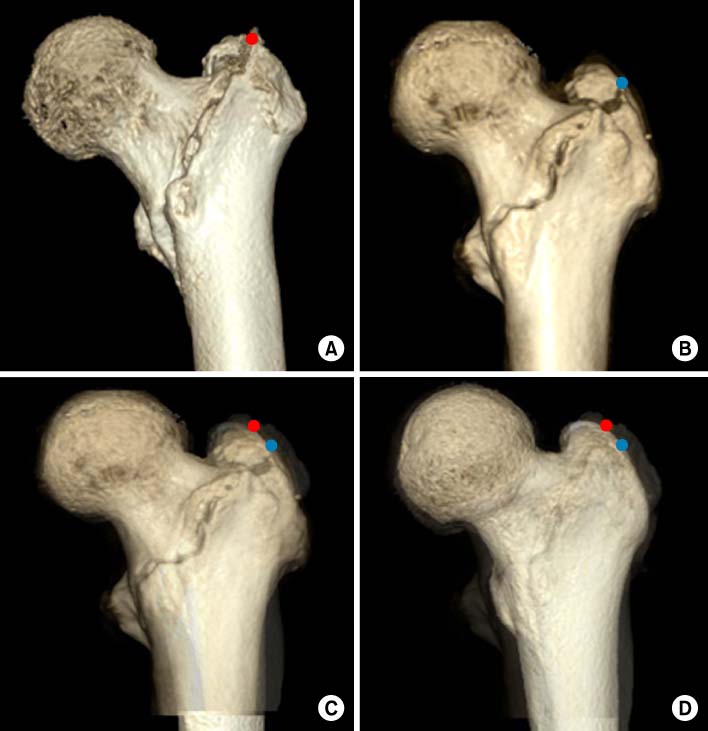
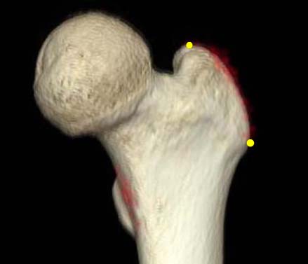
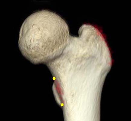
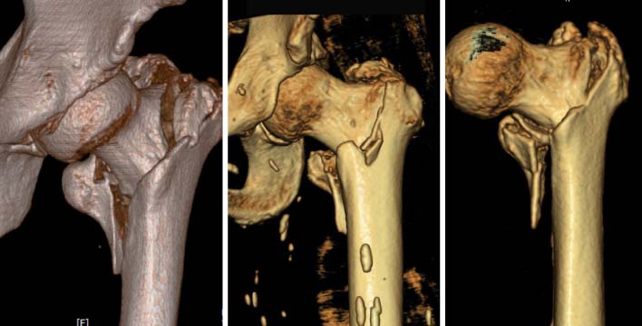
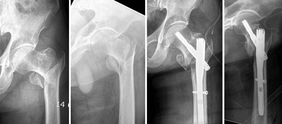
Fig. 1
Images of a trochanter fracture overlap each other (A, B) using Photoshop CS6® according to 50% opacity (C) and 20% opacity (D).
Fig. 2
Image of an overlapped trochanter fracture on a normal femur image. The lateral exit of the trochanteric fracture is located at an average distance of 10.2 mm without lateral wall fracture and not below the vastus ridge.
Fig. 3
Image of an overlapped trochanter fracture on a normal femur image. The medial exit is located at an average distance of 8.1 mm.
Fig. 4
The anteromedial cortex of the trochanter fracture was not comminuted, and however, showed several types.
Fig. 5
Immediate postoperative radiographs show that the proximal fragment of the trochanter fracture is displaced and widened inferiorly because of incorrect intramedullary nail insertion along the fracture site.
Fig. 1
Fig. 2
Fig. 3
Fig. 4
Fig. 5
Analysis of Low-Energy Trochanter Fracture Using the Multiplanar Computed Tomography Image: Application for Intramedullary Nail Fixation
Summary of Demographic Data (n=117)
Values are presented as number only or median (range).
Summary of the Outcomes (n=117)
*Vertical distance from greater trochanter to fracture line. †Vertical distance from proximal boundary of lesser trochanter to fracture line. ‡The angle between the fracture line of the distal fragment and anatomical axis of the femur.
Table 1
Summary of Demographic Data (n=117)
Values are presented as number only or median (range).
Table 2
Summary of the Outcomes (n=117)
*Vertical distance from greater trochanter to fracture line. †Vertical distance from proximal boundary of lesser trochanter to fracture line. ‡The angle between the fracture line of the distal fragment and anatomical axis of the femur.

