 E-submission
 TOTA
TOTA TOTS
TOTS
Articles
- Page Path
- HOME > J Musculoskelet Trauma > Volume 24(1); 2011 > Article
-
Surgical Technique
- Minimally Invasive Plate Osteosynthesis for the Upper Extremity Fracture Using a Lumbar Spreader: Surgical Technique
- Gu-Hee Jung, M.D., Chyul-Hyun Cho, M.D., Jae-Do Kim, M.D.
-
Journal of the Korean Fracture Society 2011;24(1):83-86.
DOI: https://doi.org/10.12671/jkfs.2011.24.1.83
Published online: January 21, 2011
Department of Orthopedic Surgery, Kosin University College fo Medicine, Busan, Korea.
*Department of Orthopedic Surgery, School of Medicine, Keimyung University, Daegu, Korea.
- Address reprint requests to: Gu-Hee Jung, M.D. Department of Orthopaedic Surgery, Gospel Hospital, Kosin University College of Medicine, 34, Amnam-dong, Seo-gu, Busan 602-702, Korea. Tel: 82-51-990-6130·Fax: 82-51-243-0181, jyujin2001@kosin.ac.kr
• Received: September 3, 2010 • Revised: September 29, 2010 • Accepted: October 25, 2010
Copyright © 2011 The Korean Fracture Society
- 839 Views
- 0 Download
Abstract
- The minimally invasive plate osteosynthesis (MIPO) which is extensively performed, is very dependent on the indirect reduction technique to prevent the exposure of fracture sites. Indirect reduction with the use of the femoral distractor is a much more efficient technique to restore the length in the fracture of lower limbs. However, the femoral distractor cannot be used for fracture of upper limbs, and other instruments for indirect reduction have not yet been reported. Therefore, we introduce the novel indirect reduction technique with the use of the lumbar spreader for the MIPO of upper limbs.
- 1. Apivatthakakul T, Arpornchayanon O, Bavornratanavech S. Minimally invasive plate osteosynthesis (MIPO) of the humeral shaft fracture. Is it possible? A cadaveric study and preliminary report. Injury, 2005;36:530-538.ArticlePubMed
- 2. Apivatthakakul T, Patiyasikan S, Luevitoonvechkit S. Danger zone for locking screw placement in minimally invasive plate osteosynthesis (MIPO) of humeral shaft fractures: a cadaveric study. Injury, 2010;41:169-172.ArticlePubMed
- 3. Borg T, Larsson S, Lindsjö U. Percutaneous plating of distal tibial fractures Preliminary results in 21 patients. Injury, 2004;35:608-614.Article
- 4. Byun YS. Minimally invasive plate osteosynthesis, MIPO. J Korean Fract Soc, 2007;20:99-114.Article
- 5. Clement H, Pichler W, Tesch NP, Heidari N, Grechenig W. Anatomical basis of the risk of radial nerve injury related to the technique of external fixation applied to the distal humerus. Surg Radiol Anat, 2010;32:221-224.ArticlePDF
- 6. Duncan SF, Weiland AJ. Minimally invasive reduction and osteosynthesis of articular fractures of the distal radius. Injury, 2001;32:Suppl 1. SA14-SA24.Article
- 7. Kregor PJ. Distal femur fractures with complex articular involvement: management by articular exposure and submuscular fixation. Orthop Clin North Am, 2002;33:153-175.
- 8. Oh CW, Oh JK, Jeon IH, et al. Minimally invasive percutaneous plate stabilization of proximal tibial fractures. J Korean Fract Soc, 2004;17:224-229.Article
- 9. Oh CW, Kyung HS, Park IH, Kim PT, Ihn JC. Distal tibia metaphyseal fractures treated by percutaneous plate osteosynthesis. Clin Orthop Relat Res, 2003;408:286-291.Article
- 10. Sen MK, Strauss N, Harvey EJ. Minimally invasive plate osteosynthesis of distal radius fractures using a pronator sparing approach. Tech Hand Up Extrem Surg, 2008;12:2-6.Article
REFERENCES
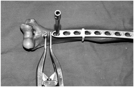


Figure & Data
REFERENCES
Citations
Citations to this article as recorded by 
 Cite
CiteMinimally Invasive Plate Osteosynthesis for the Upper Extremity Fracture Using a Lumbar Spreader: Surgical Technique

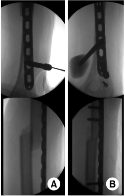
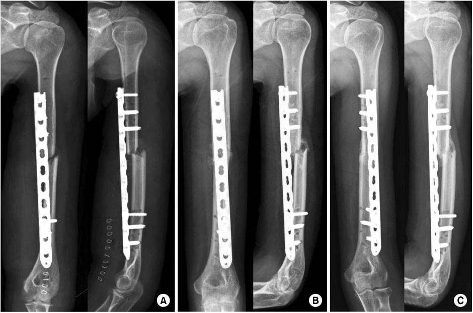
Fig. 1
The lumbar spread is placed between plate and screw to widen the interval.
Fig. 2
(A) Under the fluoroscopic guide, the plate is inserted percutanously and identified the overlapped the bone fragments.
(B) The traction is applied using the lumbar spread and the humeral length is restored.
Fig. 3
(A) Indirect reduction of the humeral fracture is achieved by distractive technique with lumbar spread.
(B) Radiographs at 5 weeks post-operatively show callus formation and (C) radiographs at 15 weeks, the radiological union.
Fig. 1
Fig. 2
Fig. 3
Minimally Invasive Plate Osteosynthesis for the Upper Extremity Fracture Using a Lumbar Spreader: Surgical Technique

