 E-submission
 TOTA
TOTA TOTS
TOTS
Articles
- Page Path
- HOME > J Musculoskelet Trauma > Volume 25(3); 2012 > Article
-
Review Article
- Treatment of Distal Humeral Fractures
- Yong-Cheol Yoon, M.D., Jong-Keon Oh, M.D., Ph.D.
-
Journal of the Korean Fracture Society 2012;25(3):223-232.
DOI: https://doi.org/10.12671/jkfs.2012.25.3.223
Published online: July 16, 2012
Department of Orthopedic Surgery, Korea University Medical Center Guro Hospital, Seoul, Korea.
- Address reprint requests to: Jong-Keon Oh, M.D., Ph.D. Division of Trauma, Department of Orthopedic Surgery, Korea University Medical Center Guro Hospital, 148, Gurodong-ro, Guro-gu, Seoul 152-703, Korea. Tel: 82-2-2626-3088, Fax: 82-2-851-3111, jkoh@korea.ac.kr
Copyright © 2012 The Korean Fracture Society
- 1,035 Views
- 4 Download
- 2 Crossref
- 1. Arnander MW, Reeves A, MacLeod IA, Pinto TM, Khaleel A. A biomechanical comparison of plate configuration in distal humerus fractures. J Orthop Trauma, 2008;22:332-336.Article
- 2. Athwal GS, Rispoli DM, Steinmann SP. The anconeus flap transolecranon approach to the distal humerus. J Orthop Trauma, 2006;20:282-285.Article
- 3. Brouwer KM, Guitton TG, Doornberg JN, Kloen P, Jupiter JB, Ring D. Fractures of the medial column of the distal humerus in adults. J Hand Surg Am, 2009;34:439-445.Article
- 4. Bryan RS, Morrey BF. Extensive posterior exposure of the elbow. A triceps-sparing approach. Clin Orthop Relat Res, 1982;(166):188-192.
- 5. Ebraheim NA, Andreshak TG, Yeasting RA, Saunders RC, Jackson WT. Posterior extensile approach to the elbow joint and distal humerus. Orthop Rev, 1993;22:578-582.
- 6. Galano GJ, Ahmad CS, Levine WN. Current treatment strategies for bicolumnar distal humerus fractures. J Am Acad Orthop Surg, 2010;18:20-30.Article
- 7. Gerwin M, Hotchkiss RN, Weiland AJ. Alternative operative exposures of the posterior aspect of the humeral diaphysis with reference to the radial nerve. J Bone Joint Surg Am, 1996;78:1690-1695.Article
- 8. Holdsworth BJ, Mossad MM. Fractures of the adult distal humerus. Elbow function after internal fixation. J Bone Joint Surg Br, 1990;72:362-365.ArticlePDF
- 9. Imatani J, Ogura T, Morito Y, Hashizume H, Inoue H. Custom AO small T plate for transcondylar fractures of the distal humerus in the elderly. J Shoulder Elbow Surg, 2005;14:611-615.Article
- 10. John H, Rosso R, Neff U, Bodoky A, Regazzoni P, Harder F. Operative treatment of distal humeral fractures in the elderly. J Bone Joint Surg Br, 1994;76:793-796.ArticlePDF
- 11. Jupiter JB. Complex fractures of the distal part of the humerus and associated complications. Instr Course Lect, 1995;44:187-198.Article
- 12. Jupiter JB, Barnes KA, Goodman LJ, Saldaña AE. Multiplane fracture of the distal humerus. J Orthop Trauma, 1993;7:216-220.Article
- 13. Kwasny O, Maier R. The significance of nerve damage in upper arm fractures. Unfallchirurg, 1991;94:461-467.
- 14. Levy JC, Kalandiak SP, Hutson JJ, Zych G. An alternative method of osteosynthesis for distal humeral shaft fractures. J Orthop Trauma, 2005;19:43-47.Article
- 15. McKee MD, Jupiter JB. A contemporary approach to the management of complex fractures of the distal humerus and their sequelae. Hand Clin, 1994;10:479-494.Article
- 16. McKee MD, Jupiter JB, Bamberger HB. Coronal shear fractures of the distal end of the humerus. J Bone Joint Surg Am, 1996;78:49-54.Article
- 17. Nauth A, McKee MD, Ristevski B, Hall J, Schemitsch EH. Distal humeral fractures in adults. J Bone Joint Surg Am, 2011;93:686-700.Article
- 18. O'Driscoll SW. The triceps-reflecting anconeus pedicle (TRAP) approach for distal humeral fractures and nonunions. Orthop Clin North Am, 2000;31:91-101.Article
- 19. O'Driscoll SW. Parallel plate fixation of bicolumn distal humeral fractures. Instr Course Lect, 2009;58:521-528.
- 20. O'Driscoll SW, Sanchez-Sotelo J, Torchia ME. Management of the smashed distal humerus. Orthop Clin North Am, 2002;33:19-33.Article
- 21. Pankaj A, Mallinath G, Malhotra R, Bhan S. Surgical management of intercondylar fractures of the humerus using triceps reflecting anconeus pedicle (TRAP) approach. Indian J Orthop, 2007;41:219-223.Article
- 22. Perry CR, Gibson CT, Kowalski MF. Transcondylar fractures of the distal humerus. J Orthop Trauma, 1989;3:98-106.Article
- 23. Remia LF, Richards K, Waters PM. The Bryan-Morrey triceps-sparing approach to open reduction of T-condylar humeral fractures in adolescents: cybex evaluation of triceps function and elbow motion. J Pediatr Orthop, 2004;24:615-619.
- 24. Ring D, Jupiter JB. Complex fractures of the distal humerus and their complications. J Shoulder Elbow Surg, 1999;8:85-97.Article
- 25. Ring D, Jupiter JB. Fractures of the distal humerus. Orthop Clin North Am, 2000;31:103-113.Article
- 26. Schwartz A, Oka R, Odell T, Mahar A. Biomechanical comparison of two different periarticular plating systems for stabilization of complex distal humerus fractures. Clin Biomech (Bristol, Avon), 2006;21:950-955.Article
- 27. Tak SR, Dar GN, Halwai MA, Kangoo KA, Mir BA. Outcome of olecranon osteotomy in the trans-olecranon approach of intra-articular fractures of the distal humerus. Ulus Travma Acil Cerrahi Derg, 2009;15:565-570.
- 28. Windolf M, Maza ER, Gueorguiev B, Braunstein V, Schwieger K. Treatment of distal humeral fractures using conventional implants. Biomechanical evaluation of a new implant configuration. BMC Musculoskelet Disord, 2010;11:172. ArticlePDF
- 29. Zalavras CG, Vercillo MT, Jun BJ, Otarodifard K, Itamura JM, Lee TQ. Biomechanical evaluation of parallel versus orthogonal plate fixation of intra-articular distal humerus fractures. J Shoulder Elbow Surg, 2011;20:12-20.Article
REFERENCES

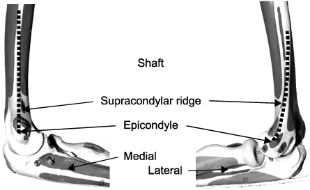

Fig. 4Bare area marked with arrow does not have articular cartilage, so bare area is in the osteotomy.
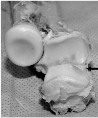
Fig. 5When hinge-opening the periarticular bone fragment (arrow) from side to side to find a coronal fragment shown in computed tomography and fix it.

Fig. 6A 3D computed tomography which displays a coronal bone fragment invading the front of trochlea (arrow). A clinical picture showing a coronal bone block fixed with a 0.9 mm K-wire. K-wire is buried in the bone, when reducing captellum and trochlea after sticking it to the fractured side and cutting it. There is an embedded K-wire to fix the coronal bone fragment in the picture taken after the surgery (arrow).
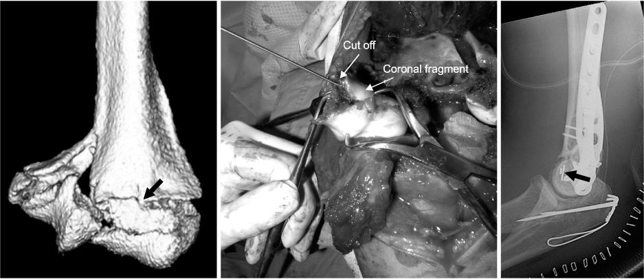
Fig. 7An example of an accurate operation of tension band wiring and plate fixation using a transosseous pin.
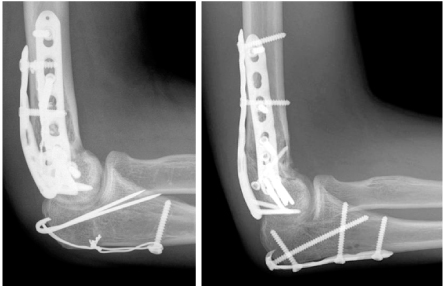
Fig. 8When K-wire is too deeply inserted into the bone without accurate reduction and compression of osteotomy part right after the surgery. Especially, inserting two k-wires into the lateral side may restrict joint movement, by causing a collision in the radial tuberosity, because they protrude too much as shown in the oblique view. Therefore, it requires a careful attention. There was an early loss of fixation as displayed in the picture taken a month later the surgery. This could be solved with plate fixation.

Fig. 9When reducing AO 13 C2 fracture with the side to side retraction approach and fixing it. It was found that there was no articular comminution or coronal bone fragment in the computed tomography, and it was possible to reduce the joint without osteotomy of olecranon, because it was separated between trochlea and capitellum.

Fig. 10A clinical image to fix a plate in the medial and posterolateral side and a radiograph which shows accurate joint reduction and joint movement during the surgery.
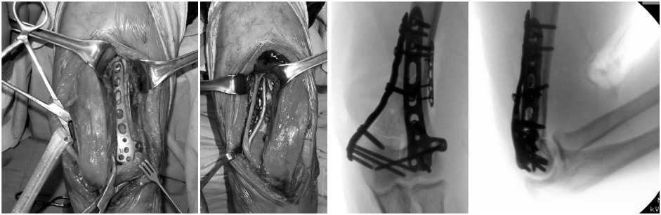
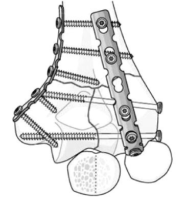
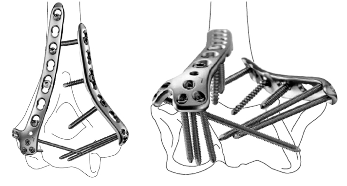
Fig. 13When inserting a screw through a screw hole of plate after reducing the periarticular bone fragment with pointed reduction forceps and fixing it rather than a separate lag screw.
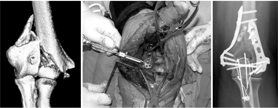


Figure & Data
REFERENCES
Citations
Citations to this article as recorded by 
- Paratricipital Approach for AO/OTA Type C2 Intra-Articular Fracture of Distal Humerus
Chul-Hyung Lee, Doo-Hun Sun, Deukhee Jung, Chung-Han An
Journal of the Korean Fracture Society.2019; 32(3): 128. CrossRef - How Difficult Is It to Surgically Treat AO-C Type Distal Humerus Fractures for Inexperienced Orthopedic Surgeons?
Seong Ho Yoo, Suk Woong Kang, Moo Ho Song, Young Jun Kim, Hyuck Bae
Journal of the Korean Fracture Society.2018; 31(2): 45. CrossRef
 Cite
CiteTreatment of Distal Humeral Fractures
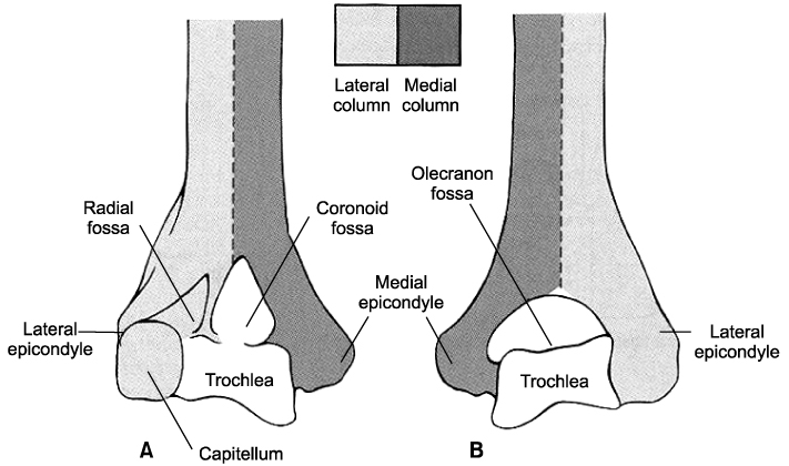

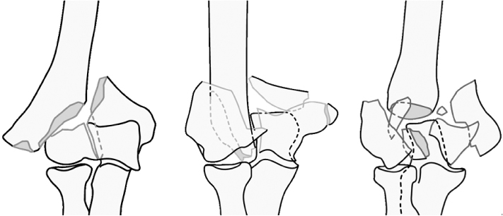

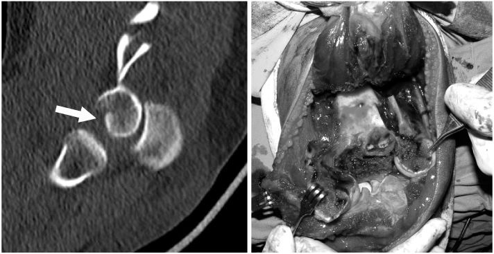


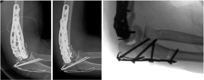
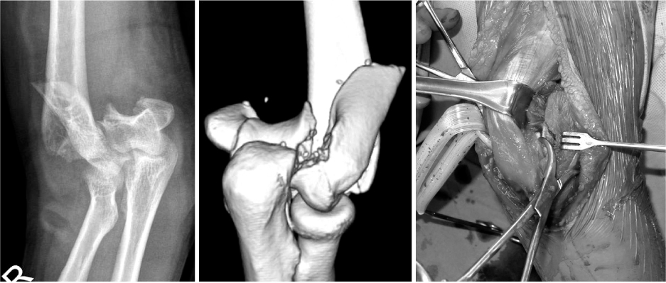


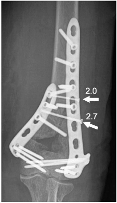
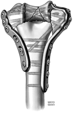
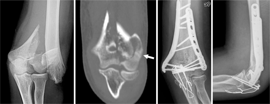
Fig. 1
(A) Anterior, (B) posterior view of column structure of the distal humerus.
Fig. 2
Alignment of medial and lateral side of the distal humerus.
Fig. 3
Illustrations show AO classification 13 C1, C2, C3.
Fig. 4
Bare area marked with arrow does not have articular cartilage, so bare area is in the osteotomy.
Fig. 5
When hinge-opening the periarticular bone fragment (arrow) from side to side to find a coronal fragment shown in computed tomography and fix it.
Fig. 6
A 3D computed tomography which displays a coronal bone fragment invading the front of trochlea (arrow). A clinical picture showing a coronal bone block fixed with a 0.9 mm K-wire. K-wire is buried in the bone, when reducing captellum and trochlea after sticking it to the fractured side and cutting it. There is an embedded K-wire to fix the coronal bone fragment in the picture taken after the surgery (arrow).
Fig. 7
An example of an accurate operation of tension band wiring and plate fixation using a transosseous pin.
Fig. 8
When K-wire is too deeply inserted into the bone without accurate reduction and compression of osteotomy part right after the surgery. Especially, inserting two k-wires into the lateral side may restrict joint movement, by causing a collision in the radial tuberosity, because they protrude too much as shown in the oblique view. Therefore, it requires a careful attention. There was an early loss of fixation as displayed in the picture taken a month later the surgery. This could be solved with plate fixation.
Fig. 9
When reducing AO 13 C2 fracture with the side to side retraction approach and fixing it. It was found that there was no articular comminution or coronal bone fragment in the computed tomography, and it was possible to reduce the joint without osteotomy of olecranon, because it was separated between trochlea and capitellum.
Fig. 10
A clinical image to fix a plate in the medial and posterolateral side and a radiograph which shows accurate joint reduction and joint movement during the surgery.
Fig. 11
Conventional orthogonal plating with congruent elbow plating system.
Fig. 12
Anatomical precontoured locking compression plate with congruent elbow plating system.
Fig. 13
When inserting a screw through a screw hole of plate after reducing the periarticular bone fragment with pointed reduction forceps and fixing it rather than a separate lag screw.
Fig. 14
An example of fixation in a small metaphyseal wedged-bone fragment using 2.7 mm / 2.0 mm screws.
Fig. 15
Parallel plating with congruent elbow plating system.
Fig. 16
A case to fix C2 fracture accompanied by a low lateral column fracture (arrow) successfully with parallel plating.
Fig. 1
Fig. 2
Fig. 3
Fig. 4
Fig. 5
Fig. 6
Fig. 7
Fig. 8
Fig. 9
Fig. 10
Fig. 11
Fig. 12
Fig. 13
Fig. 14
Fig. 15
Fig. 16
Treatment of Distal Humeral Fractures

