 E-submission
 TOTA
TOTA TOTS
TOTS
Articles
- Page Path
- HOME > J Musculoskelet Trauma > Volume 25(3); 2012 > Article
-
Review Article
- Treatment of Neglected Monteggia Fracture in Children
- Changhoon Jeong, M.D., In Park, M.D.
-
Journal of the Korean Fracture Society 2012;25(3):233-239.
DOI: https://doi.org/10.12671/jkfs.2012.25.3.233
Published online: July 16, 2012
Department of Orthopedic Surgery, The Catholic University of Korea Bucheon St. Mary's Hospital, Bucheon, Korea.
- Address reprint requests to: Changhoon Jeong, M.D. Department of Orthopedic Surgery, The Catholic University of Korea Bucheon St. Mary's Hospital, Sosa-ro 327, Wonmi-gu, Bucheon 420-717, Korea. Tel: 82-32-340-7089, Fax: 82-32-340-2671, changhoonj@naver.com
Copyright © 2012 The Korean Fracture Society
- 1,820 Views
- 52 Download
- 1 Crossref
- 1. Austin R. Tardy palsy of the radial nerve from a Monteggia fracture. Injury, 1976;7:202-204.Article
- 2. Bado JL. The Monteggia lesion. Clin Orthop Relat Res, 1967;50:71-86.
- 3. Bell Tawse AJ. The treatment of malunited anterior Monteggia fractures in children. J Bone Joint Surg Br, 1965;47:718-723.
- 4. Best TN. Management of old unreduced Monteggia fracture dislocations of the elbow in children. J Pediatr Orthop, 1994;14:193-199.Article
- 5. David-West KS, Wilson NI, Sherlock DA, Bennet GC. Missed Monteggia injuries. Injury, 2005;36:1206-1209.Article
- 6. Degreef I, De Smet L. Missed radial head dislocations in children associated with ulnar deformation: treatment by open reduction and ulnar osteotomy. J Orthop Trauma, 2004;18:375-378.
- 7. Devnani AS. Missed Monteggia fracture dislocation in children. Injury, 1997;28:131-133.Article
- 8. Evans EM. Pronation injuries of the forearm, with special reference to the anterior Monteggia fracture. J Bone Joint Surg Br, 1949;31B:578-588.
- 9. Fahmy NR. Unusual Monteggia lesions in children. Injury, 1981;12:399-404.Article
- 10. Futami T, Tsukamoto Y, Fujita T. Rotation osteotomy for dislocation of the radial head. 6 cases followed for 7 (3~10) years. Acta Orthop Scand, 1992;63:455-456.
- 11. Giustra PE, Killoran PJ, Furman RS, Root JA. The missed Monteggia fracture. Radiology, 1974;110:45-47.Article
- 12. Hirayama T, Takemitsu Y, Yagihara K, Mikita A. Operation for chronic dislocation of the radial head in children. Reduction by osteotomy of the ulna. J Bone Joint Surg Br, 1987;69:639-642.ArticlePDF
- 13. Horii E, Nakamura R, Koh S, Inagaki H, Yajima H, Nakao E. Surgical treatment for chronic radial head dislocation. J Bone Joint Surg Am, 2002;84-A:1183-1188.Article
- 14. Hudson DA, De Beer JD. Isolated traumatic dislocation of the radial head in children. J Bone Joint Surg Br, 1986;68:378-381.ArticlePDF
- 15. Hurst LC, Dubrow EN. Surgical treatment of symptomatic chronic radial head dislocation: a neglected Monteggia fracture. J Pediatr Orthop, 1983;3:227-230.
- 16. Kalamchi A. Monteggia fracture-dislocation in children. Late treatment in two cases. J Bone Joint Surg Am, 1986;68:615-619.Article
- 17. Kemnitz S, De Schrijver F, De Smet L. Radial head dislocation with plastic deformation of the ulna in children. A rare and frequently missed condition. Acta Orthop Belg, 2000;66:359-362.
- 18. Kim HT, Conjares JN, Suh JT, Yoo CI. Chronic radial head dislocation in children, Part 1: pathologic changes preventing stable reduction and surgical correction. J Pediatr Orthop, 2002;22:583-590.Article
- 19. Letts M, Locht R, Wiens J. Monteggia fracture-dislocations in children. J Bone Joint Surg Br, 1985;67:724-727.ArticlePDF
- 20. Lincoln TL, Mubarak SJ. "Isolated" traumatic radial-head dislocation. J Pediatr Orthop, 1994;14:454-457.Article
- 21. Lloyd-Roberts GC, Bucknill TM. Anterior dislocation of the radial head in children: aetiology, natural history and management. J Bone Joint Surg Br, 1977;59-B:402-407.ArticlePDF
- 22. Nakamura K, Hirachi K, Uchiyama S, et al. Long-term clinical and radiographic outcomes after open reduction for missed Monteggia fracture-dislocations in children. J Bone Joint Surg Am, 2009;91:1394-1404.Article
- 23. Nakamura T, Yabe Y, Horiuchi Y. Functional anatomy of the interosseous membrane of the forearm - dynamic changes during rotation. Hand Surg, 1999;4:67-73.Article
- 24. Olney BW, Menelaus MB. Monteggia and equivalent lesions in childhood. J Pediatr Orthop, 1989;9:219-223.Article
- 25. Ovesen O, Brok KE, Arreskøv J, Bellstrøm T. Monteggia lesions in children and adults: an analysis of etiology and long-term results of treatment. Orthopedics, 1990;13:529-534.Article
- 26. Seel MJ, Peterson HA. Management of chronic posttraumatic radial head dislocation in children. J Pediatr Orthop, 1999;19:306-312.Article
- 27. Smith FM. Monteggia fractures; an analysis of 25 consecutive fresh injuries. Surg Gynecol Obstet, 1947;85:630-640.
- 28. Speed JS, Boyd HB. Treatment of fractures of ulna with dislocation of head of radius. JAMA, 1940;115:1699-1705.Article
- 29. Stoll TM, Willis RB, Paterson DC. Treatment of the missed Monteggia fracture in the child. J Bone Joint Surg Br, 1992;74:436-440.ArticlePDF
- 30. Storen G. Traumatic dislocation of the radial head as an isolated lesion in children; report of one case with special regard to roentgen diagnosis. Acta Chir Scand, 1959;116:144-147.
- 31. Tajima T, Yoshizu T. Treatment of long-standing dislocation of the radial head in neglected Monteggia fractures. J Hand Surg Am, 1995;20:S91-S94.Article
- 32. Theodorou SD, Ierodiaconou MN, Roussis N. Fracture of the upper end of the ulna associated with dislocation of the head of the radius in children. Clin Orthop Relat Res, 1988;(228):240-249.Article
- 33. Wang MN, Chang WN. Chronic posttraumatic anterior dislocation of the radial head in children: thirteen cases treated by open reduction, ulnar osteotomy, and annular ligament reconstruction through a Boyd incision. J Orthop Trauma, 2006;20:1-5.
- 34. Wiley JJ, Pegington J, Horwich JP. Traumatic dislocation of the radius at the elbow. J Bone Joint Surg Br, 1974;56B:501-507.ArticlePDF
REFERENCES
Fig. 1
Classification of pediatric Monteggia fracture-dislocations. Redrawn from the article of Letts M, Locht R, Weins J19).
(A) Anterior plastic deformity (bending).
(B) Anterior greenstick fracture.
(C) Anterior complete fracture.
(D) Posterior.
(E) Lateral.
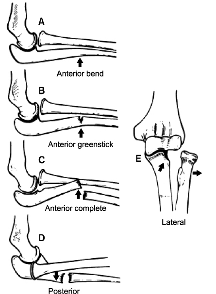
Fig. 2
Drawings of transverse cuts of proximal right radius and ulna (viewed distally) at the level of the radial head. Redrawn from the article of Seel MJ, Peterson HA (Fig. 1)26).
(A) Route of triceps tendon in Bell Tawse reconstruction. Direction of stability is posterior (large arrow).
(B) Drill hole placed obliquely to exit ulna at site of medial annular ligament attachment. Direction of stability is posteromedial (large arrow).
(C) Two drill holes exit the ulna at sites of medial and lateral annular ligament attachments. Direction of stability is anatomic (arrow).

Fig. 3Revised from the article of Hirayama T, Takemitsu Y, Yagihara K, Mikita A (Fig. 1, 2)12). (A) Ulnar bending elongation osteotomy, (B) ulnar valgus elongation osteotomy.
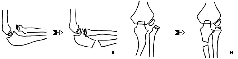
Fig. 4
Pre-operative paper tracing for ulnar bending elongation osteotomy (modified Hirayama osteotomy). Point a: Anterior portion of the spherical surface of the capitellum. Point b: Center of the radial head. Redrawn from the article of Nakamura T, Yabe Y, Horiuchi Y (Fig. 1A, B, C)23).
(A) A seven-hole plate was applied to the dorsal cortex of the ulna. The most proximal screw was positioned at the level of the coronoid process. The osteotomy line was in the center of the plate.
(B) Both the distal part of the ulna (gray) and the radius (gray) were mobilized to localize point b opposite point a. The distance between point a and point b was at 0.5 to 1.0 mm to avoid excessive pressure on the radiohumeral joint after radial head reduction.
(C) The osteotomized ulna was fixed with a plate.

Fig. 5
A 5-year-old boy reported that he had fallen from a bar 6 months earlier. At the time of the injury, the fracture had been treated nonsurgically, and the dislocation had been missed. Approximately six months after the injury, open reduction with ulnar corrective osteotomy, anular ligament repair, and transcapitellar pin fixation was performed.
(A) Pre-operative radiographs of the elbow.
(B) Operative radiograph.
(C) Post-operative radiograph.

Figure & Data
REFERENCES
Citations
Citations to this article as recorded by 
- Surgical Timing of Treating Pediatric Trauma: Urgencies/Emergencies
Chang-Wug Oh, Joon-Woo Kim, Jong-Chul Lee
Journal of the Korean Fracture Society.2015; 28(2): 146. CrossRef
 Cite
CiteTreatment of Neglected Monteggia Fracture in Children

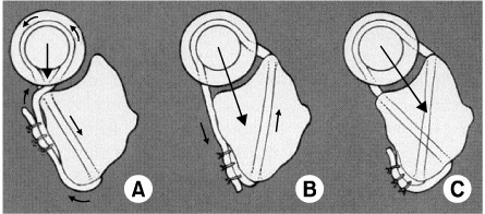

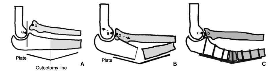
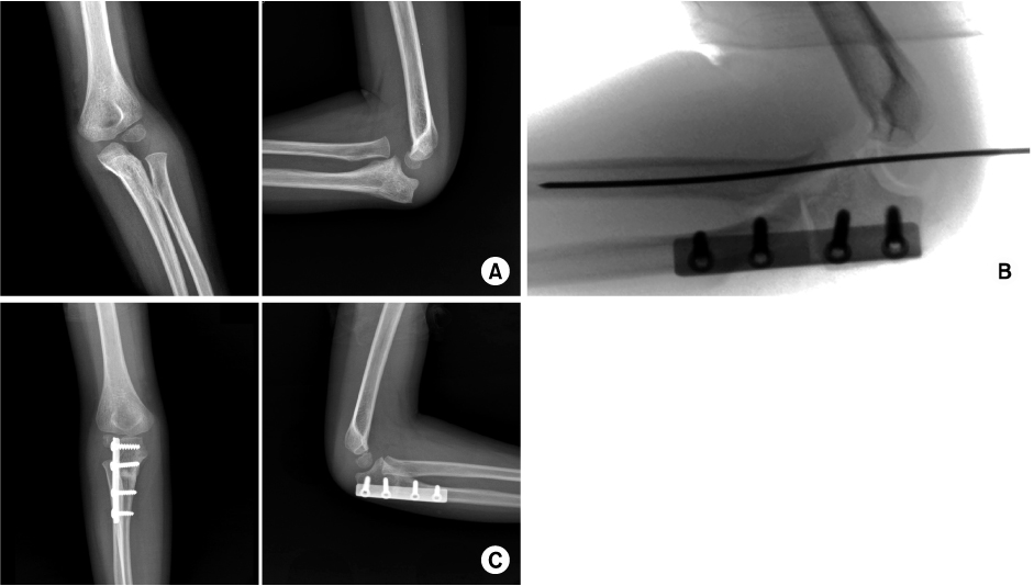
Fig. 1
Classification of pediatric Monteggia fracture-dislocations. Redrawn from the article of Letts M, Locht R, Weins J19).
(A) Anterior plastic deformity (bending).
(B) Anterior greenstick fracture.
(C) Anterior complete fracture.
(D) Posterior.
(E) Lateral.
Fig. 2
Drawings of transverse cuts of proximal right radius and ulna (viewed distally) at the level of the radial head. Redrawn from the article of Seel MJ, Peterson HA (Fig. 1)26).
(A) Route of triceps tendon in Bell Tawse reconstruction. Direction of stability is posterior (large arrow).
(B) Drill hole placed obliquely to exit ulna at site of medial annular ligament attachment. Direction of stability is posteromedial (large arrow).
(C) Two drill holes exit the ulna at sites of medial and lateral annular ligament attachments. Direction of stability is anatomic (arrow).
Fig. 3
Revised from the article of Hirayama T, Takemitsu Y, Yagihara K, Mikita A (Fig. 1, 2)12). (A) Ulnar bending elongation osteotomy, (B) ulnar valgus elongation osteotomy.
Fig. 4
Pre-operative paper tracing for ulnar bending elongation osteotomy (modified Hirayama osteotomy). Point a: Anterior portion of the spherical surface of the capitellum. Point b: Center of the radial head. Redrawn from the article of Nakamura T, Yabe Y, Horiuchi Y (Fig. 1A, B, C)23).
(A) A seven-hole plate was applied to the dorsal cortex of the ulna. The most proximal screw was positioned at the level of the coronoid process. The osteotomy line was in the center of the plate.
(B) Both the distal part of the ulna (gray) and the radius (gray) were mobilized to localize point b opposite point a. The distance between point a and point b was at 0.5 to 1.0 mm to avoid excessive pressure on the radiohumeral joint after radial head reduction.
(C) The osteotomized ulna was fixed with a plate.
Fig. 5
A 5-year-old boy reported that he had fallen from a bar 6 months earlier. At the time of the injury, the fracture had been treated nonsurgically, and the dislocation had been missed. Approximately six months after the injury, open reduction with ulnar corrective osteotomy, anular ligament repair, and transcapitellar pin fixation was performed.
(A) Pre-operative radiographs of the elbow.
(B) Operative radiograph.
(C) Post-operative radiograph.
Fig. 1
Fig. 2
Fig. 3
Fig. 4
Fig. 5
Treatment of Neglected Monteggia Fracture in Children

