 E-submission
 TOTA
TOTA TOTS
TOTS
Search
- Page Path
- HOME > Search
Review Article
- Combined acetabular and pelvic ring injuries: a reference-frame algorithm for definitive fixation sequencing
- Jeong-Hyun Koh, Seungyeob Sakong
- J Musculoskelet Trauma 2026;39(2):83-92. Published online April 9, 2026
- DOI: https://doi.org/10.12671/jmt.2026.00031
-
 Abstract
 PDF
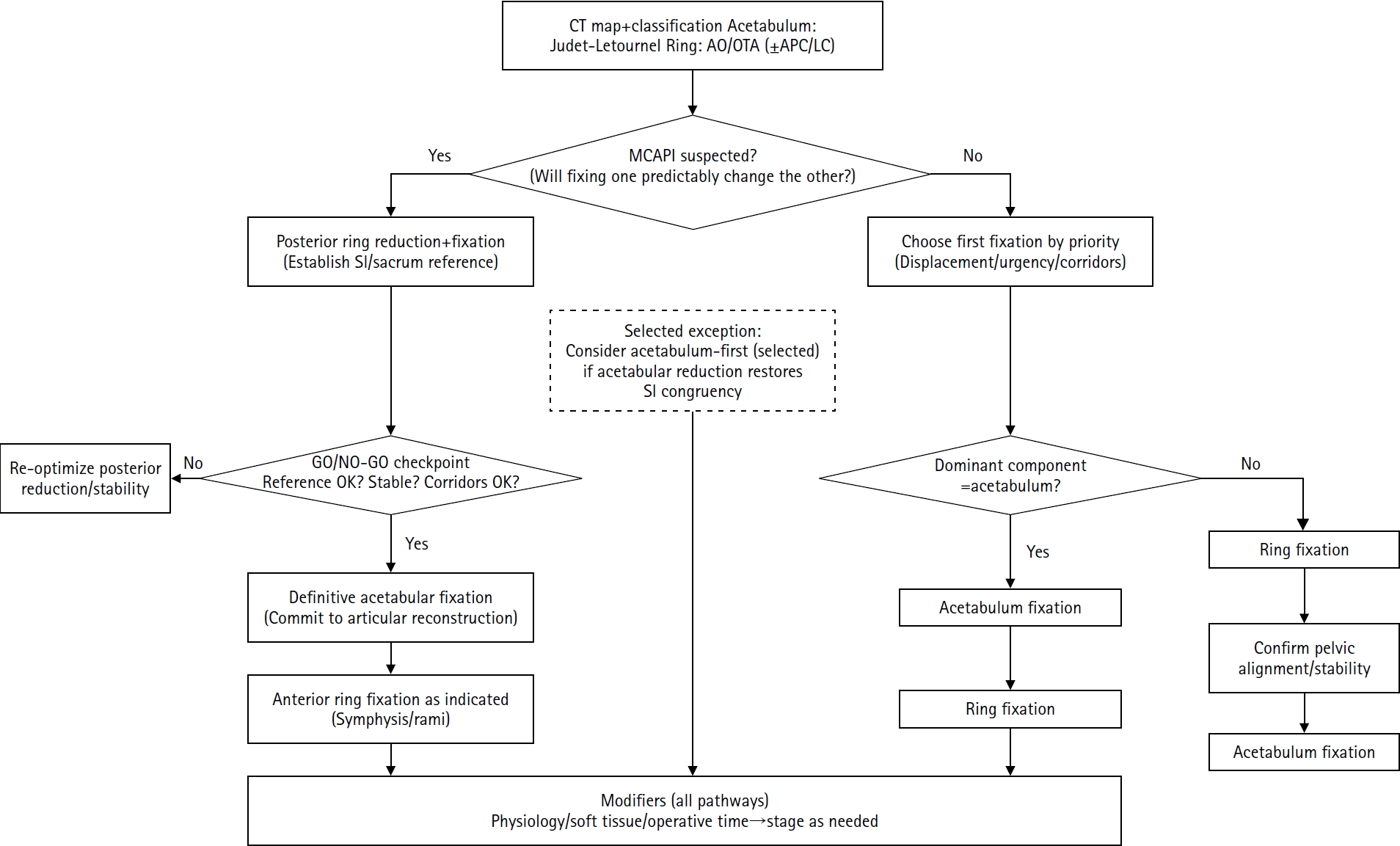
PDF - Combined acetabular and pelvic ring injuries are not simply “two fractures in one patient.” Reduction and fixation of one component can alter the alignment and reducibility of the other, rendering operative sequencing a primary decision variable rather than a secondary consideration. These injuries typically result from high-energy trauma, frequently occur in patients with polytrauma, and are further influenced by physiological tolerance and the feasibility of available operative corridors. The existing evidence base remains constrained by retrospective study designs, inconsistent definitions, variable classification systems, and heterogeneous outcome reporting, all of which limit the strength of comparative recommendations. This state-of-the-art review presents a surgeon-facing, algorithmic approach grounded in a reference-frame mindset. We emphasize computed tomography (CT)-based mapping and the use of consistent terminology to characterize acetabular morphology, pelvic ring instability, deformity vectors, suspicion of mechanical coupling, and feasible operative corridors. Mechanically connected acetabular and pelvic ring injuries (MCAPI) are introduced as a working framework for identifying patterns in which reduction or fixation of one injury predictably influences the other. In cases of suspected MCAPI, a posterior ring-based sequence is generally preferred, typically consisting of posterior ring reduction and fixation, definitive acetabular reconstruction, and subsequent anterior ring fixation. We propose an explicit intraoperative “GO/NO-GO” checkpoint (reference acceptable, stable, corridors feasible) to prevent acetabular reconstruction on a moving target. Acetabulum-first strategies may be appropriate only in selected anteroposterior compression- type configurations in which acetabular fixation plausibly restores sacroiliac congruency and posterior stabilization remains technically feasible. We summarize key outcome domains and complication patterns, highlighting hip dislocation as an important risk factor associated with both neurologic deficits and overall complications. Standardized CTbased definitions and outcome instruments, together with multicenter cohorts employing predefined decision pathways, are required to test sequencing strategies and to determine whether improved radiographic reduction translates into durable functional benefit.
- 951 View
- 23 Download

Original Article
- Comparison of the Radiological Outcomes of an Anatomical Quadrilateral Surface Plate with a Pelvic Reconstruction Plate in Acetabulum Fractures
- Sung Hyun Yoon, Hee Gon Park, Dong Uk Lee
- J Korean Fract Soc 2024;37(2):95-101. Published online April 30, 2024
- DOI: https://doi.org/10.12671/jkfs.2024.37.2.95
-
Abstract
PDF
- Purpose
This study compared the radiological outcomes of fixation using an anatomical quadrilateral surface plate with those using a traditional pelvic reconstruction plate for fractures involving the quadrilateral surface or superomedial wall of the acetabulum.
Materials and Methods
From 2015 to 2022, 47 patients who met the inclusion and exclusion criteria were analyzed retrospectively. Internal fixation of an acetabular fracture was achieved with a pelvic reconstruction plate (n=28) or an anatomical quadrilateral surface plate (n=19). The ability to achieve immediate postoperative anatomical reduction and the long-term outcomes were assessed by confirming the arthritic changes. Immediate postoperative reduction quality and long-term radiological outcomes for post-traumatic arthritis were assessed using the Matta scoring system on standard radiographs.
Results
The assessment of immediate postoperative reduction in the pelvic reconstruction plate group was satisfactory in 16 patients (57.1%) and unsatisfactory in 12 patients (42.9%). In the anatomical quadrilateral surface plate group, the results were satisfactory in 16 patients (84.2%) and unsatisfactory in 3 patients (15.8%). When evaluating over an extended follow-up period in the pelvic reconstruction plate group, 19 patients (67.9%) demonstrated satisfactory, while 9 patients (32.1%) had unsatisfactory outcomes. In the anatomical quadrilateral surface plate group, 12 patients (63.2%) achieved satisfactory, and 7 patients (36.8%) had unsatisfactory outcomes. The immediate postoperative reduction quality was superior in the anatomical quadrilateral surface plate group (p=0.03). Comparing longterm results, the anatomical quadrilateral surface plate group did not have statistically more favorable outcomes (p=0.49).
Conclusion
In this study, the anatomical quadrilateral surface plate achieved sufficiently good radiological results without significant difference from the existing pelvic reconstruction plate. It was concluded that it is a useful option that can replace the existing metal plate in the selection of surgery for acetabular fractures.
- 1,467 View
- 18 Download
Review Articles
- Total Hip Arthroplasty after Acetabular Fracture: Acute Phase and Delayed Phase
- Hwan Hee Lee, Se Won Lee, Weon Yoo Kim
- J Korean Fract Soc 2019;32(4):232-239. Published online October 31, 2019
- DOI: https://doi.org/10.12671/jkfs.2019.32.4.232
-
Abstract
PDF
- The incidence of acetabular fractures in the elderly has increased because of the increasing elderly population. To determine the treatment plan for acetabular fractures, the patient's age, gait ability, presence or absence of osteoporosis and osteoarthritis, underlying disease, and fracture pattern should be considered. The application of total hip arthroplasty for acetabular fractures with the proper indications can be expected to have a good prognosis. In this paper, the application of total hip arthroplasty as a treatment method for acetabular fractures is divided into acute and delayed phases.
-
Citations
Citations to this article as recorded by
- Effect of Korean Medicine Treatments for Pain Reduction in Patients with Hip Fracture : A Retrospective Observational Study
Nam Hoon Kim, Min Seok Oh
Journal of Physiology & Pathology in Korean Medicine.2020; 34(5): 263. CrossRef
- Effect of Korean Medicine Treatments for Pain Reduction in Patients with Hip Fracture : A Retrospective Observational Study
- 1,812 View
- 2 Download
- 1 Crossref
- Anterior Approach for the Acetabular Fractures
- Jae Youn Yoon, Jae Woo Cho, Ji Wan Kim
- J Korean Fract Soc 2019;32(3):157-164. Published online July 31, 2019
- DOI: https://doi.org/10.12671/jkfs.2019.32.3.157
-
Abstract
PDF
- In the surgical treatment of acetabular fractures, the anterior approach is used widely for anterior column fractures with or without posterior column fractures. This paper reviews the anterior approach for the anatomical reduction and rigid fixation of acetabular fractures: traditional ilioinguinal approach, modified Stoppa approach, and new Pararectal approach.
-
Citations
Citations to this article as recorded by- Adhesion of External Iliac Vessels Found in a Modified Stoppa Approach to Acetabular Fracture in a Patient with a History of Previous Abdominal Surgery
Seong-Tae Kim, Seungyup Shin, Hohyoung Lee, Seong Man Jeon
Journal of the Korean Orthopaedic Association.2022; 57(1): 68. CrossRef
- Adhesion of External Iliac Vessels Found in a Modified Stoppa Approach to Acetabular Fracture in a Patient with a History of Previous Abdominal Surgery
- 2,215 View
- 43 Download
- 1 Crossref
- Principles for Management of Periprosthetic Acetabular Fractures after Hip Arthroplasty
- Chan Woo Park, Hyoung Keun Oh, Woo Suk Lee, Youn Soo Park, Seung Jae Lim
- J Korean Fract Soc 2019;32(3):148-156. Published online July 31, 2019
- DOI: https://doi.org/10.12671/jkfs.2019.32.3.148
-
Abstract
PDF
- Periprosthetic acetabular fracture (PAF) is an uncommon complication following hip arthroplasty. However, as the number of people needing hip prostheses continues to rise, the absolute number of PAF is expected to increase as well. These fractures may occur either intraoperatively or postoperatively. Postoperative fractures can be caused by traumatic events or by pathologic conditions related to periacetabular osteolysis. The management of PAF usually depends on the degree of displacement and the stability of the acetabular component. While most of non-displaced fractures can be managed nonoperatively by protected weight bearing, displaced fractures with unstable implants require surgical intervention, which is often technically challenging. This review summarized the latest findings on the epidemiology, the diagnosis, the classification, and the treatment of PAF.
-
Citations
Citations to this article as recorded by- Treatment of Periprosthetic Femoral Fractures after Hip Arthroplasty
Jung-Hoon Choi, Jong-Hyuk Jeon, Kyung-Jae Lee
Journal of the Korean Fracture Society.2020; 33(1): 43. CrossRef
- Treatment of Periprosthetic Femoral Fractures after Hip Arthroplasty
- 2,571 View
- 54 Download
- 1 Crossref
- Pelvis/Acetabular Fractures in the Elderly: When and How to Fix?
- Kyeong Hyeon Park, Chang Wug Oh, Joon Woo Kim
- J Korean Fract Soc 2018;31(3):102-113. Published online July 31, 2018
- DOI: https://doi.org/10.12671/jkfs.2018.31.3.102
-
Abstract
PDF
- Owing to the increase in life expectancy, the incidence of osteoporotic fracture of the pelvis and acetabulum is increasing. Fractures in the elderly population is different from those in younger patients. Pelvic ring and acetabular fractures in geriatric patients are more likely the result of low-energy trauma, but the outcomes are generally poorer than those of the younger population. Multiple management options are available, but no intervention has become the standard of care for these fractures in the elderly. A treatment strategy should be established depending on the state of the individual patient. Regardless of whether nonsurgical or surgical treatment is selected, early ambulation should be considered to avoid the complications associated with prolonged immobilization.
-
Citations
Citations to this article as recorded by- Effect of Korean Medicine Treatments for Pain Reduction in Patients with Hip Fracture : A Retrospective Observational Study
Nam Hoon Kim, Min Seok Oh
Journal of Physiology & Pathology in Korean Medicine.2020; 34(5): 263. CrossRef
- Effect of Korean Medicine Treatments for Pain Reduction in Patients with Hip Fracture : A Retrospective Observational Study
- 919 View
- 7 Download
- 1 Crossref
Case Report
- Reduction Technique of Dome Impaction Using the Modified Stoppa Approach: A Technical Note
- Ji Wan Kim, Yong Min Seo, Hyo Seok Jang
- J Korean Fract Soc 2017;30(3):131-136. Published online July 31, 2017
- DOI: https://doi.org/10.12671/jkfs.2017.30.3.131
-
Abstract
PDF
- In elderly acetabular fractures, central dislocation of the femoral head and impacted superior dome of the acetabulum is common. Unreduced dome impaction can lead to degenerative arthritis and results in poor results. Herein, we present a case of operative reduction and fixation performed via the modified Stoppa approach in acetabular fracture with superior dome impaction.
-
Citations
Citations to this article as recorded by- Surgical outcomes of acetabular fracture of elderly patients with superomedial dome impaction
Eic Ju Lim, Hyun-Chul Shon, Jae-Young Yang, Joosuk Ahn, Jung Jae Kim, Ji Wan Kim
Scientific Reports.2023;[Epub] CrossRef - Anterior Approach for the Acetabular Fractures
Jae Youn Yoon, Jae-Woo Cho, Ji Wan Kim
Journal of the Korean Fracture Society.2019; 32(3): 157. CrossRef
- Surgical outcomes of acetabular fracture of elderly patients with superomedial dome impaction
- 1,211 View
- 24 Download
- 2 Crossref
Original Articles
- Modified Stoppa Approach in Acetabular Fractures
- Ji Wan Kim, Young Chang Kim
- J Korean Fract Soc 2014;27(4):274-280. Published online October 31, 2014
- DOI: https://doi.org/10.12671/jkfs.2014.27.4.274
-
Abstract
PDF
- PURPOSE
The purpose of this study is to evaluate the clinical results of modified Stoppa approach in acetabular fractures.
MATERIALS AND METHODS
Twelve patients who underwent surgery using the modified Stoppa approach for acetabular fractures were enrolled. There were 10 cases of isolated acetabular fracture, two cases of acetabular fracture combined with pelvic ring injury. There were two cases of anterior column fracture, nine cases of both column fracture, and one case of T-type fracture according to Letournel classification. The clinical outcomes were evaluated from Harris hip score (HHS) at postoperative one year and complications. The radiologic result was evaluated according to Matta criteria; anatomical, imperfect, and poor.
RESULTS
According to the radiological results, there were eight cases of anatomical, three cases of imperfect, and one case of poor reduction. The average HHS was 82.5 and 10 patients had excellent or good results. The other two patients had poor results due to lumbosacral plexopathy and poor reduction, respectively. The complication included one case of incomplete sciatic nerve palsy, which was recovered at postoperative three months.
CONCLUSION
Internal fixation of acetabular fractures using the modified Stoppa approach had satisfactory clinical and radiological outcomes. The modified Stoppa approach can be a useful option for acetabular fractures with appropriate indication and anatomical information. -
Citations
Citations to this article as recorded by- Adhesion of External Iliac Vessels Found in a Modified Stoppa Approach to Acetabular Fracture in a Patient with a History of Previous Abdominal Surgery
Seong-Tae Kim, Seungyup Shin, Hohyoung Lee, Seong Man Jeon
Journal of the Korean Orthopaedic Association.2022; 57(1): 68. CrossRef - Anterior Approach for the Acetabular Fractures
Jae Youn Yoon, Jae-Woo Cho, Ji Wan Kim
Journal of the Korean Fracture Society.2019; 32(3): 157. CrossRef - Reduction Technique of Dome Impaction Using the Modified Stoppa Approach: A Technical Note
Ji Wan Kim, Yong Min Seo, Hyo-Seok Jang
Journal of the Korean Fracture Society.2017; 30(3): 131. CrossRef - Biological fixation of pelvic ring and acetabular fractures: a pilot study with anatomical validation
Abdelfattah Mohamed Fathy Saoud, Ahmed Mohamed Sallam, Ahmed Mohamed Morsey
Current Orthopaedic Practice.2017; 28(3): 303. CrossRef - Cerclage Clamping Using Cerclage Passer for Reduction of Anterior and Posterior Column Fracture
Ki Chul Park, Hyun Joong Cho, Hun Chul Kim, Kyung-Sik Min, Hae Won Jeong
Journal of the Korean Orthopaedic Association.2016; 51(6): 486. CrossRef
- Adhesion of External Iliac Vessels Found in a Modified Stoppa Approach to Acetabular Fracture in a Patient with a History of Previous Abdominal Surgery
- 1,514 View
- 17 Download
- 5 Crossref
- Surgical Treatment of Posterior Wall Fractures of the Acetabulum
- Young Soo Byun, Se Ang Chang, Young Ho Cho, Dae Hee Hwang, Sung Rak Lee, Sang Hee Kim
- J Korean Fract Soc 2007;20(2):123-128. Published online April 30, 2007
- DOI: https://doi.org/10.12671/jkfs.2007.20.2.123
-
Abstract
PDF
- PURPOSE
To evaluate the results of surgical treatment of posterior wall fractures of the acetabulum and to determine the factors affecting the results.
MATERIALS AND METHODS
Thirty-one posterior wall fractures were reviewed; 7 type A1-1, 19 type A1-2 and 5 type A1-3 by AO classification. Postoperatively, the accuracy of the reduction was evaluated. At the final follow-up, clinical and radiographic results were evaluated with medical records and radiographs. The factors affecting the results were determined.
RESULTS
The reduction was graded as anatomical in 22 patients, imperfect in seven and poor in two. The clinical result was excellent in 21 hips, good in six, fair in three and poor in one. The quality of the reduction was strongly associated with the clinical result. The radiographic result was excellent in 22 hips, good in five, fair in two and poor in two. The clinical result was related closely to the radiographic result. Complications were osteoarthritis in three patients, osteonecrosis of the femoral head in one, heterotopic ossification in one, penetration of a screw into the joint in one and iatrogenic sciatic nerve injury in one. The factors affecting the clinical results were fracture patterns, the surgeon's experience, the accuracy of the reduction and late complications.
CONCLUSION
In this present series of posterior wall fractures, as their prognosis depends on the severity of the injury and the accuracy of the reduction, satisfactory result can be obtained by anatomical reduction with thorough preoperative planning and the surgeon's experience.
- 800 View
- 4 Download
- Triradiate Approach in Surgical Treatment of Complex Fracture of Acetabulum
- Kang Il Kim, Kyung Hoi Koo, Bun Joong Kang, Hyung Bin Park, Sun Chul Hwang, Soon Taek Jeong, Hae Ryong Song, Se Hyun Cho
- J Korean Soc Fract 2001;14(4):616-622. Published online October 31, 2001
- DOI: https://doi.org/10.12671/jksf.2001.14.4.616
-
Abstract
PDF
- PURPOSE
To determine the advantages of triradiate approach in complex acetabular fractures, the results were reviewed for 24 patients who had open reduction and internal fixation of complex acetabular fractures with a triradiate approach.
MATERIALS AND METHODS
Twenty four patients were followed for a mean of 3 years after the operation. All patients with complex fractures of the acetabulum were treated with open reduction and internal fixation using Y-shaped triradiate incision, osteotomy of the greater trochanter, and arthrotomy of the hip joint. In 13 patients the fracture was fixed with reconstruction plates and in I 1 patients the fracture was fixed with the plates and wires.
RESULTS
All fractures united and no patient required subsequent total hip replacement arthroplasty. Four patients had heterotopic ossification without serious limitation of motion of the hip and one patient had grade IV lesion as defined by Brooker et al, which limited motion of the hip enough to impair function. Six patients showed posttraumatic arthritis at the latest radiograph. The overall clinical result was excellent for 7 hips, good for 13, and fair for 4 as defined by d' Aubigne and Postel. The radiological result was excellent for 13 hips, and good for 6 as defined by Matta. One femoral head necrosis was observed at the latest follow-up.
CONCLUSION
A triradiate approach provides a good extra and intraarticular access to complex fracture of the acetabulum, which facilitates an accurate reduction, rigid fixation, removal of loose osteochondral fragments and management of labial injury, without increased morbidity of the hip joint.
- 686 View
- 10 Download
- Internal fixation with pelvic plate for displaced Acetabular Fracture
- Taek Rim Yoon, Sung Nam Jung, Sang Jin Park, Eun Kyoo Song
- J Korean Soc Fract 2000;13(4):733-740. Published online October 31, 2000
- DOI: https://doi.org/10.12671/jksf.2000.13.4.733
-
Abstract
PDF
- PURPOSE
The purpose of this study was to review the clinical and radiographic results after plate fixation for displaced acetabular fracture.
MATERIALS AND METHODS
A clinical analysis was performed on 47 cases of displaced unstable acetabular fracture which had been fixed with plates and screws. Clinical and radiographical results were analyzed according to Epstein criteria.
RESULTS
In 44 cases, internal fixation was performed using only plate and screws. In three cases, the fixation was augmented with cerclage wiring. The fracture type included posterior wall or posterior column fracture in 37 cases(78.7%). Satisfactory results were achieved in 39 cases(86.7%) on clinical grade and 40 cases(88.9%) on radiographic grade. The complications were deep wound infection in two cases, avascular necrosis of femoral head in one case, post traumatic arthritis in 2 cases, and malunion with partial ankylosis in one case. Total hip arthroplasty were needed in two cases with avascular necrosis and infection.
CONCLUSION
Early surgical treatment including accurate anatomical reduction and stable internal fixation is emphasized for better clinical and radiographic results. The plate and screw fixation is well indicated for posterior wall and/or posterior column fracture of acetabulum. -
Citations
Citations to this article as recorded by- Cerclage Clamping Using Cerclage Passer for Reduction of Anterior and Posterior Column Fracture
Ki Chul Park, Hyun Joong Cho, Hun Chul Kim, Kyung-Sik Min, Hae Won Jeong
Journal of the Korean Orthopaedic Association.2016; 51(6): 486. CrossRef - Comparative Results of Acetabular Both Column Fracture According to the Fixation Method
Kyung-Jae Lee, Byung-Woo Min, Eun-Seok Son, Hyuk-Jun Seo, Jin-Hyun Park
Hip & Pelvis.2011; 23(2): 131. CrossRef - Treatment of Acetabular Column Fractures with Limited Open Reduction and Screw Fixation
Jung-Jae Kim, Hyoung Keun Oh, Sung-Yoon Kim
Journal of the Korean Fracture Society.2007; 20(1): 26. CrossRef
- Cerclage Clamping Using Cerclage Passer for Reduction of Anterior and Posterior Column Fracture
- 1,000 View
- 0 Download
- 3 Crossref
- Operative Treatment of Displaced Acetabular Fractures: Prognostic Significance of Accurate Reduction
- Hong Geun Jung, Jong Bum Lim, Myung Ho Kim
- J Korean Soc Fract 2000;13(4):713-723. Published online October 31, 2000
- DOI: https://doi.org/10.12671/jksf.2000.13.4.713
-
Abstract
PDF
- PURPOSE
The purpose of this study was to clinically evaluate the series of displaced acetabular fractures and also to verify that the accuracy of reduction is one of the important prognostic factors for good clinical outcome. MATERIAL AND METHODS: The study is based on retrospective review on 23 patients with displaced acetabular fractures who had undertaken open reduction and internal fixation during the period of June 1st, 1994 to December 31st, 1997. Follow up evaluation of the patients was done for average 25.1 months(15-45 months). According to Letournel and Judet classification, 15 of 23 hips hips were classified as elementary types and 8 hips as complex types. Average age at operation was 43.4(22-66years) years old. Twenty one of 23 fractures were caused by traffic accidents. Twenty of 23 hips were combined with hip dislocation, 18 of which were posterior type. Twenty-one of 23 hips were operated on by single operative approach (Kocher-Langenbeck or iliofemoral approach), while 2 cases were approached by anterior and posterior approach in one stage. Functional evaluations and Radiographic evaluations for the postoperative status of 34 patients were done with the criteria by Matta.
RESULTS
Overall clinical results for 14(60.9%) hips of total 23 hips were excellent or good. According to radiographic criteria, 13(56.5%) hips were classified as excellent or good. Postoperative hip joint congruity was found in 13(56.5%) hips, 11(84.6%) of which were included in good or excellent categories of clinical as well as radiographic results.
CONCLUSION
These findings indicated that for most displaced acetabular fractures, the good results with patient satisfaction can be achieved, if the hip joint were congruous post-operatively. Therefore the accuracy of reduction was verified as very important prognostic factor for good clinical and radiographic results.
- 511 View
- 0 Download
- Three-Dimensional Computed Tomography of Acetabular Fractures
- Poong Taek Kim, Joo Chul Ihn, Chang Wug Oh, Seung Hoon Oh
- J Korean Soc Fract 2000;13(1):46-51. Published online January 31, 2000
- DOI: https://doi.org/10.12671/jksf.2000.13.1.46
-
Abstract
PDF
- PURPOSE
In the evaluation of acetebular fractures, conventional radiography is limited by distortion, magnification, and overlap of fracture fragments. Computed tomography(CT) has already been shown to be superior in this field. The purpose of this paper was to use 3D reformations for classification of acetabular fractures and planning of operation.
MATERIALS AND METHODS
From July 1994 to December 1998, we reviewed 40 acetabular fractures. We evaluated fractures as plain X-ray(inlet & outlet view, AP view, obturator foramen & illiac wing view), axial CT with 3 mm slices, and 3D reformations. We classified fractures by classification of Letournel.
RESULTS
32 cases of 40 cases were displaced fractures, We recognized fracture easily in 3D reformations. 12 cases were posteior wall fracture. 9 cases were both column frctures. We interpretated both column fractures difficultly in plain X-ray, but we had many informations about rotation & displacement of fracture fragment by 3D reformations. Undisplaced fracture was 8 cases. We interpretated undisplaced fracture difficultly in 3D reformations and distinguished difficultly from normall 3D reformations.
CONCLUSION
3D reformations were useful for analysis of complex displaced fracture but not useful for analysis of undisplaced fracture. Acetabular internal oblique view was useful for analysis of quadrilateral space & posterior wall fractures. Acetabular external view was useful for decision of surgical approach.
- 705 View
- 0 Download
- Results of Treatment for Acetabular Fracture involving Posterior Wall
- Choong Hee Won, Yong Min Kim, Kyoung Jin Park, Kyoung Il Jeong, Sin No Lee
- J Korean Soc Fract 1999;12(4):754-760. Published online October 31, 1999
- DOI: https://doi.org/10.12671/jksf.1999.12.4.754
-
Abstract
PDF
- The purpose of this study is to analyze the results of treatment of posterior wall fracture of acetabulum, which were treated at our hospital from September 1994 to December 1996. Among 24 posterior wall fractures, 15 cases were confirmed as isolated posterior wall fractures and nine fractures were associated with other acetabular fracture(4 transverse fracture, 3 both column fracture, and 2 posterior column fracture). Clinical follow-up was performed for a minimum of 2 years. The posterior wall fracture was classified according to fracture size(type 1<25%, type 2: 25-50%, type 3: 50-75%, type 4: >75%) and comminution (A: without comminution, B: with comminution, C: impacted) on standard roentgenogram and CT scan. Fourteen among 24 posterior wall fractures were followed for a minimum of 2 years, and the mean Harrif hip score was 91.2. Dislocation of hip occurred in 12 hips(50%). There was no definite difference of Harris hip score in regard to fracture size and comminution of posterior wall. Fractures with posterior hip dislocation had poor result compared with fractures without posterior hip dislocation. Anatomical reduction showed better clinical result than imperfect and poor reductions.
-
Citations
Citations to this article as recorded by- Surgical Treatment of Posterior Wall Fractures of the Acetabulum
Young-Soo Byun, Se-Ang Chang, Young-Ho Cho, Dae-Hee Hwang, Sung-Rak Lee, Sang-Hee Kim
Journal of the Korean Fracture Society.2007; 20(2): 123. CrossRef
- Surgical Treatment of Posterior Wall Fractures of the Acetabulum
- 988 View
- 1 Download
- 1 Crossref
Case Report
- Stress fractures in calcaneus and juxtatectal region of the acetabulum : case report
- Soon Man Hong, Hong Tae Kim, Young Soo Byun, Sang Chul Shin, Kyoung Hoon Hyun, Soo Yeol Jeon
- J Korean Soc Fract 1999;12(4):749-753. Published online October 31, 1999
- DOI: https://doi.org/10.12671/jksf.1999.12.4.749
-
Abstract
PDF
- We have experienced a fatigue fracture occurred in the calcaneus of 49-year-old man and an insufficiency fracture occurred in the juxtatectal region of acetabulum in 70 -year-old woman. Both cases healed successively after rest. We suggest these fractures must be considered in differential diagnosis.
- 587 View
- 0 Download
Original Articles
- Hooked Spring Plate : Its uses in paterior wall acetabular fracture fixation
- Joo Chul Ihn, Poong Taek Kim, Dong Kyu Shin
- J Korean Soc Fract 1994;7(2):606-615. Published online November 30, 1994
- DOI: https://doi.org/10.12671/jksf.1994.7.2.606
-
Abstract
PDF
- For fixation of small osteochondral fracture fragment which was difficult to fix with ordinary fixation device in comminuted acetabular posterior wall fracture, we employed a modified 3.5mm one thired tubular plate that was shaped into so-called hooked spring plate. During operation, one end of a plate with two to five hole is flattened for a lenght of 1cm. The flattened end is fashioned into two spikes by trimming the end to the adjacent screw holes with a wire cutter. The resultant spikes are bent to 90 with respect to the plate. The residual proteion of the plates is contoured with convex bow with respect to the surface of the bone. The hooks are placed either through the capsule and around the edge of the fragment or they are embedded into the fragment itself. Six consecutive patients undergoing Kocher-Langenbeck approach for open reduction with internal fixation of posterior wall acetabular fracture(7/91-1.93) were reviewed. There were five simple type and one associated type, as transverse and posterior wall. In two cases application of spring plate in isolation was done. In four cases application of spring plate as part of a reconstruction plate assembly was done All six fractures progressed to union without any loss of the reduction of fixation. In conclusion, the application of spring plate is mechanically sound, valuable for fixation of difficult small osteochondral fractures with avoidance of intaarticular penetration of metal. This method eliminates the need to employ screws in the immediate juxta-articular portion of the plate and promotes early rehabilitation.
-
Citations
Citations to this article as recorded by- The Surgical Outcomes of Isolated Greater Tuberosity Fractures of the Proximal Humerus Fixed with the Spring Plate
Dong-Ju Shin, Young-Soo Byun, Se-Ang Chang, Hee-Min Yun, Ho-Won Park, Jae-Young Park
Journal of the Korean Fracture Society.2009; 22(3): 159. CrossRef
- The Surgical Outcomes of Isolated Greater Tuberosity Fractures of the Proximal Humerus Fixed with the Spring Plate
- 945 View
- 2 Download
- 1 Crossref
- Surgical treatment of Unstable Acetabular Fracture Clinical analysis of 28 cases with consideration of surgical problems and complications
- I Kim, Y K Woo, Y S Kim, S W Song, S Y Kwon, S A Park
- J Korean Soc Fract 1994;7(2):444-456. Published online November 30, 1994
- DOI: https://doi.org/10.12671/jksf.1994.7.2.444
-
Abstract
PDF
- Authors reviewed total 28 cases of acetabular fracture with operative management followed up over 1 year. The clinical results were retrospectively analyzed with consideration of surgical problems and complications. The overall results were as follows: 1. According to the classification by Judet and Letoumel(1974), 20 cases were elementary fractures and 8 cases were associated fractures. The posterior wall fractures were most common in 9 case. 2. Kocher-Langenbeck approach in 18 cases, ilioinguinal in 4 cases, iliofemoral in 4 cases and triradiate transtrochanteric approach in 2 cases were used. 3. The devices for internal fixation were as follows screw only in 8 cases. plate and screw in 14 cases, plate and screw with circumferential wiring In 4 cases, wire and staple only in 1 each case. 4. The early and late complications occurred postoperatively as follows : incomplete sciatic nerve palsy 2 cases, wound infection 2 cases as early complications and posttraumatic arthritis 6 cases. avascular necrofis of femoral head 2 cases, heterotropic ossification 1 case as late complications. Two cases of sciatic nerve palsy were spontaneously recovered and 2 cases of wound infection were controlled by adequate drainage and antibiotic therapy. And then, the total hip arthroflasty were carried out for 2 cases of avascular necrosis of femoral head, and 6 cases of posttaumatic arthritis and 1 case of heterotopic ossification were under observation. 5. Postoperatively, the causes of inadequate reduction and insufficient fixation were radiographically analyzed with immediate]y and lastly checked plain films, of which causes in 9 cases were as follows : inappropriate approach for exposure in 4 cases, delayed operation due to major associated injury over 3 weeks in 3 cases and severe comminution in 2 cases. As a result, we reached to put an emphasis on an importatnce of preoperative planning, including the evaluation of individual fracture personality, the choice of surgical approach and the method of internal fixation.
- 558 View
- 1 Download
 First
First Prev
Prev

