 E-submission
 TOTA
TOTA TOTS
TOTS
Articles
- Page Path
- HOME > J Musculoskelet Trauma > Volume 20(2); 2007 > Article
-
Original Article
- Surgical Treatment of Posterior Wall Fractures of the Acetabulum
- Young-Soo Byun, M.D., Se-Ang Chang, M.D., Young-Ho Cho, M.D., Dae-Hee Hwang, M.D., Sung-Rak Lee, M.D., Sang-Hee Kim, M.D.
-
Journal of the Korean Fracture Society 2007;20(2):123-128.
DOI: https://doi.org/10.12671/jkfs.2007.20.2.123
Published online: June 14, 2016
Department of Orthopedic Surgery, Daegu Fatima Hospital, Daegu, Korea.
- Address reprint requests to: Se-Ang Chang, M.D. Department of Orthopedic Surgery, Daegu Fatima Hospital, 576-31 Sinam-dong Dong-gu, Daegu 701-600, Korea. Tel: 82-53-940-7320, Fax: 82-53-954-7417, fatimaos@unitel.co.kr
Copyright © The Korean Fracture Society. All rights reserved
- 800 Views
- 4 Download
Abstract
-
Purpose
- To evaluate the results of surgical treatment of posterior wall fractures of the acetabulum and to determine the factors affecting the results.
-
Materials and Methods
- Thirty-one posterior wall fractures were reviewed; 7 type A1-1, 19 type A1-2 and 5 type A1-3 by AO classification. Postoperatively, the accuracy of the reduction was evaluated. At the final follow-up, clinical and radiographic results were evaluated with medical records and radiographs. The factors affecting the results were determined.
-
Results
- The reduction was graded as anatomical in 22 patients, imperfect in seven and poor in two. The clinical result was excellent in 21 hips, good in six, fair in three and poor in one. The quality of the reduction was strongly associated with the clinical result. The radiographic result was excellent in 22 hips, good in five, fair in two and poor in two. The clinical result was related closely to the radiographic result. Complications were osteoarthritis in three patients, osteonecrosis of the femoral head in one, heterotopic ossification in one, penetration of a screw into the joint in one and iatrogenic sciatic nerve injury in one. The factors affecting the clinical results were fracture patterns, the surgeon's experience, the accuracy of the reduction and late complications.
-
Conclusion
- In this present series of posterior wall fractures, as their prognosis depends on the severity of the injury and the accuracy of the reduction, satisfactory result can be obtained by anatomical reduction with thorough preoperative planning and the surgeon's experience.
- 1. Baumgaertner MR. Fractures of the posterior wall of the acetabulum. J Am Acad Orthop Surg, 1999;7:54-65.Article
- 2. Brooker AF, Bowerman JW, Robinson RA, Riley LH Jr. Ectopic ossification following total hip replacement. Incidence and a method of classification. J Bone Joint Surg Am, 1973;55:1629-1632.
- 3. Calkins MS, Zych G, Latta L, Borja FJ, Mnaymneh W. Computed tomography evaluation of stability in posterior fracture dislocation of the hip. Clin Orthop Relat Res, 1988;227:152-163.Article
- 4. Epstein HC. Posterior fracture-dislocations of the hip; longterm follow-up. J Bone Joint Surg Am, 1974;56:1103-1127.
- 5. Kebaish A, Roy A, Rennie W. Displaced acetabular fractures: long-term follow-up. J Trauma, 1991;31:1539-1542.
- 6. Keith JE Jr, Brashear HR Jr, Guiford WB. Stability of posterior fracture-dislocations of the hip. Quantitative assessment using computed tomography. J Bone Joint Surg Am, 1988;70:711-714.Article
- 7. Letournel E. Acetabulum fractures: classification and management. Clin Orthop Relat Res, 1980;151:81-106.Article
- 8. Letournel E, Judet R. Fractures of the acetabulum. 2nd ed. New York: Springer-Verlag; 1993. p. 535-563.
- 9. Matta JM. Fracture of the acetabulum: accuracy of reduction and clinical results in patients managed operatively within three weeks after the injury. J Bone Joint Surg Am, 1996;78:1632-1645.Article
- 10. Matta JM, Merritt PO. Displaced acetabular fractures. Clin Orthop Relat Res, 1988;230:83-97.Article
- 11. Middlebrooks ES, Sims SH, Kellam JF, Bosse MJ. Incidence of sciatic nerve injury in operatively treated acetabular fractures without somatosensory evoked potential monitoring. J Orthop Trauma, 1997;11:327-329.Article
- 12. Min BW, Kang CS, Pyun YS, Song KS, Kang CH, Lee SH. Surgical treatment for posterior fracture-dislocations of the hip with fracture of acetabulum. J Korean Soc Fract, 1994;7:530-537.Article
- 13. Moed BR, Carr SE, Watson JT. Open reduction and internal fixation of posterior wall fractures of the acetabulum. Clin Orthop Relat Res, 2000;377:57-67.Article
- 14. Olson SA, Bay BK, Pollak AN, Sharkey NA, Lee T. The effect of variable size posterior wall acetabular fractures on contact characteristics of the hip joint. J Orthop Trauma, 1996;10:395-402.Article
- 15. Pantazopoulos T, Nicolopoulos CS, Babis GC, Theodoropoulos T. Surgical treatment of acetabular posterior wall fractures. Injury, 1993;24:319-323.Article
- 16. Saterbak AM, Marsh JL, Brandser E, Nepola JV, Turbett T. Outcome of surgically treated posterior wall acetabular fractures. Orthop Trans, 1997;21:627.
- 17. Siebenrock KA, Gautier E, Ziran BH, Ganz R. Trochanteric flip osteotomy for cranial extension and muscle protection in acetabular fracture fixation using a Kocher-Langenbeck approach. J Orthop Trauma, 1998;12:387-391.Article
- 18. Tile M, Helfet DL, Kellam JF. Fractures of the pelvis and acetabulum. 3rd ed. Philadelphia: Lippincott Williams & Wilkins; 2003. p. 427-475. p. 547-558.
- 19. Vailas JC, Hurwitz S, Wiesel SW. Posterior acetabular fracture-dislocations: fragment size, joint capsule, and stability. J Trauma, 1989;29:1494-1496.
- 20. Won CH, Kim YM, Park KJ, Jeong KI, Lee SN. Results of treatment for acetabular fracture involving posterior wall. J Korean Soc Fract, 1999;12:754-760.Article
- 21. Yang KH, Han DY, Park SJ, Jung JM. Anatomical results according to fracture pattern after surgical treatment of acetabular fractures. J Korean Soc Fract, 2000;13:754-760.Article
REFERENCES
Fig. 1
(A) Preoperative radiographs and CT scan show displaced fracture fragments of the posterior wall with dislocation of the femoral head.
(B) Postoperative radiographs and CT scan show anatomical reduction of the fracture and fixation with a lag screw, a spring plate and a reconstruction plate, but a small fracture gap due to comminution of the fracture.
(C) Follow-up radiographs 2 years after operation show the normal hip joint without complication. Both clinical and radiographic results are excellent.
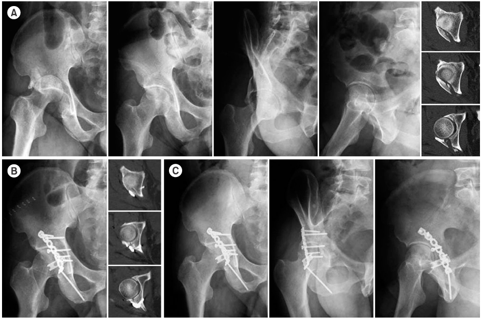
Type A1-2 posterior wall fracture by a traffic accident in a 51-year-old man.
Fig. 2
(A) Preoperative radiographs and CT scan show a displaced fracture of the posterior wall with an undisplaced transverse fracture, intra-articular loose body, a fracture of the femoral head, and separation of the symphysis pubis.
(B) Postoperative radiographs and CT scan show anatomical reduction and stable fixation of the posterior wall and femoral head fractures.
(C) Follow-up radiographs 1.5 years after operation show minor spur at the margin of the femoral head and Grade I heterotopic ossification. The clinical result is good.

Type A1-2 posterior wall fracture by a traffic accident in a 24-year-old man.
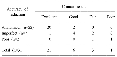
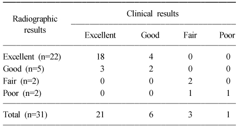
Figure & Data
REFERENCES
Citations
Citations to this article as recorded by 
 Cite
CiteSurgical Treatment of Posterior Wall Fractures of the Acetabulum

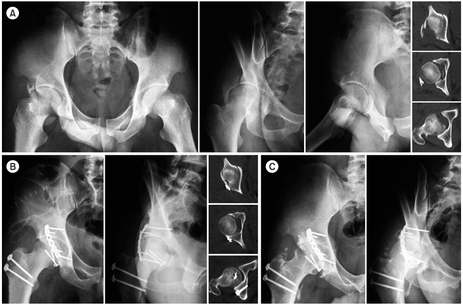
Fig. 1
Type A1-2 posterior wall fracture by a traffic accident in a 51-year-old man.
(A) Preoperative radiographs and CT scan show displaced fracture fragments of the posterior wall with dislocation of the femoral head.
(B) Postoperative radiographs and CT scan show anatomical reduction of the fracture and fixation with a lag screw, a spring plate and a reconstruction plate, but a small fracture gap due to comminution of the fracture.
(C) Follow-up radiographs 2 years after operation show the normal hip joint without complication. Both clinical and radiographic results are excellent.
Fig. 2
Type A1-2 posterior wall fracture by a traffic accident in a 24-year-old man.
(A) Preoperative radiographs and CT scan show a displaced fracture of the posterior wall with an undisplaced transverse fracture, intra-articular loose body, a fracture of the femoral head, and separation of the symphysis pubis.
(B) Postoperative radiographs and CT scan show anatomical reduction and stable fixation of the posterior wall and femoral head fractures.
(C) Follow-up radiographs 1.5 years after operation show minor spur at the margin of the femoral head and Grade I heterotopic ossification. The clinical result is good.
Fig. 1
Fig. 2
Surgical Treatment of Posterior Wall Fractures of the Acetabulum
Correlation of the accuracy of the reduction and the clinical results
Correlation of the clinical results and the radiographic results
Table 1
Correlation of the accuracy of the reduction and the clinical results
Table 2
Correlation of the clinical results and the radiographic results

