 E-submission
 TOTA
TOTA TOTS
TOTS
Articles
- Page Path
- HOME > J Musculoskelet Trauma > Volume 30(3); 2017 > Article
-
Technical Note
- Reduction Technique of Dome Impaction Using the Modified Stoppa Approach: A Technical Note
-
Ji Wan Kim, M.D., Ph.D.
 , Yong Min Seo, M.D., Hyo-Seok Jang, M.D.
 -
Journal of the Korean Fracture Society 2017;30(3):131-136.
DOI: https://doi.org/10.12671/jkfs.2017.30.3.131
Published online: July 21, 2017
Department of Orthopedic Surgery, Haeundae Paik Hospital, Inje University College of Medicine, Busan, Korea.
- Correspondence to: Ji Wan Kim, M.D., Ph.D. Department of Orthopedic Surgery, Inje University Haeundae Paik Hospital, 875, 875 Haeun-daero, Haeundae-gu, Busan 48108, Korea. Tel: +82-51-797-0668, Fax: +82-51-797-0669, bakpaker@hanmail.net
• Received: March 20, 2017 • Revised: May 24, 2017 • Accepted: May 31, 2017
Copyright © 2017 The Korean Fracture Society. All rights reserved.
This is an Open Access article distributed under the terms of the Creative Commons Attribution Non-Commercial License (http://creativecommons.org/licenses/by-nc/4.0) which permits unrestricted non-commercial use, distribution, and reproduction in any medium, provided the original work is properly cited.
- 1,288 Views
- 24 Download
- 2 Crossref
Abstract
- In elderly acetabular fractures, central dislocation of the femoral head and impacted superior dome of the acetabulum is common. Unreduced dome impaction can lead to degenerative arthritis and results in poor results. Herein, we present a case of operative reduction and fixation performed via the modified Stoppa approach in acetabular fracture with superior dome impaction.
- 1. Anglen JO, Burd TA, Hendricks KJ, Harrison P. The “Gull Sign”: a harbinger of failure for internal fixation of geriatric acetabular fractures. J Orthop Trauma, 2003;17:625-634.PubMed
- 2. Kim JW, Herbert B, Hao J, Min W, Ziran BH, Mauffrey C. Acetabular fractures in elderly patients: a comparative study of low-energy versus high-energy injuries. Int Orthop, 2015;39:1175-1179.ArticlePubMedPDF
- 3. Collinge CA, Lebus GF. Techniques for reduction of the quadrilateral surface and dome impaction when using the anterior intrapelvic (modified Stoppa) approach. J Orthop Trauma, 2015;29:Suppl 2. S20-S24.Article
- 4. Casstevens C, Archdeacon MT, d'Heurle A, Finnan R. Intrapelvic reduction and buttress screw stabilization of dome impaction of the acetabulum: a technical trick. J Orthop Trauma, 2014;28:e133-e137.PubMed
- 5. Laflamme GY, Hebert-Davies J. Direct reduction technique for superomedial dome impaction in geriatric acetabular fractures. J Orthop Trauma, 2014;28:e39-e43.ArticlePubMed
- 6. Garner MR, Sagi HC. Isolated anterior intrapelvic approach for operative reduction and fixation of an anterior column acetabulum fracture with superior impaction. J Orthop Trauma [Internet], 2016;cited 2016 Jul 12. Available from: http://journals.lww.com/jorthotrauma/Documents/Sept-2016-JOT7994.pdf
- 7. Kim JW, Kim YC. Modified Stoppa approach in acetabular fractures. J Korean Fract Soc, 2014;27:274-280.Article
- 8. Jung GH, Lee Y, Kim JW, Kim JW. Computational analysis of the safe zone for the antegrade lag screw in posterior column fixation with the anterior approach in acetabular fracture: a cadaveric study. Injury, 2017;48:608-614.ArticlePubMed
- 9. Moon SW, Kim JW. Usefulness of intraoperative three-dimensional imaging in fracture surgery: a prospective study. J Orthop Sci, 2014;19:125-131.ArticlePubMed
- 10. Gary JL, Paryavi E, Gibbons SD, et al. Effect of surgical treatment on mortality after acetabular fracture in the elderly: a multicenter study of 454 patients. J Orthop Trauma, 2015;29:202-208.PubMed
- 11. Jouffroy P; Bone and Joint Trauma Study Group (GETRAUM). Indications and technical challenges of total hip arthroplasty in the elderly after acetabular fracture. Orthop Traumatol Surg Res, 2014;100:193-197.ArticlePubMed
- 12. Tosounidis TH, Stengel D, Giannoudis PV. Anteromedial dome impaction in acetabular fractures: issues and controversies. Injury, 2016;47:1605-1607.ArticlePubMed
- 13. Sagi HC, Afsari A, Dziadosz D. The anterior intra-pelvic (modified rives-stoppa) approach for fixation of acetabular fractures. J Orthop Trauma, 2010;24:263-270.ArticlePubMed
- 14. Scolaro JA, Routt ML Jr. Reduction of osteoarticular acetabular dome impaction through an independent iliac cortical window. Injury, 2013;44:1959-1964.ArticlePubMed
- 15. Ma K, Luan F, Wang X, et al. Randomized, controlled trial of the modified Stoppa versus the ilioinguinal approach for acetabular fractures. Orthopedics, 2013;36:e1307-e1315.ArticlePubMed
- 16. Hammad AS, El-Khadrawe TA. Accuracy of reduction and early clinical outcome in acetabular fractures treated by the standard ilio-inguinal versus the Stoppa/iliac approaches. Injury, 2015;46:320-326.ArticlePubMed
- 17. Kim JW, Shon HC, Park JH. Injury of the obturator nerve in the modified Stoppa approach for acetabular fractures. Orthop Traumatol Surg Res, 2017;[epub].Article
- 18. Zhuang Y, Lei JL, Wei X, Lu DG, Zhang K. Surgical treatment of acetabulum top compression fracture with sea gull sign. Orthop Surg, 2015;7:146-154.ArticlePubMedPMC
REFERENCES
Fig. 1

A 65-year-old man with acetabular factures. (A) Radiograph of right acetabular fracture involving both columns, with a medially displaced quadrilateral plate and superior dome impaction. (B) Coronal reconstruction computed tomography (CT) showing impacted fragment at the acetabular dome. (C) Sagittal reconstruction CT showing 2 pieces of impacted fragment. (D) Immediate postoperative pelvis anteroposterior image, showing a good reduction through the modified Stoppa approach. (E, F) Postoperative CT images, showing well-reduced dome impaction and congruent joint.
Fig. 2

(A) Good function at the 6-month postoperative follow-up. (B-D) Pelvis x-ray at 10 months after surgery.
Fig. 3
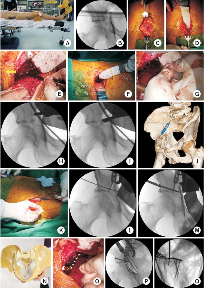
Surgical technique. (A) Patient positioning. (B) Intraoperative fluoroscopy image showing widening of the joint space. (C) Longitudinal incision of rectus abdominis. (D) Deep blunt dissection of the retropubic area. (E) Deep dissection of the quadrilateral plate (arrow: obturator nerve). (F) Confirmation of the position of impacted fragments with a probe under fluoroscopic image. (G) Reduction of impacted fragments. (H, I) Fluoroscopic images of impacted fragments, before and after reduction, respectively. (J) Indirect reduction of the anterior column with a 3.5 mm locking compression plate. (K) Insertion of a K-wire (guide wire). (L) Advance to the quadrilateral surface of the guide wire after reduction of the posterior column. (M) Insertion of a 4.0 cannulated screw. (N) Preparing of a pre-bended reconstruction plate. (O) Passing the long reconstruction plate along the pelvic brim. (P) Insertion of two screws to fix the posterior column after insertion of the screws to the posterior ilium and reduction of the posterior column with a collinear clamp. (Q) Insertion of screws to the pubis.
Figure & Data
REFERENCES
Citations
Citations to this article as recorded by 
- Surgical outcomes of acetabular fracture of elderly patients with superomedial dome impaction
Eic Ju Lim, Hyun-Chul Shon, Jae-Young Yang, Joosuk Ahn, Jung Jae Kim, Ji Wan Kim
Scientific Reports.2023;[Epub] CrossRef - Anterior Approach for the Acetabular Fractures
Jae Youn Yoon, Jae-Woo Cho, Ji Wan Kim
Journal of the Korean Fracture Society.2019; 32(3): 157. CrossRef
 Cite
CiteReduction Technique of Dome Impaction Using the Modified Stoppa Approach: A Technical Note
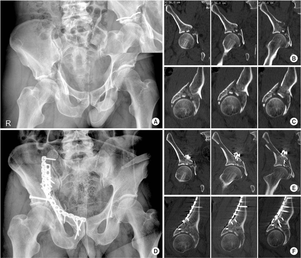
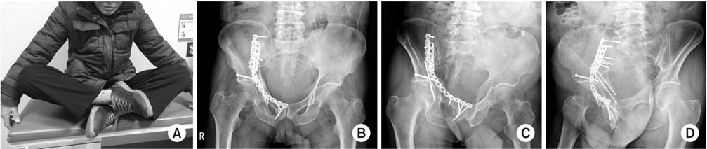

Fig. 1
A 65-year-old man with acetabular factures. (A) Radiograph of right acetabular fracture involving both columns, with a medially displaced quadrilateral plate and superior dome impaction. (B) Coronal reconstruction computed tomography (CT) showing impacted fragment at the acetabular dome. (C) Sagittal reconstruction CT showing 2 pieces of impacted fragment. (D) Immediate postoperative pelvis anteroposterior image, showing a good reduction through the modified Stoppa approach. (E, F) Postoperative CT images, showing well-reduced dome impaction and congruent joint.
Fig. 2
(A) Good function at the 6-month postoperative follow-up. (B-D) Pelvis x-ray at 10 months after surgery.
Fig. 3
Surgical technique. (A) Patient positioning. (B) Intraoperative fluoroscopy image showing widening of the joint space. (C) Longitudinal incision of rectus abdominis. (D) Deep blunt dissection of the retropubic area. (E) Deep dissection of the quadrilateral plate (arrow: obturator nerve). (F) Confirmation of the position of impacted fragments with a probe under fluoroscopic image. (G) Reduction of impacted fragments. (H, I) Fluoroscopic images of impacted fragments, before and after reduction, respectively. (J) Indirect reduction of the anterior column with a 3.5 mm locking compression plate. (K) Insertion of a K-wire (guide wire). (L) Advance to the quadrilateral surface of the guide wire after reduction of the posterior column. (M) Insertion of a 4.0 cannulated screw. (N) Preparing of a pre-bended reconstruction plate. (O) Passing the long reconstruction plate along the pelvic brim. (P) Insertion of two screws to fix the posterior column after insertion of the screws to the posterior ilium and reduction of the posterior column with a collinear clamp. (Q) Insertion of screws to the pubis.
Fig. 1
Fig. 2
Fig. 3
Reduction Technique of Dome Impaction Using the Modified Stoppa Approach: A Technical Note

