 E-submission
 TOTA
TOTA TOTS
TOTS
Articles
- Page Path
- HOME > J Musculoskelet Trauma > Volume 23(3); 2010 > Article
-
Original Article
- Open Reduction and Internal Fixation with AO Calcaneal Plate for Displaced Intra-articular Calcaneal Fracture
- Myung Jin Lee, M.D., Sung Keun Sohn, M.D., Kyu Yeol Lee, M.D., Sung Soo Kim, M.D., Min Soo Kang, M.D., Hyeon Jun Kim, M.D, Sang Kyu Sun, M.D.
-
Journal of the Korean Fracture Society 2010;23(3):303-309.
DOI: https://doi.org/10.12671/jkfs.2010.23.3.303
Published online: July 31, 2010
Department of Orthopedic Surgery, College of Medicine, Dong-A University, Busan, Korea.
*Department of Orthopedic Surgery, Dong-Eui Medical Center, Busan, Korea.
- Address reprint requests to: Hyeon Jun Kim, M.D. Department of Orthopedic Surgery, College of Medicine, Dong-A University, 1, Dongdaesin-dong 3-ga, Seo-gu, Busan 602-715, Korea. Tel: 82-51-240-5167, Fax: 82-51-254-6757, campbellkim@naver.com
• Received: November 19, 2009 • Revised: March 4, 2010 • Accepted: April 7, 2010
Copyright © 2010 The Korean Fracture Society
- 1,409 Views
- 3 Download
- 2 Crossref


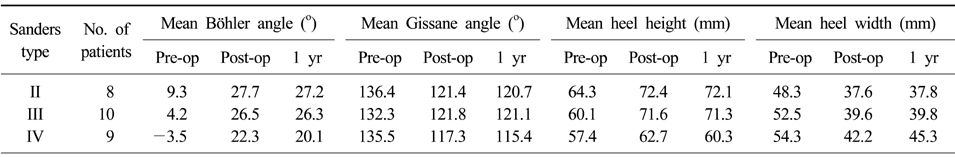
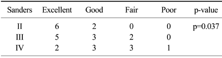
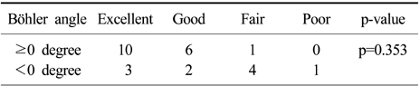
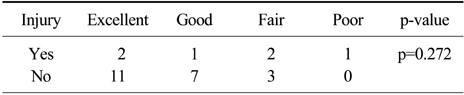
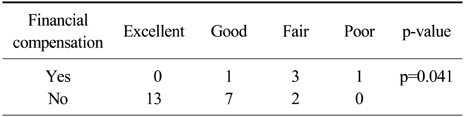
Figure & Data
REFERENCES
Citations
Citations to this article as recorded by 
- Surgical Treatment of Calcaneal Fractures of Sanders Type II and III by A Minimally Invasive Technique with 6.5 mm Cancellous Screw
Yong Seung Oh, Kyung Ho Lee, Jung Ho Kim, Myoung Jin Lee
Journal of Korean Foot and Ankle Society.2019; 23(3): 116. CrossRef - Usefulness of Treatment with 6.5 mm Cancellous Screw and Steinmann Pin Fixation for Calcaneal Joint Depression Fracture
Gi-Soo Lee, Chan Kang, Deuk-Soo Hwang, Chang-Kyun Noh, Gi-Young Lee
Journal of Korean Foot and Ankle Society.2015; 19(1): 11. CrossRef
 Cite
CiteOpen Reduction and Internal Fixation with AO Calcaneal Plate for Displaced Intra-articular Calcaneal Fracture
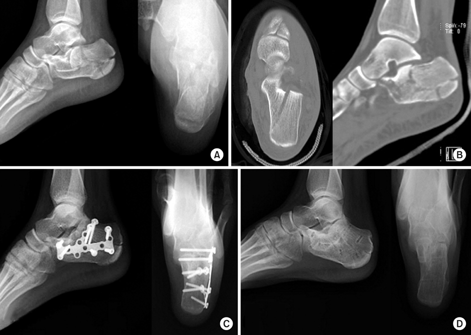
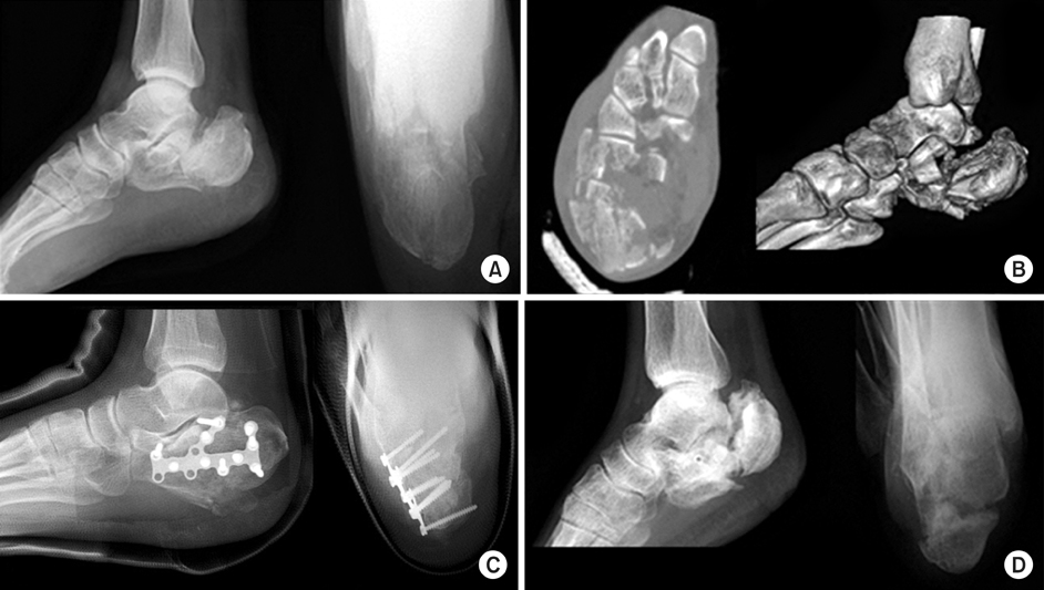
Figure 1
A 21-year-old female patient sustained left intra-articular calcaneal fracture by slip down.
(A) Preoperative X-ray shows tongue type intra-articular calcaneal fracture.
(B) Preoperative axial and sagittal CT scans show Sanders type IIb calcaneal fracture.
(C) Postoperative X-ray shows restoration of Böhler angle and heel width.
(D) At follow-up of 13months, X-ray shows no subtalar joint arthritis.
Figure 2
A 45-year-old male patient sustained right open comminuted intra-articular calcaneus fracture by fall down from 6 meter height.
(A) Preoperative X-ray shows joint depression type intra-articular calcaneal fracture with negative Böhler angle.
(B) Preoperative semicoronal and 3D CT scan shows Sanders type IV calcaneal fracture.
(C) Postoperative X-ray shows restoration of Böhler angle and stable fixation.
(D) Postoperative X-ray shows varus change of heel and malunion of calcaneus and losses of corrections.
Figure 1
Figure 2
Open Reduction and Internal Fixation with AO Calcaneal Plate for Displaced Intra-articular Calcaneal Fracture
Summary of radiologic parameters
The relationship between Sanders type and Maryland foot score (Linear by linear association)
The relationship between preoperative Böhler angle and Maryland foot score (Fisher's exact test)
The relationship between associated injury and Maryland foot score (Fisher's exact test)
The relationship between financial compensation and Maryland foot score (Fisher's exact test)
Table 1
Summary of radiologic parameters
Table 2
The relationship between Sanders type and Maryland foot score (Linear by linear association)
Table 3
The relationship between preoperative Böhler angle and Maryland foot score (Fisher's exact test)
Table 4
The relationship between associated injury and Maryland foot score (Fisher's exact test)
Table 5
The relationship between financial compensation and Maryland foot score (Fisher's exact test)

