 E-submission
 TOTA
TOTA TOTS
TOTS
Search
- Page Path
- HOME > Search
Review Article
- Complications of hand fractures: strategies for prevention and management
- Jong Woo Kang
- J Musculoskelet Trauma 2026;39(1):1-11. Published online January 25, 2026
- DOI: https://doi.org/10.12671/jmt.2025.00304
-
 Abstract
 PDF
PDF - Various complications can occur after hand fractures. Among them, joint stiffness and malunion are the most common and significant complications, which are often accompanied by tendon adhesions and joint contracture. Careful evaluations of injury characteristics, such as fracture patterns, alignment, and soft tissue injury, are the first step to select appropriate management strategies and prevent complications of hand fractures. Close observation of its clinical prognosis is also essential for early detection and preemptive management of complications. Management of complications includes immobilization, rehabilitation, and various surgical techniques such as tenolysis or capsular release for joint stiffness, corrective osteotomy for malunion, and revisional fixation with bone graft for nonunion. The authors discuss prevention, early recognition, and management strategies for complications of hand fractures in this review.
- 3,206 View
- 81 Download

Original Article
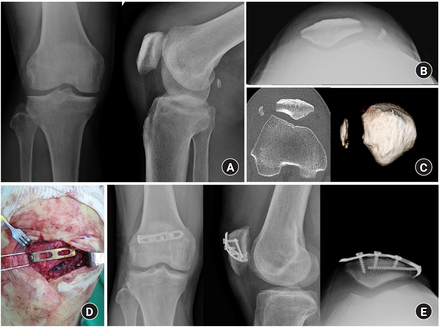
- Lateral marginal fractures of the patella and patellofemoral pain
- Jae-Ang Sim, Chul-Ho Kim, Ji Wan Kim
- J Musculoskelet Trauma 2025;38(3):152-159. Published online July 22, 2025
- DOI: https://doi.org/10.12671/jmt.2025.00171
-
Abstract
PDF
- Background
This study investigated the characteristics of lateral marginal fractures of the patella and evaluated the clinical outcomes.
Methods
We retrospectively reviewed all patients with lateral marginal fractures of the patella, defined as a vertical fracture line within 15 mm of the lateral patellar border, from 2008 to 2020. In total, 41 patients were included. Patient characteristics, radiologic findings, and clinical outcomes, including the Lysholm score at 1 year postoperation, were evaluated.
Results
The injury mechanisms were direct in 34 cases and indirect in seven. Furthermore, 85% of patients had a skyline view of the patella at the initial visit, and one medial subluxation of the patella was found. Forty of the 41 patients underwent surgery. Anatomical and nonanatomical (>1-mm displacement or excision) reductions were carried out in 36 cases (88%) and five cases (12%), respectively. The average Lysholm score was 89.1 (range, 67–99). The nonanatomical reduction group had a poorer functional score (79.8 vs. 90.4; P=0.010). Lateral patellar compression syndrome occurred in two patients with nonanatomical reduction.
Conclusions
Lateral marginal fractures of the patella affected patellofemoral stability. Anatomical reduction showed good functional outcomes, while nonanatomical reduction was associated with patellofemoral stability and pain. Therefore, surgeons should perform anatomical reduction with any appropriate fixation method. Level of Evidence: IV
- 2,635 View
- 40 Download
Review Article
- Atypical femoral fractures: an update
- Won-Tae Cho, Jeong-Hyun Koh, Seungyeob Sakong, Jung-Taek Kim
- J Musculoskelet Trauma 2025;38(2):41-52. Published online March 28, 2025
- DOI: https://doi.org/10.12671/jmt.2025.00031
-
Abstract
PDF
- This narrative review provides an up-to-date overview of atypical femoral fractures (AFFs), emphasizing diagnostic criteria, epidemiology, pathophysiology, risk factors, and evaluation with screening strategies. AFFs are rare but significant complications associated with prolonged bisphosphonate (BP) therapy for osteoporosis. Although the pathogenesis of AFFs has not been fully elucidated, its primary mechanism is thought to involve impaired bone remodeling, leading to unhealed microfractures that progress to stress fractures under repetitive loading. AFFs can occur in various regions of the femur, influenced by femoral geometry and the lower limb axis. Other risk factors include prolonged steroid use, arthroplasty, genetic predispositions, and metabolic bone disorders. The diagnosis of AFFs is based on criteria established by the American Society for Bone and Mineral Research. Key radiographic features include lateral cortical transverse fracture lines and localized cortical thickening, typically with minimal or no comminution on the medial cortex. Dual-energy X-ray absorptiometry for screening tests and magnetic resonance imaging as an advanced imaging modality enable the early detection of incomplete fractures. This multi-modal approach facilitates the prompt identification of prodromal cortical changes, reducing the risk of complete fractures in high-risk populations, particularly patients undergoing prolonged BP therapy. Level of Evidence: V
-
Citations
Citations to this article as recorded by
- Clinical Images: Bisphosphonate‐associated atypical femoral fracture with contralateral cortical beaking
Andreina Martinez Paulino, Valentin Marian
ACR Open Rheumatology.2026;[Epub] CrossRef - Atypical femoral fracture: The periprosthetic variant about two cases without bisphosphonate use
Guillaume Auberger, Thomas Aubert, Younes Kerroumi, Philippe Leclerc, Simon Marmor
SICOT-J.2026; 12: 41. CrossRef - Atypical Femur Fractures Without Bisphosphonate Exposure (AFFwB): A Retrospective Report of 21 Cases
Lorenzo Lucchetta, Carmelinda Ruggiero, Samuele Berardi, Alice Franceschi, Michele Bisaccia, Giuseppe Rinonapoli
Journal of Clinical Medicine.2025; 15(1): 25. CrossRef
- Clinical Images: Bisphosphonate‐associated atypical femoral fracture with contralateral cortical beaking
- 65,535 View
- 643 Download
- 3 Crossref
Original Article
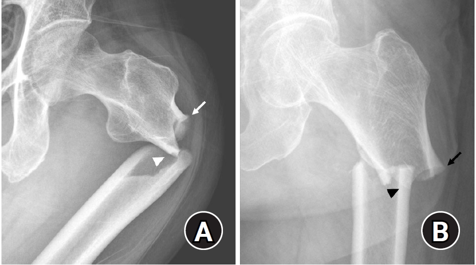
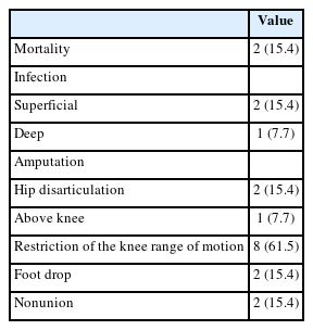
- Acute Compartment Syndrome of Thigh: Ten-Year Experiences from a Level I Trauma Center
- Hyung Keun Song, Won-Tae Cho, Wan-Sun Choi, Seung-Yeob Sakong, Sumin Im
- J Musculoskelet Trauma 2024;37(4):171-174. Published online October 25, 2024
- DOI: https://doi.org/10.12671/jmt.2024.37.4.171
-
Abstract
PDF
- Purpose
To assess the demographics, injury mechanisms, treatments, and outcomes of traumatic acute compartment syndrome in the thigh.
Materials and Methods
Patients diagnosed with thigh compartment syndrome were analyzed retrospectively at the authors’ level I trauma center from March 2012 to February 2022. Data were collected from medical and radiological records, focusing on demographics, injury details, treatment timelines, and clinical outcomes.
Results
The cohort included 13 patients (11 males and 2 females) with a mean age of 46 years. Injuries primarily resulted from falls (6 patients) and vehicle accidents (5 patients). Fractures were noted in 11 patients, with seven involving the lower extremities and seven having open fractures; three of these were severe enough to be classified as Gustilo–Anderson type IIIc with associated femoral artery injuries. Time from the injury to fasciotomy ranged from within six hours to more than 24 hours. Fasciotomies were mainly single-sided (10 patients), targeting primarily the anterior compartments, and bilateral in three cases. Wound closures were performed using delayed primary closure (four patients) and partial- thickness skin grafts (five patients). Two patients died from multi-organ failure; other complications included infections (three patients), amputations (three patients), and long-term disabilities like drop foot (two patients), sensory deficits, joint stiffness (eight patients), and fracture non-unions requiring additional surgery (two patients).
Conclusion
Thigh-compartment syndrome, though infrequent, poses significant risks of mortality and chronic disability. This underscores the importance of prompt diagnosis and intervention.
- 2,613 View
- 62 Download
Review Articles
- Hip Fractures in the Elderly: Perioperative Management and Prevention of Medical Complications
- Keong-Hwan Kim
- J Korean Fract Soc 2023;36(1):39-44. Published online January 31, 2023
- DOI: https://doi.org/10.12671/jkfs.2023.36.1.39
-
Abstract
PDF
- Elderly patients with hip fractures are at an increased risk of developing medical complications with higher mortality rates. Most patients require surgical treatment, and an early surgical intervention can reduce complications and lower mortality risk. A restrictive red blood cell transfusion strategy is usually applied, and the amount of transfusion can be reduced through medications such as tranexamic acid. Delirium can be prevented using non-pharmacological methods. In addition, it is necessary to prevent venous thromboembolism through mechanical or chemical prophylaxis. A multidisciplinary approach using the ERAS (Enhanced Recovery After Surgery) protocol and orthogeriatric care can help to reduce medical complications and mortality.
-
Citations
Citations to this article as recorded by- Comparison of Operation Time, Vital Signs, Bleeding Tendency, and Recovery Time Based on Anesthesia Methods in Patients Undergoing Hip Fracture Surgery
Je Bog Yoo, Woo Young In, Chang Ok Pyo, Jeung Hee Kwon, Min Ji Lee, Kwang Hee Kim, Kyoung Ok Kim, Mi Yu
Journal of PeriAnesthesia Nursing.2026; 41(3): 591. CrossRef - Treatment of Incompletely Displaced Femoral Neck Fractures Using Trochanteric Fixation Nail-Advanced in Patients Older Than 50 Years of Age
Jee Young Lee, Gyu Min Kong
Journal of Orthopaedic Trauma.2025; 39(7): 352. CrossRef
- Comparison of Operation Time, Vital Signs, Bleeding Tendency, and Recovery Time Based on Anesthesia Methods in Patients Undergoing Hip Fracture Surgery
- 1,992 View
- 46 Download
- 2 Crossref
- Fixation Options of Unstable Posterior Pelvic Ring Disruption: Ilio-Sacral Screw Fixation, S2AI Fixation, Posterior Tension Band Plate Fixation, and Spino-Pelvic Fixation
- Dong Hee Kim, Jae Hoon Jang, Myungji Shin, Gu Hee Jung
- J Korean Fract Soc 2019;32(4):240-247. Published online October 31, 2019
- DOI: https://doi.org/10.12671/jkfs.2019.32.4.240
-
Abstract
PDF
- The fixation methods that can be used for unstable posterior pelvic ring injuries have undergone many innovative changes due to the recent development of surgical and imaging techniques. After understanding the appropriate indications of first and second sacroiliac screw fixation and spinopelvic fixation, innovative methods, including the trans-sacral screw fixation, posterior tension-band plate fixation, and the S2AI screw, would be chosen and applied. Considering the anatomical complexity and proximity to the surrounding vessels and nerves in the posterior fixation, the safe zone according to the fixation options should be well understood in preoperative planning. Moreover, the functional reduction of the posterior pelvic ring through the reduction and fixation of the anterior lesion should be achieved before placing the implant to reduce the number of malposition-related complications.
-
Citations
Citations to this article as recorded by- Clinical Research through Computational Anatomy and Virtual Fixation
Ju Yeong Kim, Dong-Geun Kang, Gu-Hee Jung
Journal of the Korean Orthopaedic Association.2023; 58(4): 299. CrossRef
- Clinical Research through Computational Anatomy and Virtual Fixation
- 1,786 View
- 19 Download
- 1 Crossref
- Locked Plating in Elderly Patients with Distal Femur Fracture: How to Avoid Complications?
- Chul Young Jang, Je Hyun Yoo
- J Korean Fract Soc 2019;32(2):112-119. Published online April 30, 2019
- DOI: https://doi.org/10.12671/jkfs.2019.32.2.112
-
Abstract
PDF
- Distal femur fractures in elderly patients with osteoporosis are complicated because poor bone quality makes screw purchase and fixation less secure, presenting many clinical challenges to the orthopedic surgeon. Minimally invasive locked plating using an angularly stable locking compression plate has become an integral tool for achieving secure fixation in osteoporotic distal femur fractures with improved biomechanical performance. On the other hand, complications, such as implant failure and periplate fracture, have still occurred. This paper describes the principles of internal fixation in minimally invasive lateral locked plating in elderly patients with osteoporotic distal femur fractures as well as how to avoid complications.
- 1,742 View
- 33 Download
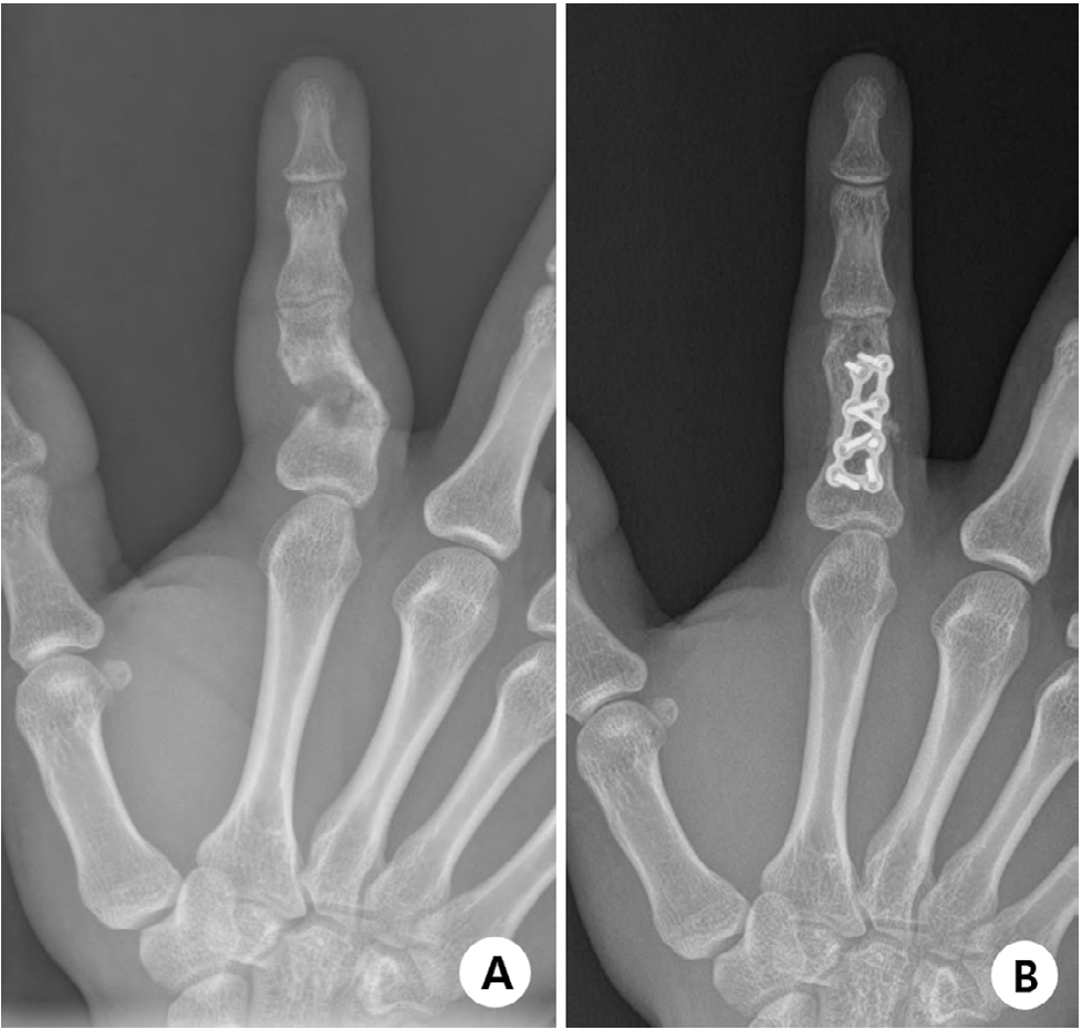
- Hand Fractures
- Seokwon Yang, Jong Pil Kim
- J Korean Fract Soc 2018;31(2):61-70. Published online April 30, 2018
- DOI: https://doi.org/10.12671/jkfs.2018.31.2.61
-
Abstract
PDF
- Hand fractures are the second most common fracture in the upper extremities after the distal radius, and patients with these injuries may be experienced in hand surgery clinics. On the other hand, during the treatment of hand fractures, complications can occur due to complex functions of the hand and small-sized injuries to the bone and soft tissues. This review focused on the principles of management of these fractures, including injury mechanism, evaluations and recent treatment options. Minimally invasive surgery in various types of hand fractures, including the phalanx and metacarpal bone, is preferred because early mobilization after surgery has been emphasized to reduce complications, such as stiffness.
-
Citations
Citations to this article as recorded by- A novel finger brace for preventing finger stiffness after trauma or surgery: a preliminary report with a case series
Dae-Geun Kim, Hyo Jun Park
Archives of Hand and Microsurgery.2023; 28(4): 239. CrossRef
- A novel finger brace for preventing finger stiffness after trauma or surgery: a preliminary report with a case series
- 981 View
- 3 Download
- 1 Crossref
- Fracture of the Talus
- Tae Jung Bang, Sun Kyu Kim, Hyung Jin Chung
- J Korean Fract Soc 2016;29(3):213-220. Published online July 31, 2016
- DOI: https://doi.org/10.12671/jkfs.2016.29.3.213
-
Abstract
PDF
- Although talus fractures are uncommon, proper management is important because they are often associated with severe complications. Talar neck and body fractures occupy most of the talar fractures. It remains controversial whether talar neck fractures require emergent or elective treatment. Elective definitive fixation, however, may reduce risks of wound complications. Many surgeons recommend dual surgical approaches—anteromedial and anterolateral—to allow accurate visualization and anatomic reduction. Although there are various methods of fixation, the use of plates is necessary in comminuted talar fractures. Outcomes may vary and will be dependent on the degree of the initial fracture displacement. It is necessary to restore articular congruency and axial alignment for normalizing hindfoot function. Common complications include posttraumatic arthritis, avascular necrosis, malunion, and nonunion.
- 1,654 View
- 27 Download
Original Articles
- Clinical Outcomes of Fasciotomy for Acute Compartment Syndrome
- Ji Yong Park, Young Chang Kim, Ji Wan Kim
- J Korean Fract Soc 2015;28(4):223-229. Published online October 31, 2015
- DOI: https://doi.org/10.12671/jkfs.2015.28.4.223
-
Abstract
PDF
- PURPOSE
The purpose of this study is to evaluate clinical outcomes and complications after fasciotomy in acute compartment syndrome.
MATERIALS AND METHODS
Seventeen cases diagnosed as compartment syndrome and underwent fasciotomy from January 2011 to February 2015 were evaluated retrospectively. We investigated the causes and regions of acute compartment syndrome, the methods of wound management, the necessity of skin graft, and the complications including amputation and infection.
RESULTS
According to the causes of acute compartment syndrome, there were 7 fractures, 1 traumatic hematoma, 6 reperfusion injury, and 3 rhabdomyolysis. The regions of acute compartment syndrome were 3 cases of thigh, 10 cases of leg, and 3 cases of foot. One case had acute compartment syndrome involving thigh, leg, and foot. Of 17 cases, 3 cases died due to reperfusion injury and one case with severe necrosis of soft tissues underwent amputation. Among the 13 cases excluding 4 cases with death or amputation, 3 cases underwent split thickness skin graft. Shoelace technique and/or vacuum-assisted closure (VAC) was used for 9 cases, and wound closure without skin graft was achieved in all except one case, while 2 cases required skin graft among 4 cases without shoelace technique or VAC. There were 2 cases of infection.
CONCLUSION
Acute compartment syndrome caused by reperfusion injury had poor outcomes. Shoelace technique and/or VAC were useful for management of wound after fasciotomy.
- 1,061 View
- 9 Download
- Neurologic Injury within Pelvic Ring Injuries
- Ji Wan Kim, Dong Hoon Baek, Jae Hyun Kim, Young Chang Kim
- J Korean Fract Soc 2014;27(1):17-22. Published online January 31, 2014
- DOI: https://doi.org/10.12671/jkfs.2014.27.1.17
-
Abstract
PDF
- PURPOSE
To evaluate the incidence of neurologic injury in pelvic ring injuries and to assess the risk factors for neurologic injury related to pelvic fractures.
MATERIALS AND METHODS
Sixty-two patients with the pelvic ring injury were enrolled in the study from March 2010 to May 2013. When the neurologic injury was suspected clinically, the electro-diagnostic tests were performed. Combined injuries, fracture types, and longitudinal displacements were examined for correlations with the neurologic injury.
RESULTS
There were 7 cases of AO/OTA type A, 37 cases of type B, and 18 cases of type C. Among them, 25 patients (40%) had combined spine fractures, and the average of longitudinal displacement was 7 mm (1-50 mm). Of the 62 patients, 13 (21%) had neurologic injury related with pelvic fractures; 5 with lumbosacral plexus injury, 5 with L5 or S1 nerve injury, 2 with obturator nerve injury, and 1 case of lateral femoral cutaneous nerve injury. There were no relationships between the neurologic injuries and fracture types (p=0.192), but the longitudinal displacements of posterior ring and combined spine fractures were related to the neurologic injury within pelvic ring injury (p=0.006, p=0.048).
CONCLUSION
The incidence of neurologic injury in pelvis fracture was 21%. In this study, the longitudinal displacements of posterior ring and combined spine fractures were risk factors for neurological injury in pelvic ring injury. -
Citations
Citations to this article as recorded by- Surgical Outcome of Posterior Pelvic Fixation Using S1, S2 Screws in Vertically Unstable Pelvic Ring Injury
Kwang Hee Yeo, Nam Hoon Moon, Jae Min Ahn, Jae Yoon Jeong, Jae Hoon Jang
Journal of the Korean Fracture Society.2018; 31(1): 9. CrossRef
- Surgical Outcome of Posterior Pelvic Fixation Using S1, S2 Screws in Vertically Unstable Pelvic Ring Injury
- 1,151 View
- 8 Download
- 1 Crossref
- Complications of Femoral Pertrochanteric Fractures Treated with Proximal Femoral Nail (PFN)
- Kee Byoung Lee, Byung Taek Lee
- J Korean Fract Soc 2007;20(1):33-39. Published online January 31, 2007
- DOI: https://doi.org/10.12671/jkfs.2007.20.1.33
-
Abstract
PDF
- PURPOSE
We analyzed the complications of femoral pertrochanteric fractures treated with proximal femoral nail (PFN®) to reduce the its complications.
MATERIALS AND METHODS
We evaluated the complications among 198 patients who were treated with PFN® from June 2001 to August 2005 in our hospital.
RESULTS
The complications were presented in 28 cases (14.1%). Cut-out of lag screw was in 1 case, cut-out of lag screw and antirotation screw were in 3 cases, cut-out of antirotation screw in 3 cases, of these femoral head fracture was in 1 case. Femoral neck fracture in 1 case, Osteonecrosis of femoral head in 1 case, cortical fracture during the insertion of distal interlocking screw in 1 case, breakage of drill bit intraoperatively in 1 case, fibrous union in 2 case, thigh skin irritation due to screw back-out in 3 cases, periprosthetic fractures in 2 cases, varus collapse more than 10 degrees in 4 cases, superficial and deep infections in 3 cases, breakage of nail in 1 case, varus collapse after PFN removal in 1 case, persistent thigh pain in 1 case. Of all these cases, 9 cases (4.5%) were required reoperation with general or spinal anesthesia. Complications related with screws or fracture reduction were 19 cases (9.6%) and, of these, 17 cases (89.5%) showed increased TAD (tip apex distance) or nonanatomical reduction.
CONCLUSION
To reduce the complications of PFN®, we need to exact surgical technique and anatomical reduction and consider the modification of implant design to prevent of cut-out of screws. -
Citations
Citations to this article as recorded by- Intraoperative Abdominal Penetration of the Lag Screw: A Rare Complication During Proximal Femoral Nail Anti‐Rotation Surgery
Mohammad Javad Dehghani Firoozabadi, Ramin Bozorgmehr, Fatemeh Bastan, Maryam Rashidian
Clinical Case Reports.2026;[Epub] CrossRef - Proximal Femoral Nail Mechanical Failure: A Case Report and Biomechanical Study
Dimitrios Papanikolopoulos, Christos Kalligeros, Apostolos Polyzos, Vasileios Spitas, Vasileios Soranoglou
Cureus.2022;[Epub] CrossRef - Clinical and radiological outcomes of patients treated with the talon distalfix proximal femoral nail for intertrochanteric femur fractures
Furkan Yapici, Hanifi Ucpunar, Yalkin Camurcu, Necati Emirhan, Oguzhan Tanoglu, Ismail Tardus
Injury.2020; 51(4): 1045. CrossRef - Implant Fracture Analysis of the TFNA Proximal Femoral Nail
Anton Lambers, Bertram Rieger, Alan Kop, Peter D’Alessandro, Piers Yates
Journal of Bone and Joint Surgery.2019; 101(9): 804. CrossRef - Radiographic Outcomes of Osteosynthesis Using Proximal Femoral Nail Antirotation (PFNA) System in Intertrochanteric Femoral Fracture: Has PFNA II Solved All the Problems?
Won Chul Shin, Jung Dong Seo, Sang Min Lee, Nam Hoon Moon, Jung Sub Lee, Kuen Tak Suh
Hip & Pelvis.2017; 29(2): 104. CrossRef - Avascular necrosis of the femoral head following trochanteric fractures in adults: A systematic review
Antonio Barquet, Gabriel Mayora, Joao Matheus Guimaraes, Roberto Suárez, Peter V. Giannoudis
Injury.2014; 45(12): 1848. CrossRef - Anatomical Measurement of Normal Korean Proximal Femur Using Plain Radiography: A Problem when using Proximal Femoral Nail Anti-rotation
Jong-Seok Park, Woo-Jong Kim, Jae-Wan Soh, Byung-Woong Jang, Tae-Heon Kim, You-Sung Suh
Hip & Pelvis.2011; 23(4): 303. CrossRef - PFNA and PFN in Intertrochanteric Fractures - Comparison Study of Sliding -
Suk Kyu Choo, Hyoung Keun Oh, Jun Young Choi
Hip & Pelvis.2010; 22(1): 79. CrossRef
- Intraoperative Abdominal Penetration of the Lag Screw: A Rare Complication During Proximal Femoral Nail Anti‐Rotation Surgery
- 2,231 View
- 21 Download
- 8 Crossref
- The Correlation between Surgical Timing and Perioperative Complications in the Treatment of Displaced Supracondylar Humeral Fractures of Children
- Soo Hong Han, Duck Yun Cho, Hyung Ku Yoon, Byung Soon Kim, Jae Hwa Kim, Hyung Kun Park, Se Hyen Kim
- J Korean Soc Fract 2003;16(2):278-283. Published online April 30, 2003
- DOI: https://doi.org/10.12671/jksf.2003.16.2.278
-
Abstract
PDF
- PURPOSE
Even though emergent percutaneous pinning after closed reduction is the popularized treatment of the displaced type II and type III pediatric supracondylar fractures of the humerus, the timing of pinning still presents controversy. The purpose of this study is to suggest an appropriate surgical time without significant perioperative complications.
MATERIALS AND METHODS
From April 1995 to January 2002, 179 consecutive patients who had undergone surgical treatment were selected. They were divided to 5 groups [A group: 8 hours or less following injury (24 cases), B group: from 9 to 16 hours (63 cases), C group: from17 hours to 24 hours (63 cases), D group: from 25 hours to 48 hours (18 cases), and E group: from 49 hours to 72 hours (11 cases)] and reviewed retrospectively to analyze perioperative complications and operation time.
RESULTS
There was no significant difference between each group with respect to surgical wound infection, iatrogenic ulnar nerve injury, VIC, operation time and the necessity of reoperation (p>0.05).
CONCLUSION
Within the parameters outlined in our study, we could not find the any meaningful correlation between surgical timing and occurrence of perioperative complications and also, we think that the timing of percutaneous pinning can be delayed to the time when a surgeon considers it appropriate.
- 654 View
- 0 Download
- Neurologic Complications of Elbow Fractures in Children
- Suk Kyu Choo, Gyu Won Park
- J Korean Soc Fract 2002;15(4):595-600. Published online October 31, 2002
- DOI: https://doi.org/10.12671/jksf.2002.15.4.595
-
Abstract
PDF
- PURPOSE
We analyzed neurologic complications of the elbow fractures in children and evaluated clinical results of type of fractures, frequency of nerve injuries and displacement of fracture fragments and spontaneous recovery of each nerve injuries.
MATERIALS AND METHODS
We analyzed 17 child-patients (20cases) with nerve injuries who were treated conservatively and follewed up for at least 1 year since December 1999. and we analyzed type of fractures, differences between fracture type and nerve injuries, frequency of each nerve injuries and periods of spontaneous recovery of each nerves.
RESULTS
There were all 148 elbow fractures in children. Children with neurologic complications were 17(20 nerves) and 14 in supracondylar and 3 in medial epicondylar fractures. There were 6 in radial nerve, 8 in ulnar nerve, 3 in median nerve and 3 in anterior interosseous nerve. Both ulnar and median nerve injuries were 3 patients. They were recovered spontaneously and mean periods of recovery was 7.3 weeks, 6.5 weeks in radial nerves, 7.0 in median nerves, 7.6 in anterior interosseous nerves, 7.8 in ulnar nerves and radial nerve recovery was most fast than any others. One patient with ulnar nerve injury who was diagnosed medial epicondylar fracture recovered 2 weeks after excision of nonuioned fragment. Among 14 supracondylar fractures, there was 11 posteromedial displacement, 1 posterolateral and 2 posterior. Most of them was displaced posteromedially.
CONCLUSION
All nerve injuries happened in supracondylar and medial condylar fractures and almost recovered. Nerve injuries in the supracondylar fractures was displaced fractures than nondisplaced simple fractures and displacement of fracture fragment and nerve injuries was not agreed with previous published books or papers. We recommand that observation is the appropriate way to manage these nerve injuries in most cases than immediate operation for excision.
- 720 View
- 1 Download
- Complications and Affecting Factors for Intracapsular Femoral Neck Fractures Treated by Multiple Pinning
- Sung Jung Kim, Shin Yoon Kim, Gi Bong Cha, Chang Wug Oh, Il Hyung Park, Joo Chul Ihn
- J Korean Soc Fract 2002;15(2):201-208. Published online April 30, 2002
- DOI: https://doi.org/10.12671/jksf.2002.15.2.201
-
Abstract
PDF
- PURPOSE
To investigate the relationship between the complications of intracapsular femoral neck fractures treated by multiple pinning and several affecting factors.
MATERIALS AND METHODS
Sixty-eight patients with intracapsular femoral neck fractures were treated by multiple pinning from March 1993 to January 2000 and followed at more than one year. Relationship between the complications such as failure of union, collapse of femoral head due to osteonecrosis of femoral head and several affecting factors including displacement of fracture according to Garden stage, state of reduction, position of screws, time interval from injury to operation, and fracture level were analyzed. The Fisher exact test, chi-square test, and multivariate logistic regression analysis were used to find the relevant factors influencing incidence of complications. Statistical significance was set at p < 0.05.
RESULTS
Position of screw was the most important single factor affecting the results of treatment of intracapsular femoral neck fracture (p=0.046). Moreover, the Garden stage and position of screw were revealed affecting the incidence of complications together with other factors (each p value was 0.028 and 0.027).
CONCLUSION
We considered that satisfactory position of screw was important to reduce complications after multiple pinning for intracapsular femoral neck fracture. And the results of operation also seemed to closely relate with multiple factors including Garden stage and status of reduction. -
Citations
Citations to this article as recorded by- Factors Predicting Complications after Internal Fixation of Femoral Neck Fractures
Tae-Ho Kim, Jong-Oh Kim, Sung-Sik Kang
Journal of the Korean Fracture Society.2009; 22(2): 79. CrossRef
- Factors Predicting Complications after Internal Fixation of Femoral Neck Fractures
- 874 View
- 0 Download
- 1 Crossref
- The Complications during Treatment of Femoral Neck Fracture in Children - Coxa Vara, SCFE and Avascular Necrosis
- Myung Rae Cho, Won Jae Song, Jeong Hwan Son, Jeong Ho Park
- J Korean Soc Fract 2002;15(1):1-6. Published online January 31, 2002
- DOI: https://doi.org/10.12671/jksf.2002.15.1.1
-
Abstract
PDF
- Femur neck fractures in children are relatively rare by comparison with the incidence in adults and can occur by high-energy trauma. The common complications of femoral neck fracture are avascuar necrosis, coxa vara, premature physeal closure and nonunion. The femoral neck fracture by traffic accident in 7-years-old girl was reduced closely and fixed internally. After 1 year later, varus deformity, slipped capital femoral epiphysis and avascular necrosis occurred. Valgus osteotomy and epiphysiodesis were done. There are many series of complications after femoral neck fractures in children, but the case followed by varus deformity, slipped capital femoral epiphysis and avascular necrosis as complications is rare. We report the clinical and radiologic finding of this case with pertinent literature.
- 851 View
- 2 Download
- Complications after Surgical Treatment in Fracture of The Neck of Humerus
- Ho Jung Kang, Sang Jin Shin, Dae Eui Lim, Eung Shick Kang
- J Korean Soc Fract 2001;14(1):91-98. Published online January 31, 2001
- DOI: https://doi.org/10.12671/jksf.2001.14.1.91
-
Abstract
PDF
- PURPOSE
The causes and risk factors of complications following operative treatment of fracuture of neck of humerus were analysis. MATERIALS & METHODS: From 1995 to 1998, 32 cases of fracture of neck of humerus on which operative treatment have been taken were reviewed. The average age was 48.3 years. There were 13 cases of two part fracture, 11 cases of three part fracture and 8 cases of four part fracture, with 4 cases associated with comminution. Closed reduction and pinning was performed in 11 cases. An external fixator was applied in 1 case. Other 18 cases underwent open reduction using various fixation method including 4 K-wires, 2 cannulated screws, 5 plates, 1 Ender nail and 6 tension band wirings combined with screws each. 2 cases were underwent hemiarthroplasty.
RESULTS
Thirteen patients (41%) had postoperative complications. There were 3 nonunion, 2 pin site infection, 2 inferior subluxation of humeral head, 3 impingement syndrome, 1 hardware failure, 1 avascular necrosis of humeral head and 1 glenoid rim erosion. The incidence of postoperative complication was high in ages older than 40 years and the four part and comminuted fractures. The insufficient fixation due to osteoporosis, incomplete reduction, surgical technique and use of inappropriate implant were considered as related causative factures.
CONCLUSION
The patient's age, the quality of bone, severity of fracture and methods of fixation are all important contributing factors for postoperative complications.
- 791 View
- 6 Download
- The Problems of Locked Intramedullary Nailing in the Proximal Shaft Fractures of the Tibia
- Jin Woo Kwon, Kyoung Tae Sohn, Seung Ho Shin, Jae Il Kim
- J Korean Soc Fract 1999;12(1):76-82. Published online January 31, 1999
- DOI: https://doi.org/10.12671/jksf.1999.12.1.76
-
Abstract
PDF
- Proximal shaft fractures of the tibia have a high incidence of complication and often result in poor outcomes. Plate fixation and locked intramedullary nailing are the most common methods of treatment, but now the latter is more popular because of soft tissue problem, osteomyelitis etc.. The purpose of this study is to evaluate the results of locked intramedullary nailing in the treatment of proximal shaft fractures of the tibia and to draw a conclusion that what type of fracture patterns are the appropriate indication of nailing. We analyzed 18 proximal shaft fractures of the tibia which were treated by locked intramedullary nailing from October 1991 to March 1997 and followed more than 12 months. The results were as follows ; The complications were occurred in 12 cases(66.6%); 4 cases of delayed or non-union, 8 of angular deformity, 1 of leg length discrepancy. Delayed or non-unions were caused by fracture site comminution and bone defect. 5 anterior angular deformities were due to the pulling of the knee extensor mechanism and 3 valgus deformities were due to medially located entry portal. In conclusion, since locked intramedullary nailing in proximal tibial fractures causes a high incidence of complications, it is recommended in transverse or undisplaced fractures. And plate fixation and bone graft will be recommended in comminuted or displaced oblique fractures, if soft tissue condition is permitted.
- 637 View
- 2 Download
- Treatment and Complications of Lateral Humeral Condylar Fractures in Children
- Young Bae Pyo, Sang Ho Ha, Byoung Ho Lee, Gyoo Bum Cho
- J Korean Soc Fract 1997;10(1):188-194. Published online January 31, 1997
- DOI: https://doi.org/10.12671/jksf.1997.10.1.188
-
Abstract
PDF
- In dealing with lateral humeral condylar injuries, the chance of having a poor functional result with inappropriate management is much greater. Therefore, careful attention in treatment is required in order to reduce additional damage caused by excessive manipulation and firm internal fixation with accurate anatomical reduction is recommended for the prevention of complications even if displacement is not severe. The authors analyzed 29 fractures of the lateral condyle of humerus in children who were treated from Jan. 1990 to Dec. 1994. The results were as follows ; 1. All of fractures were Milch type II and Jakobs stage II was most common in 14 cases(48.3%). 2. They were treated with cast immobilization in 5 cases(17.2%), with percutaneous K-wires pinning in 5 cases(17.2%) and with open reduction and internal fixation in 19 cases(65.5%). 3. The complications were 12 cases of bony spur, 8 cases of bony overgrowth. 2 cases of premature epiphyseal fusion, 1 case of cubitus valgus with extension limitation, 1 case of pin site infection. 4. According to the criteria of Hardacre, we obtained excellent result in 10 cases(34.5%), good result in 17 cases(58.6%) and poor result in 2 case(6.9%).
- 668 View
- 0 Download
- Complications in Femur Shaft Fracture treated with Interlocking Intramedullary Nailing
- Ho Geun Chang, Woon Hwa Jeong, Ung Joo Lee, Seo Joong Choi, Jun Dong Chang, Won Ho Cho, Chang Ju Lee
- J Korean Soc Fract 1996;9(4):891-898. Published online October 31, 1996
- DOI: https://doi.org/10.12671/jksf.1996.9.4.891
-
Abstract
PDF
- The interlocking IM nailing is one of the most commonly used treatment method of the femoral shaft fracture. Althrough it has many advantages, but various complications is reported including delayed union, nonunion, malunion, interlocking nail or screw breakage, joint stiffness and infection. In order to evaluate the complications we reviewed 38 cases in treated with interlocking IM nailing in femoral shaft fracture from january, 1991 to december, 1995 in Hangang Sacred-Heart hospital. The average follow up duration was 14 months (range ; 6 months to 38 months). The following results are obtained 1. There were 1 cases of nonunion. Nonunion was of no analytic significance in sex, age, the fracture site and associated fracture (P=0.186), but significant in segmental fracture (P=0.02). Because of the small number group, we considered that it would be necessary more clinical expenence. 2. There were 3 cases interlocking screw breakage, all of which were occured in proximal screw. Partial weight bearing was permitted average postoperation 4.3 months. Average duration between operation and screw breakage was 1.3 months, between operation and bone union was 11.7 months. 3. There was 1 case of interlocking nail breakage. we considered that it was resulted from usage of relatively small nail(11 mm) and postoperative early weight bearing(8 weeks).
- 824 View
- 3 Download
- The Treatment of Trochanteric Fractures by Intramedullary Nail: Concerning Perioperative Complications
- J H Wang, K J Roh, Y H Yun, D J Kim, D W Kim, S W Kim
- J Korean Soc Fract 1996;9(2):384-391. Published online April 30, 1996
- DOI: https://doi.org/10.12671/jksf.1996.9.2.384
-
Abstract
PDF
- Trochanteric fractures of the femur are usually seen in the elderly with osteoporosis and in young adult by high energy trauma. These fractures need firm internal fixation and early weight bearing, and Gamma interlocking nail or intramedullary hip screw is commonly used for this purpose at present. We analysed 23 patients with trochanteric fracture, who were treated with Gamma-locking nail or intramedullary hip screw from July 1992 to June 1994 with minimum 1 year follow-up. The mean age was 70 years-old. The intertrochanteric fracture was seen in 21 cases and subtrochanteric fracture in 2 cases. The final outcome was achieved with less surgical time, less blood loss and earlier rehabilitation. In addition, in biomechanical point of view, the advantages of using these nails are in short lever arm and in short lever arm and in lower bending moment. However, complications were developed in 10 patients. Intraoperative complications included the problem due to mismatching of the femoral component of the nail in 7 cases, problems of distal target screw in 2 cases, femoral shaft fracture in 1 case and cortical fractures in 2 cases. Bursitis in 2 cases, progressive coxa vara in 2 cases, thigh and knee pain in 1 case were developed postoperatively. Although using a smaller modified nail reduced these complications and obtained better result, we conclude that with careful surgjcal technique, choice of accurate indication and suitable modified femoral component, the Gamma nail and intramedullary hip screw are an advance in the treatment of peritrochanteric fractures.
- 627 View
- 1 Download
Note
- Complications and Its Treatment of Ankle Fractures
- In Kim, Seung Ko Rhee, Soon Yong Kwon, Ki Won Kim, Yong Keun Cho, Han Chang, Won Jong Bahk, Nam Kee Lee, Seung Ki Kim
- J Korean Soc Fract 1995;8(4):736-746. Published online October 31, 1995
- DOI: https://doi.org/10.12671/jksf.1995.8.4.736
-
Abstract
PDF
- We have investigated total 294 cases of ankle fractures, which were treated and followed for average 17 months after treatment at St. Marys hospital since 1980, to detect the complications and to define their provoking factors. The results were as follows; 1. Twenty-six cases out of total 294 cases of ankle fracture(8.8%) were complicated clinically and radiologically. 2. Their complications are osteoarthritis(8/26, 31%), diastasis of distal tibio-fibular syndesmosis(9/26, 34.6%), varus ankle deformity(5/26, 19.2%), malunion(6/26, 23%), non-union and ankle instability(each 2/26, 7.7%) in its order, but 14 cases of the 26 cases complained painful limited ankle motion and limp. So, clinical symptoms are not closely related with radiologic changes in complications of ankle fracture. 3. The complications are common in elderly patients over 50 of their ages(12.26, 46%) and in younger patients under 16 of their ages(5/26, 20%). 4. The complications are frequently found in pronation-external rotation injuries(6/61, 1O%), pronation-dorsiflexion(9/14, 64%) and supination-external rotation injuries(8/165, 4.8%) in orders. 5. Malpractice with misuse of instrument(12/26, 46%), mistakes in preoperative evaluation and neglect any ankle fracture or diastasis of syndesmosis(8/26, 30.7%) and severity of injuries(6.26, 23%) are common causes of complications of ankle fractures. 6. Varus ankle deformity due to early epiphyseal closure are shown in 5 cases(5/28, 20%) and three of them are treated with supramalleolar corrective osteotomies and Langenskiolds physolysis In conclusion, the complications of ankle fracture could be reduced by accurate pre-operative evaluation to detect the hidden soft tissue injuries or fracture mechanism and by also anatomic reduction, rigid internal fixation and early ankle motions. childrens ankle fracture will induce angular deformity and limb length discrepancy due to frequent epjphyseal damage, so long-term follow up should be kept in mind until their skeletal growth are ceased.
- 570 View
- 0 Download
Original Articles
- Problems and Complications after Interlocking Intramedullary Nailing for Femoral Fracture
- Young Bae Pyo, Sang Hong Lee, Young Hyun Jeon
- J Korean Soc Fract 1995;8(3):505-512. Published online July 31, 1995
- DOI: https://doi.org/10.12671/jksf.1995.8.3.505
-
Abstract
PDF
- Interlocking nailing is the best method available in treatment of femoral shaft fracture dui to its many advantages compared with other method. However, we have encountered many problems during the operative procedure and postoperative periods due to technical complexities. After we have evaluated problems during and after the procedure of interlocking nailing for the femoral fractures in 56 cases, we suggest the means to avoid these errors and complications. 1. Among the 54 patients,39 complications and technical errors have been reported in 19 patients. 2. Intraoperative complications and technical errors were encountered in ; 9 cases of improper portal of entry, 3 cases of new fragmentation,3 cases of angulation,2 cases of new fracture line,2 cases of failure of distal locking screw insertion,2 cases of distraction of fracture site, 1 case of proximal protrusion of nail and 1 case of rotation of nail. 3. Postoperative complications were encountered ,6 cases of delayed union,2 cases of nonunion,2 cases of limb shortening,2 cases of deep infection,2 cases of loosening of distal locking screw, 1 case of breakage of distal locking screw and 1 case of failure of nail.
- 659 View
- 0 Download
- Case Analysis for Complications in Fracture Treatment
- I H Chung, Y Y Won, C H Jeon, J I Ahn, B M Park, B S Bim
- J Korean Soc Fract 1995;8(2):399-406. Published online April 30, 1995
- DOI: https://doi.org/10.12671/jksf.1995.8.2.399
-
Abstract
PDF
- The authors analysed 220 patients(225 cases) of complications including implant failure and refracture in fracture treatment which had been requested for medical consulta!ion by the Korean Automobile, Fire & Marine Insurance Co., Ltd. from July 1984 to December 1992. The results were as follows: 1. The patients were consisted of 178(81%) males and 42(19%) females. 2. The average age of patients was 36.9 years. 3. There were 140(60%) cases of femur,63(29%) cases of tibia and 12(5%) cases of humerus. 4. The most common previous mode of treatment was plate and screw fixation (183 cases,83%). 5. There were 171(78%) cases of metal flilure,14 cases of secondary factlue and 14 cases of efracture. 6. Among the causes of complications, improper postoperative care (156 cases,71%) was the most common causes of complications. 7. It is recommended that in the operative treatment of fracture, orthopaedic surgeon should obey the AO principles and proper postoperative care and also consider the conservative treatment as a good counter part of the operative treatment.
- 605 View
- 0 Download
- Pitfalls and Complications in the Use of Interlocking Intramedullary Nailing for the Femoral Fractures
- Sung Kwan Hwang, Jong Seon Yoon
- J Korean Soc Fract 1990;3(1):71-78. Published online May 31, 1990
- DOI: https://doi.org/10.12671/jksf.1990.3.1.71
-
Abstract
PDF
- The introduction of percutaneously inserted transfixing interlocking screws increase the stabization potential of the intramedullary nail. The use of interlocking system extend the indications for closed intramedullary nailing. However, the added technical complexity, related with the locking screws, introduces a new errors and pitfalls. Among 32 patients who had closed interlocking I.M. nailing for 32 femoral fractures, 7 patients showed intra-or-postoperative complications. Six patients had intraoperative complication : four patients(new fracture near the fracture site) ; one patient(femur neck fracture), and one patient(failure of distal locking). Two patients experienced postoperative complication :one deep infection and another, proximal migration of nail. All 7 patients had pitfalls and complications related with operative techniques for interlocking I.M nailing. After careful analysis of the pitfalls and complications, following suggestions were considered. 1. To reduce the new fracture near the fracture site, insert the nail through an entry portal of trochanteric fossa centered over the axis of the femoral medullary canal. 3. Avoid repeated awling and placing the entry portal too far medially, which can result in iatorogenic fracture of the femur neck. 4. After distal screw insertion, the correct position of the screw in the screw hole should be confirmed on AP and lateral view. 5. Adequate preoperative and postoperative use of antibiotics were seriously considered in case of open fractures.
- 662 View
- 4 Download
 First
First Prev
Prev

