 E-submission
 TOTA
TOTA TOTS
TOTS
Articles
- Page Path
- HOME > J Musculoskelet Trauma > Volume 20(1); 2007 > Article
-
Original Article
- Complications of Femoral Pertrochanteric Fractures Treated with Proximal Femoral Nail (PFN)
- Kee-Byoung Lee, M.D., Byung-Taek Lee, M.D.
-
Journal of the Korean Fracture Society 2007;20(1):33-39.
DOI: https://doi.org/10.12671/jkfs.2007.20.1.33
Published online: June 14, 2016
Department of Orthopedic Surgery, Hallym University Sacred Heart Hospital, College of Medicine, Anyang, Korea.
- Address reprint requests to: Byung-Taek Lee, M.D. Department of Orthopedic Surgery, Hallym University Sacred Heart Hospital, 896, Pyeongchon-dong, Anyang 431-070, Korea. Tel: 82-31-380-1814, Fax: 82-31-382-1814, btleemd@naver.com
Copyright © The Korean Fracture Society. All rights reserved
- 2,231 Views
- 21 Download
- 8 Crossref
Abstract
-
Purpose
- We analyzed the complications of femoral pertrochanteric fractures treated with proximal femoral nail (PFN®) to reduce the its complications.
-
Materials and Methods
- We evaluated the complications among 198 patients who were treated with PFN® from June 2001 to August 2005 in our hospital.
-
Results
- The complications were presented in 28 cases (14.1%). Cut-out of lag screw was in 1 case, cut-out of lag screw and antirotation screw were in 3 cases, cut-out of antirotation screw in 3 cases, of these femoral head fracture was in 1 case. Femoral neck fracture in 1 case, Osteonecrosis of femoral head in 1 case, cortical fracture during the insertion of distal interlocking screw in 1 case, breakage of drill bit intraoperatively in 1 case, fibrous union in 2 case, thigh skin irritation due to screw back-out in 3 cases, periprosthetic fractures in 2 cases, varus collapse more than 10 degrees in 4 cases, superficial and deep infections in 3 cases, breakage of nail in 1 case, varus collapse after PFN removal in 1 case, persistent thigh pain in 1 case. Of all these cases, 9 cases (4.5%) were required reoperation with general or spinal anesthesia. Complications related with screws or fracture reduction were 19 cases (9.6%) and, of these, 17 cases (89.5%) showed increased TAD (tip apex distance) or nonanatomical reduction.
-
Conclusion
- To reduce the complications of PFN®, we need to exact surgical technique and anatomical reduction and consider the modification of implant design to prevent of cut-out of screws.
- 1. Ahn SJ, Park JH. Proximal femoral nail (PFN) for the treatment of the femoral trochanteric fracture. J Korean Fract Soc, 2004;17:7-12.Article
- 2. Al-yassari G, Langstaff RJ, Jones JW, Al-Lami M. The AO/ASIF proximal femoral nail (PFN) for the treatment of unstable trochanteric femoral fracture. Injury, 2002;33:395-399.Article
- 3. Baumgaertner MR, Curtin SL, Lindskog DM. Intramedullary versus extramedullary fixation for the treatment of intertrochanteric hip fractures. Clin Orthop Relat Res, 1998;348:87-94.Article
- 4. Bellabarba C, Herscovici D Jr, Ricci WM. Percutaneous treatment of peritrochanteric fractures using the gamma nail. Clin Orthop Relat Res, 2000;375:30-42.Article
- 5. Boldin C, Seibert FJ, Fankhauser F, Peicha G, Grechenig W, Szyszkowitz R. The proximal femoral nail (PFN) - a minimal invasive treatment of unstable proximal femoral fractures: a prospective study of 55 patients with a follow-up of 15 months. Acta Orthop Scand, 2003;74:53-58.Article
- 6. Bridle SH, Patel AD, Bircher M, Calvert PT. Fixation of intertrochanteric fractures of the femur. A randomized prospective comparison of the gamma nail and the dynamic hip screw. J Bone Joint Surg Br, 1991;73:330-334.
- 7. Butt MS, Krikler SJ, Nafie S, Ali MS. Comparison of dynamic hip screw and gamma nail: a prospective, randomized, controlled trial. Injury, 1995;26:615-618.Article
- 8. Curtis MJ, Jinnah RH, Wilson V, Cunningham BW. Proximal femoral fractures: a biomechanical study to compare intramedullary and extramedullary fixation. Injury, 1994;25:99-104.Article
- 9. Domingo LJ, Cecilia D, Herrera A, Resines C. Trochanteric fractures treated with a proximal femoral nail. Int Orthop, 2001;25:298-301.ArticlePDF
- 10. Gadegone WM, Salphale YS. Proximal femoral nail - an analysis of 100 cases of proximal femoral fractures with an average follow up of 1 year. Int Orthop, 2006;Epub ahead of print.ArticlePDF
- 11. Harrington P, Nihal A, Singhania AK, Howell FR. Intramedullary hip screw versus sliding hip screw for unstable intertrochanteric femoral fractures in the elderly. Injury, 2002;33:23-28.Article
- 12. Haynes RC, Poll RG, Miles AW, Weston RB. Failure of femoral head fixation: a cadaveric analysis of lag screw cut-out with the gamma locking nail and AO dynamic hip screw. Injury, 1997;28:337-341.Article
- 13. Herrera A, Domingo LJ, Calvo A, Martinez A, Cuenca J. A comparative study of trochanteric fractures treated with the gamma nail or the proximal femoral nail. Int Orthop, 2002;26:365-369.ArticlePDF
- 14. Kim BS, Lew S, Ko SH, Cho SD, Yang JH, Park MS. Treatment of femoral intertrochanteric fracture with proximal femoral nail. J Korean Fract Soc, 2004;17:1-6.Article
- 15. Kukla C, Pichl W, Prokesch R, et al. Femoral neck fracture after removal of the standard gamma interlocking nail: a cadaveric study to determine factors influencing the biomechanical properties of the proximal femur. J Biomech, 2001;34:1519-1526.Article
- 16. Mahomed N, Harrington I, Kellam J, Maistrelli G, Hearn T, Vroemen J. Biomechanical analysis of the gamma nail and sliding hip screw. Clin Orthop Relat Res, 1994;304:280-288.Article
- 17. Moon DH, Choi JS, Kim GB, Kim JW, Kim KT. Treatment of unstable intertrochanteric femoral fracture with the AO/ASIF proximal femoral nail (PFN). J Korean Soc Fract, 2003;16:136-142.Article
- 18. Moon YW, Suh DH, Kang ST, Kwon DJ, Ji YN, Lee KB. The proximal femoral nail for intertrochanteric fracture of the femur. J Korean Soc Fract, 2003;16:29-36.Article
- 19. Osnes EK, Lofthus CM, Falch JA, et al. More postoperative femoral fractures with the gamma nail than sliding screw plate in the treatment of trochanteric fractures. Acta Orthop Scand, 2001;72:252-256.
- 20. Papasimos S, Koutsojannis CM, Panagopoulos A, Megas P, Lambiris E. A randomized comparison of AMBI, TGN and PFN for treatment of unstable trochanteric fractures. Arch Orthop Trauma Surg, 2005;125:462-468.ArticlePDF
- 21. Saudan M, Lubbeke A, Sadowski C, Riand N, Stern R, Hoffmeyer P. Pertrochanteric fractures: is there an advantage to an intramedullary nail?: a randomized, prospective study of 206 patients comparing the dynamic hip screw and proximal femoral nail. J Orthop Trauma, 2002;16:386-393.
- 22. Shin DK, Kwun KW, Kim SK, Lee SW, Choi CH, Kim KM. Proximal femoral nail (PFN) for femur intertrochanteric fracture. J Korean Soc Fract, 2002;15:328-335.Article
- 23. Simmermacher RK, Bosch AM, Van der Werken C. The AO/ASIF-proximal femoral nail (PFN): a new device for the treatment of unstable proximal femoral fractures. Injury, 1999;30:327-332.Article
- 24. Werner-Tutschku W, Lajtai G, Schmiedhuber G, Lang T, Pirkl C, Orthner E. Intra- and perioperative complications in the stabilization of per- and subtrochanteric femoral fractures by means of PEN. Unfallchirurg, 2002;105:881-885.
- 25. Yang KH, Choi YW, Won JH. Femoral neck fracture after removal of the intramedullary nail for the fixation of an intertrochanteric fracture: report of 2 cases. J Korean Orthop Assoc, 2003;38:768-770.ArticlePDF
REFERENCES
Fig. 1
(B) Immediate postoperative AP radiograph showed that antirotation
screw placed higher than greater trochanter.
(C) Antirotation screw showed Z-effect and penetrated the hip
joint in the oblique radiograph.
(D) We removed the antirotation screw.
(E) Femoral head fracture was occurred after fall down.
(F) Bipolar hemiarthroplasty was performed.
(A) 59 years old male with intertrochanteric fracture, preoperative AP radiogrpah.
Fig. 2
(B) Immediate postoperative AP and lateral radiograph showed anatomical reduction.
(C) Postoperative 8 months, osteonecrosis of femoral head had developed.
(D) Femoral head had been replaced.

(A) AP radiograph showed relatively extracapsular basicervical fracture.
Fig. 3
(B) In the lateral radiograph, fracture line was observed easily near the inappropriate drilling hole which had been made intraoperatively.

(A) Periprosthetic fracture was developed but fracture line was not seen definitely in the AP radiogrpah.
Fig. 4
(C) Reverse Z-effect phenomenon was shown at postoperative 8 months.

(A, B) Postoperative radiogrpah showed non-anatomical reduction in the lateral plane.
Fig. 5
(B) Antirotation screw showed Z-effect phenomenon and penetrated hip joint at the postoperative 6 weeks.
(C) We removed antirotation screw.
(D) Lag screw migrated proximally and penetrated hip joint again.
(A) Immediate postoperative AP radiograph showed that antirotation screw placed higher than greater trochanter.
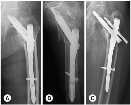
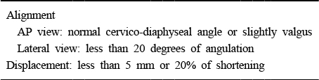
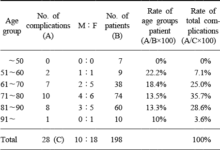
Figure & Data
REFERENCES
Citations
Citations to this article as recorded by 
- Intraoperative Abdominal Penetration of the Lag Screw: A Rare Complication During Proximal Femoral Nail Anti‐Rotation Surgery
Mohammad Javad Dehghani Firoozabadi, Ramin Bozorgmehr, Fatemeh Bastan, Maryam Rashidian
Clinical Case Reports.2026;[Epub] CrossRef - Proximal Femoral Nail Mechanical Failure: A Case Report and Biomechanical Study
Dimitrios Papanikolopoulos, Christos Kalligeros, Apostolos Polyzos, Vasileios Spitas, Vasileios Soranoglou
Cureus.2022;[Epub] CrossRef - Clinical and radiological outcomes of patients treated with the talon distalfix proximal femoral nail for intertrochanteric femur fractures
Furkan Yapici, Hanifi Ucpunar, Yalkin Camurcu, Necati Emirhan, Oguzhan Tanoglu, Ismail Tardus
Injury.2020; 51(4): 1045. CrossRef - Implant Fracture Analysis of the TFNA Proximal Femoral Nail
Anton Lambers, Bertram Rieger, Alan Kop, Peter D’Alessandro, Piers Yates
Journal of Bone and Joint Surgery.2019; 101(9): 804. CrossRef - Radiographic Outcomes of Osteosynthesis Using Proximal Femoral Nail Antirotation (PFNA) System in Intertrochanteric Femoral Fracture: Has PFNA II Solved All the Problems?
Won Chul Shin, Jung Dong Seo, Sang Min Lee, Nam Hoon Moon, Jung Sub Lee, Kuen Tak Suh
Hip & Pelvis.2017; 29(2): 104. CrossRef - Avascular necrosis of the femoral head following trochanteric fractures in adults: A systematic review
Antonio Barquet, Gabriel Mayora, Joao Matheus Guimaraes, Roberto Suárez, Peter V. Giannoudis
Injury.2014; 45(12): 1848. CrossRef - Anatomical Measurement of Normal Korean Proximal Femur Using Plain Radiography: A Problem when using Proximal Femoral Nail Anti-rotation
Jong-Seok Park, Woo-Jong Kim, Jae-Wan Soh, Byung-Woong Jang, Tae-Heon Kim, You-Sung Suh
Hip & Pelvis.2011; 23(4): 303. CrossRef - PFNA and PFN in Intertrochanteric Fractures - Comparison Study of Sliding -
Suk Kyu Choo, Hyoung Keun Oh, Jun Young Choi
Hip & Pelvis.2010; 22(1): 79. CrossRef
 Cite
CiteComplications of Femoral Pertrochanteric Fractures Treated with Proximal Femoral Nail (PFN)

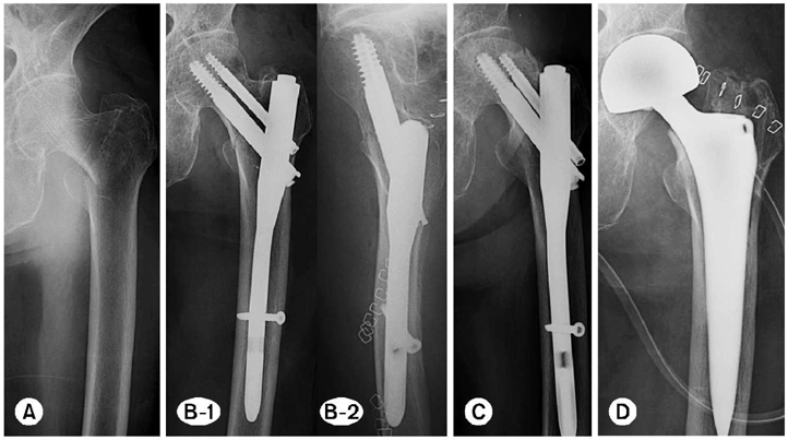
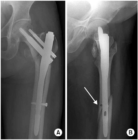
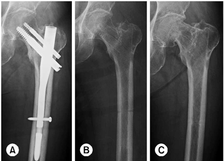


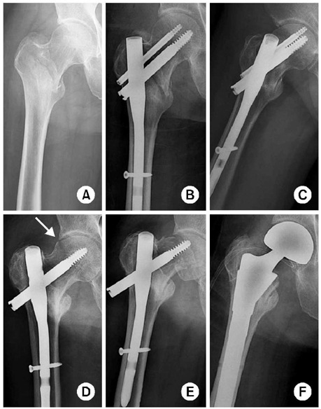
Fig. 1
(A) 59 years old male with intertrochanteric fracture, preoperative AP radiogrpah.
(B) Immediate postoperative AP radiograph showed that antirotation
screw placed higher than greater trochanter.
(C) Antirotation screw showed Z-effect and penetrated the hip
joint in the oblique radiograph.
(D) We removed the antirotation screw.
(E) Femoral head fracture was occurred after fall down.
(F) Bipolar hemiarthroplasty was performed.
Fig. 2
(A) AP radiograph showed relatively extracapsular basicervical fracture.
(B) Immediate postoperative AP and lateral radiograph showed anatomical reduction.
(C) Postoperative 8 months, osteonecrosis of femoral head had developed.
(D) Femoral head had been replaced.
Fig. 3
(A) Periprosthetic fracture was developed but fracture line was not seen definitely in the AP radiogrpah.
(B) In the lateral radiograph, fracture line was observed easily near the inappropriate drilling hole which had been made intraoperatively.
Fig. 4
(A, B) Postoperative radiogrpah showed non-anatomical reduction in the lateral plane.
(C) Reverse Z-effect phenomenon was shown at postoperative 8 months.
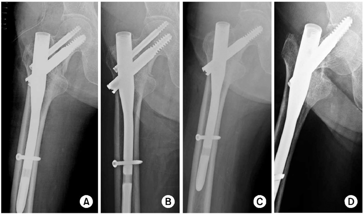
Fig. 5
(A) Immediate postoperative AP radiograph showed that antirotation screw placed higher than greater trochanter.
(B) Antirotation screw showed Z-effect phenomenon and penetrated hip joint at the postoperative 6 weeks.
(C) We removed antirotation screw.
(D) Lag screw migrated proximally and penetrated hip joint again.
Fig. 6
(A, B) Postoperative radiogrpah showed non-anatomical reduction in the lateral plane.
(C) Reverse Z-effect phenomenon was shown at postoperative 8 months.
Fig. 1
Fig. 2
Fig. 3
Fig. 4
Fig. 5
Fig. 6
Complications of Femoral Pertrochanteric Fractures Treated with Proximal Femoral Nail (PFN)
Anatomical reduction criteria
Analysis of complications according to age and sex.
Table 1
Anatomical reduction criteria
Table 2
Analysis of complications according to age and sex.

