 E-submission
 TOTA
TOTA TOTS
TOTS
Search
- Page Path
- HOME > Search
Original Article
- Relationship of lateral malleolar fracture patterns to posterior malleolar fracture morphology in supination-external rotation ankle fractures in Korea: a retrospective cohort study
- Jong-Eun Kim, Chan-Jin Park, Jun-Young Lee, Keun-Bae Lee, Gun-Woo Lee
- J Musculoskelet Trauma 2025;38(4):212-220. Published online October 24, 2025
- DOI: https://doi.org/10.12671/jmt.2025.00234
-
 Abstract
 PDF
PDF - Background
Posterior malleolar fractures frequently accompany rotational ankle fractures. However, the morphological relationship between lateral and posterior malleolar fractures in supination-external rotation (SER) ankle fractures remains unclear. This study aimed to classify lateral malleolar fracture patterns in SER type 3 and 4 ankle fractures and investigated their associations with posterior malleolar fracture morphology.
Methods
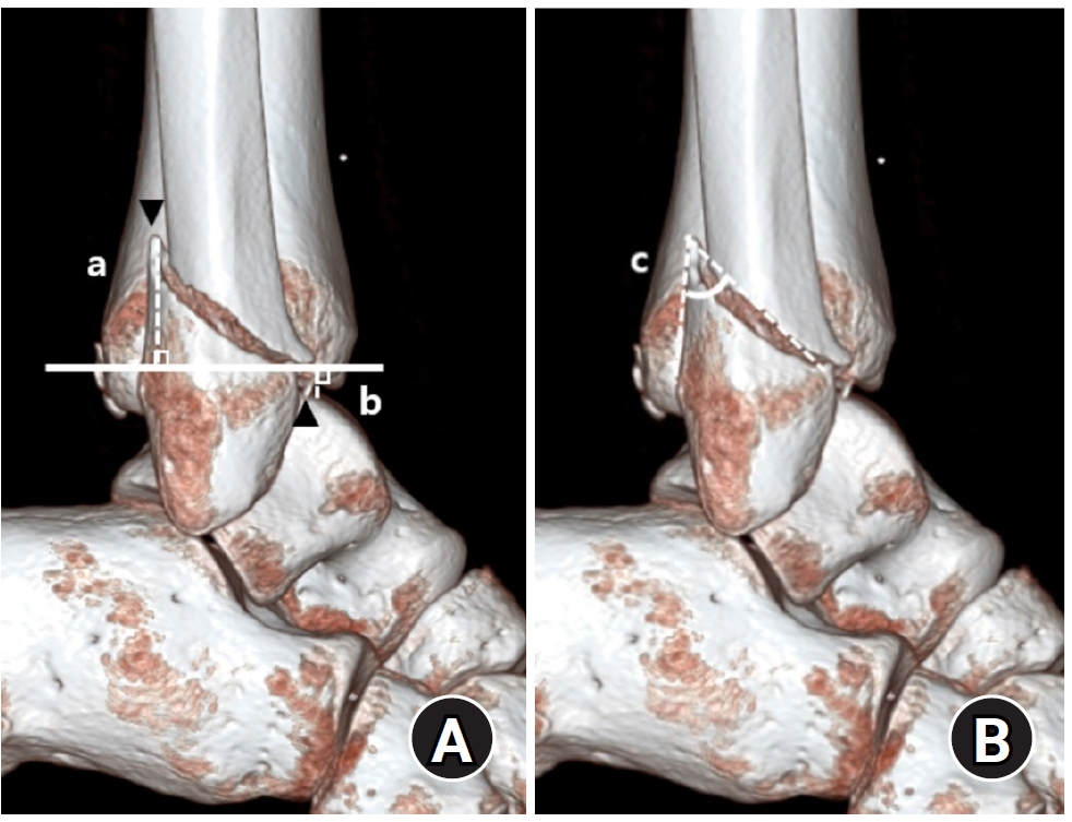
We retrospectively reviewed 132 patients with SER type 3 or 4 ankle fractures and concurrent posterior malleolar fractures between January 2016 and December 2021. Lateral malleolar fractures were categorized as fibular fractures extending <4.5 cm proximal to the ankle joint (102 ankles) or fibular fractures extending ≥4.5 cm proximal to the ankle joint (30 ankles) based on posterior cortex height measured using three-dimensional computed tomography (3D-CT). Posterior malleolar fracture morphology was assessed using the Haraguchi and Bartonicek classifications. Quantitative parameters—including fracture height, angle, and articular involvement—were analyzed using 3D-CT imaging.
Results
Fibular fractures extending ≥4.5 cm proximal to the ankle joint were associated with a significantly higher frequency of Haraguchi type II and Bartonicek types 3 and 4 posterior malleolar fractures. This group also exhibited greater articular involvement (19.2% vs. 12.0%) and posterior cortical height (55.4 mm vs. 24.8 mm) compared to the <4.5 cm group (all P<0.001).
Conclusions
In SER type 3 and 4 ankle fractures, a fibular fracture extending ≥4.5 cm proximal to the ankle joint may be associated with posterior malleolar fractures exhibiting greater articular involvement and medial extension. Preoperative evaluation of the lateral malleolar fracture pattern may provide useful insights into posterior malleolar morphology and assist in surgical planning. However, these findings should be interpreted with caution due to inherent study limitations. Level of evidence: IV
- 1,461 View
- 33 Download

Case Report
- Irreducible Ankle Fracture Dislocation due to Dislocated Tibialis Posterior Tendon - A Case Report -
- Seungyup Shin, Bum-Soo Kim, Ji-Won Lee, Euisun Yoon
- J Korean Fract Soc 2023;36(2):52-56. Published online April 30, 2023
- DOI: https://doi.org/10.12671/jkfs.2023.36.2.52
-
Abstract
PDF
- An irreducible ankle dislocation is a rare injury. The cause is a dislocation of the distal fibula anteriorly or posteriorly or the insertion of soft tissue, such as the deltoid ligament or posteromedial tendon. The tibialis posterior tendon can be dislocated through distal tibiofibular diastasis and prevent reduction of the ankle joint. The authors experienced anterolateral ankle fracture dislocation with a diastasis of the distal tibiofibular joint, and reduction was impossible because of impingement of the tibialis posterior tendon dislocated anteriorly through the distal tibiofibular diastasis. This paper reports the treatment of this injury.
- 770 View
- 15 Download
Original Articles
- Analysis of Clinical and Functional Outcomes according to the Blood Sugar Control Status at the Time of Ankle Fractures Resulting from Rotational Injuries
- Jun Young Lee, Dong Seop Lim, Seung Hyun Lee, Seo Jin Park
- J Korean Fract Soc 2022;35(4):135-141. Published online October 31, 2022
- DOI: https://doi.org/10.12671/jkfs.2022.35.4.135
-
Abstract
PDF
- Purpose
Patients with diabetes are known to have poor clinical outcomes due to the high incidence of complications after ankle joint fracture surgery. This study reports the clinical and functional outcomes based on glycemic control status among patients with ankle joint fractures who underwent surgical treatment.
Materials and Methods
Among patients who underwent surgical treatment due to ankle joint fractures from January 2015 to October 2019, 253 patients with a minimum follow-up of 12 months were identified. We divided them into 3 groups: 195 patients with no diabetes (Group A), 26 patients with well-controlled diabetes (Group B), and 32 patients with uncontrolled diabetes (Group C). In addition, patients with lateral, medial malleolar, bimalleolar, and trimalleolar fractures were identified using radi-ography. The functional outcome measures used for evaluation were the Revised Foot Function Index (FFI), Short Musculoskeletal Function Assessment (SMFA), and the Foot and Ankle Outcome Score (FAOS).
Results
Bone union at 3 months after surgery was high in Group A, showing significant differences compared to the other groups. There was a significant difference between the groups in the incidence of arthropathy and one or more complications. However, the FFI, SMFA, and FAOS did not show significant differences between the groups.
Conclusion
The incidence of complications was high in patients with uncontrolled diabetes compared to the patients with well-controlled diabetes and those with no diabetes. However, functional outcomes showed no significant difference.
- 853 View
- 6 Download
- Radiological Assessment for Morphological Diversity of Distal Fibula
- Su Young Bae, Jin Hee Yoo
- J Korean Fract Soc 2014;27(1):1-9. Published online January 31, 2014
- DOI: https://doi.org/10.12671/jkfs.2014.27.1.1
-
Abstract
PDF
- PURPOSE
The purpose of this study is to determine whether the morphological consistency of distal fibula could be defined by measurement through radiological assessment as there was doubt regarding the adequacy of anatomical distal fibular plates.
MATERIALS AND METHODS
Plain radiographs and computed tomography (CT) images of 300 cases from 2009 to 2012 were reviewed. The distance from the lateral vertex to the tip of the distal fibula and to the lateral margin of the shaft was measured, respectively, in order to understand the shape of the lateral curve of the distal fibula on plain radiographs. The neutral ridge was defined as a point of the lateral ridge located in the center of the antero-posterior diameter and the distance from the tip of the distal fibula to the neutral ridge was measured for determining the shape of the ridge on CT images. The angle of the lateral and posterior surface of the fibular incisura at the level of the neutral ridge was also measured.
RESULTS
A statistically significant difference in the lateral vertex and margin of the fibular shaft on plain radiographs and distance from the tip of the distal fibula to the neutral ridge, angle of the fibular lateral surface on CT images was observed between male and female. The mean distance from the lateral vertex to the tip of distal fibula was 12.2+/-3.0 mm, to the lateral margin of the fibular shaft was 5.6+/-1.7 mm, distance from tip of the distal fibula to the neutral ridge was 54.9+/-6.4 mm, the fibular lateral surface angle was 52.2degrees+/-9.1degrees, and the fibular posterior surface angle was 32.5degrees+/-9.3degrees.
CONCLUSION
Based on the various radiologic parameters, it was concluded that there was a wide morphological diversity of shape of lateral curve and fibular ridge.
- 625 View
- 3 Download
- Treatment of the Trimalleolar Fracture Using Posterolateral Approach: Minimum 2-year Follow Up Results
- Gwang Chul Lee, Jun Young Lee, Sang Ho Ha, Jae Won You, Sang Hong Lee, Hong Moon Sohn, Ki Young Nam, Kwang Hyo Seo
- J Korean Fract Soc 2011;24(4):328-334. Published online October 31, 2011
- DOI: https://doi.org/10.12671/jkfs.2011.24.4.328
-
Abstract
PDF
- PURPOSE
To analyze the long term follow up results of treatment with posterolateral approach and to investigate its usefulness in the patients of trimalleolar fracture with posterior fragment which is above 25% of articular involvement.
MATERIALS AND METHODS
There were 34 cases of trimalleolar fracture in our hospital from May 2004 to April 2008. We investigated 20 patients who underwent operation with the posterolateral approach and over-2 years follow up cases. The mean follow up period was 34 (24~58) months. Preoperative posterior malleolar fragment involved above 25% of articular surface in all cases and displaced more than 2 mm in 11 cases. We analyzed the radiologic type of posterior malleolar fragments and evaluated the function and pain through AOFAS score and complications.
RESULTS
All cases showed primary union at mean 13.1 weeks. The complications are that partial ankylosis result of soft tissue contracture is seen in 2 cases (10%) and post-traumatic arthritis is seen in 1 cases (5%) and 17 cases (85%) of all patients are showed excellent AOFAS score.
CONCLUSION
The posterolateral approach is a valuable method because that it enables us to easily reduction and internal fixation of the posterior malleolus and lateral malleolus at one time and the results are satisfied for a long time follow up. -
Citations
Citations to this article as recorded by
- Outcomes of Immediate Operative Treatment of Ankle Trimalleolar Open Fractures
Jun-Young Lee, Yong-Jin Cho, Sin-Wook Kang, Yung-Min Cho, Hyun-Bai Choi
Journal of Korean Foot and Ankle Society.2020; 24(1): 25. CrossRef
- Outcomes of Immediate Operative Treatment of Ankle Trimalleolar Open Fractures
- 863 View
- 3 Download
- 1 Crossref
- Treatment of the Posterior Malleolar Fracture Using Posterior Approach
- Hyun Wook Chung, Dong Hwan Kim, Si Hoon Yoo, Jin Soo Suh
- J Korean Fract Soc 2010;23(1):50-56. Published online January 31, 2010
- DOI: https://doi.org/10.12671/jkfs.2010.23.1.50
-
Abstract
PDF
- PURPOSE
For fixation of the large posterior malleolar fracture fragment, indirect anterior fixation with cannulated screw has been widely used, but the anatomical reduction is not always obtained. The purpose of this article is to evaluate the clinical result of posterior malleolar fractures treated with anatomical reduction and internal fixation using posterior approach.
MATERIALS AND METHODS
We have analyzed the 15 patients with posterior malleolar fractures, treated with posterior approach from August 2005 to August 2008. The mean follow up period was 17.6 months, We have reviewed the perioperative joint integrity, method of operation, postoperative care, bony union and complication. A clinical outcome was evaluated by AOFAS (American orthopedic foot and ankle society) scaling system and Olerud & Molander scoring system.
RESULTS
Among 15 cases, posterolateral approach and posteromedial approach were chosen in 9 cases and 6 cases respectively. The radiologic unions were achieved at 12.4 (12~18) weeks. Mean AOFAS score was 90.3 (72~98), and Olerud & Molander score was "excellent" in 5 cases, "good" in 7 cases, "fair" in 1 case and "poor" in 2 cases. Postoperative complications in 2 cases revealed a posttraumatic arthritis and a scar band contracture respectively.
CONCLUSION
In posterior malleolar fracture of ankle joint, the integrity of joint has closely affected clinical outcomes. We suggest that a posterior approach for posterior malleolar fracture with especially incarcerated fragments and comminuted fractures, can be a useful method for anatomical reduction and stable fixation, and satisfactory clinical results. -
Citations
Citations to this article as recorded by- Single lateral approach for open reduction and internal fixation of posterior malleolar fragment in Weber B rotational ankle fracture
Jaehyung Lee, Hwan Ryu, Jae Yong Park
Medicine.2023; 102(3): e32725. CrossRef - Posterior Malleolus Fractures in Trimalleolar Ankle Fractures: Malleolus versus Transyndesmal Fixation
Bilgehan Tosun, Ozgur Selek, Umit Gok, Halil Ceylan
Indian Journal of Orthopaedics.2018; 52(3): 309. CrossRef - Single Oblique Posterolateral Approach for Open Reduction and Internal Fixation of Posterior Malleolar Fractures With an Associated Lateral Malleolar Fracture
Jun Young Choi, Ji Hoon Kim, Hyeong Tak Ko, Jin Soo Suh
The Journal of Foot and Ankle Surgery.2015; 54(4): 559. CrossRef
- Single lateral approach for open reduction and internal fixation of posterior malleolar fragment in Weber B rotational ankle fracture
- 952 View
- 3 Download
- 3 Crossref
- Radiologic Analysis and Treatment of Posterior Malleolar Fractures of the Ankle
- Jae Sung Lee, Soo Yong Kang, Han Jun Lee, Young Bong Ko
- J Korean Fract Soc 2009;22(2):98-103. Published online April 30, 2009
- DOI: https://doi.org/10.12671/jkfs.2009.22.2.98
-
Abstract
PDF
- PURPOSE
The purpose of this study was to classify posterior malleolar fractures according to the position of fragments and to analyze radiologic features of each type.
MATERIALS AND METHODS
We analyzed forty-six patients of ankle fractures involving a posterior malleolus who were treated between January 2004 and December 2007. The posterior malleolar fractures were categorized into three types (posterolateral, posteromedial, shell) based on the major fracture line. In each type, we analyzed amount of displacement, involvement of articular surface, existence of subluxation and osteochondral impacted fragments.
RESULTS
The forty-six patients were categorized into three types: Posterolateral (PL) type (33 cases, 72%), Posteromedial (PM) type (8 cases, 17%), shell type (5 cases, 11%). Of the 8 cases with PM type, 7 cases showed displacement more than Grade II, 4 cases showed subluxation of ankle joint, and 3 cases showed osteochondral impacted fragment. Average involvement of articular surface of PM type is 35% (15~65%).
CONCLUSION
Posterior malleolar fractures with medial extension tended to have adverse effect on ankle stability and Preoperative CT scan is essential for evaluation of fracture type and determination of appropriate surgical approach. -
Citations
Citations to this article as recorded by- Treatment of Isolated Posterior Malleolus Fracture in the Ankle
Ji Hoon Kim, Seong Mu Cha, Dae Yeon Jo, Jin Soo Suh
Journal of the Korean Orthopaedic Association.2014; 49(1): 29. CrossRef
- Treatment of Isolated Posterior Malleolus Fracture in the Ankle
- 886 View
- 2 Download
- 1 Crossref
- Nonoperative Treatment of Isolated Lateral Malleolar Fracture
- Woo Chun Lee, Jong Ho Ahn
- J Korean Fract Soc 2005;18(3):291-293. Published online July 31, 2005
- DOI: https://doi.org/10.12671/jkfs.2005.18.3.291
-
Abstract
PDF
- PURPOSE
To evaluate the results of conservative treatment for isolated lateral malleolus fracture without medial ankle injury.
MATERIALS AND METHODS
From March 1999 to February 2003, 25 ankles in 25 patients were treated for isolated lateral malleolus fracture and followed for more than one year. Mean age was 46.9 years (range, 20~71 years). Cases without any swelling or tenderness on the deltoid area, or cases with minimal pain, swelling or tenderness on the deltoid area and medial clear space 1 mm or less on stress radiograph were included for the study. Immediate weight bearing was allowed with below-knee cast immobilization in all cases.
RESULTS
All were supinatin-external rotation stage II injury and mean duration of cast immobilization was 6.3+/-1.6 weeks after injury. There was no case which showed widening of medial clear space during routine radiographic follow-up. There was no change in the degree of displacement in spite of immediate weight bearing with short leg cast on.
CONCLUSION
Because the lateral malleolus fracture without medial injury can be managed nonoperatively, we need to differentiate this type of fracture to avoid unnecessary surgery, and for early return to normal daily activity. -
Citations
Citations to this article as recorded by- Posterior Plating in Distal Fibular Fracture
Choong-Hyeok Choi, Young-A Cho, Jae-Hoon Kim, Il-Hoon Sung
Journal of the Korean Fracture Society.2007; 20(2): 161. CrossRef
- Posterior Plating in Distal Fibular Fracture
- 1,160 View
- 5 Download
- 1 Crossref
Case Report
- Treatment of Peterson classification Type VI of Physeal Injury in Ankle Joint: 2 cases report
- Byung Il Lim, Tai Seung Kim, Kuhn sung Whang, Il Hoon Sung
- J Korean Soc Fract 2000;13(4):1061-1066. Published online October 31, 2000
- DOI: https://doi.org/10.12671/jksf.2000.13.4.1061
-
Abstract
PDF
- Peterson classification type VI, which has been reported newly on physeal injury classification, is defined as partial missing of the metaphysis and epiphysis with a portion of the physis. It has not been reported in the Republic of Korea to our knowledge. Because this is an open fracture, immediate surgery is needed in all cases. Angular deformity and leg length discrepancy occurs as a result of the formation of the physeal bar. Additional reconstuctive operation, therefore, should be necessary. We report two cases of Peterson classification type VI, both cases were open fracture at the level of ankle joint owing to pedestrian traffic accident. In our experience, Peterson classification type VI required multiple operations because progression of angular deformity with growth, and must be followed up until maturity.
-
Citations
Citations to this article as recorded by- Changes in Oxygen Saturation and Walk in Relation to Smoking and Types of Shoes
Jea-Cheol Park, Jong-Man Han, Woon-Soo Cho, Yong-Nam Kim
The Journal of Korean Physical Therapy.2015; 27(1): 55. CrossRef
- Changes in Oxygen Saturation and Walk in Relation to Smoking and Types of Shoes
- 885 View
- 4 Download
- 1 Crossref
Original Articles
- Radiologic Evaluation of the Ankle Joint: Comparison of Different criteria & its A vailability of Clinical Practice
- Deuk Soo Hwang, Seung Ho Yune, Kwang Jin Rhee, June Kyu Lee, Je Taek Jeong
- J Korean Soc Fract 1998;11(4):880-885. Published online October 31, 1998
- DOI: https://doi.org/10.12671/jksf.1998.11.4.880
-
Abstract
PDF
- Generally it is Known that the best clinical results in treatment of injuries of the ankle are obtained by anatomical restoration of the joint. For objective measurements of tibiotalar joint, some investigators ued different criteria and defined the specific reference points under variable angle of internally rotated anteroposterior projection. But, occasionally we didn't acquire the accurate roentgenographic finding that was suggested by investigators. So, we check the variable angle of internal rotation film in addition to angle suggested by investigators and compare the criteria between them. The purpose of this study is to evaluate availability of internally rotated mortise view and its criteria in clinical practice. Following results was acquired. First, there was no significant difference in measuring the medical clear space on depand on variability of rotation angle. Second, the overlapping distance of tibiofibular syndesmosis decreased by increasing internal rotation angle, but was not under 1mm (ie, index of injury). A third, to measure the Weber's 3 criteria, we need to check the variable internal rotation angle, if necessary. Finally, we acquired the normal range of measurement about Tile's 2 criteria by variable internal rotation angle.
- 440 View
- 0 Download
- Compression Arthrodesis of Ankle Joint using Autocompression Angle Plate
- Eun Sun Moon, Eun Kyoo Song, Yong Gi Choi
- J Korean Soc Fract 1989;2(2):174-178. Published online November 30, 1989
- DOI: https://doi.org/10.12671/jksf.1989.2.2.174
-
Abstract
PDF
- We performed the compression arthrodesis in ankle joint of 4 cases, using autocompression angle plate. In all cases, we made successful union within 3 months, without any specific complications. This method gives several advantages, such as short period of external support, good cosmetic effect, early union and early rehabilitation.
- 485 View
- 0 Download
 First
First Prev
Prev

