 E-submission
 TOTA
TOTA TOTS
TOTS
Articles
- Page Path
- HOME > J Musculoskelet Trauma > Volume 25(1); 2012 > Article
-
Original Article
- Treatment of Distal Femur Fracture with Minimally Invasive Locking Compression Plate Osteosynthesis
- Ki-Chul Park, M.D., Kyu-Sung Chung, M.D., Joon-Ki Moon, M.D.
-
Journal of the Korean Fracture Society 2012;25(1):13-19.
DOI: https://doi.org/10.12671/jkfs.2012.25.1.13
Published online: January 31, 2012
Department of Orthopedic Surgery, Guri Hospital, Hanyang University College of Medicine, Guri, Korea.
- Address reprint requests to: Ki-Chul Park, M.D. Department of Orthopedic Surgery, Guri Hospital, Hanyang University College of medicine, 249-1, Gyomoon-dong, Guri 471-701, Korea. Tel: 82-31-560-2318, Fax: 82-31-557-8781, kcpark@hanyang.ac.kr
• Received: July 21, 2011 • Revised: October 30, 2011 • Accepted: November 15, 2011
Copyright © 2012 The Korean Fracture Society
- 1,469 Views
- 21 Download
- 4 Crossref
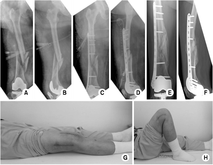
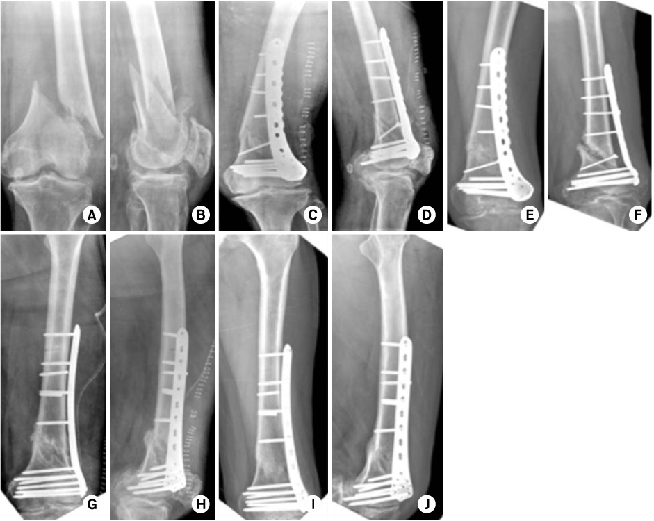
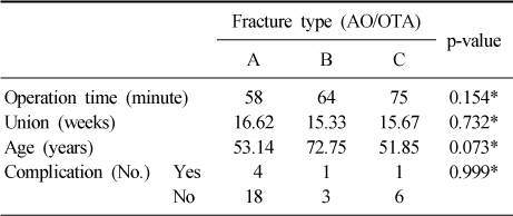

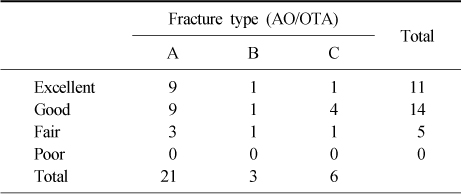
Figure & Data
REFERENCES
Citations
Citations to this article as recorded by 
- Surgical Treatment of AO/OTA 33-C Intra-Articular Distal Femoral Fractures through Parapatellar Approach
Suk Kyu Choo, Sung Tan Cho, Hyoung Keun Oh
Journal of the Korean Fracture Society.2022; 35(1): 1. CrossRef - Comparing Outcomes of Retrograde Intramedullary Nail and Locking Plate Fixation in Distal Femoral Fractures
Byung-Ho Yoon, Bo Kwon Hwang, Hyoung-Keun Oh, Suk Kyu Choo, Jong Min Sohn, Yerl-Bo Sung
Journal of the Korean Fracture Society.2021; 34(4): 131. CrossRef - Incidence of nonunion after surgery of distal femoral fractures using contemporary fixation device: a meta‐analysis
Byung-Ho Yoon, In Keun Park, Youngwoo Kim, Hyoung-Keun Oh, Suk Kyu Choo, Yerl-Bo Sung
Archives of Orthopaedic and Trauma Surgery.2021; 141(2): 225. CrossRef - Minimally Invasive Plate Osteosynthesis with Locking Compression Plate for Distal Femur Fracture
Sung Won Cho, Sang Ho Ha, Gwang Chul Lee, Woong Hee Kim
Journal of the Korean Fracture Society.2013; 26(3): 205. CrossRef
 Cite
CiteTreatment of Distal Femur Fracture with Minimally Invasive Locking Compression Plate Osteosynthesis


Fig. 1
(A, B) A 66-year-old woman with communited, distal femur fracture by traffic accident (AO-OTA classification 32-C1).
(C, D) Radiographs show a postoperative state which is reduced and internal fixation.
(E, F) Follow-up radiographs after 6 months show bony union with good alignment.
(G, H) Clinical photographs after 6 months show nearly full range of motion on knee joint.
Fig. 2
(A, B) A 57-year-old man with extraarticular metaphyseal fragmented wedge, distal femur fracture injured by traffic accident (AO-OTA classification 33-A2).
(C, D) Radiographs show a postoperative state which is reduced and internal fixation.
(E, F) Follow-up radiographs after 20 weeks show a nonunion state.
(G, H) Radiographs show a second operation state via removal of previous implant, autogenous iliac bone graft and reimplantation.
(I, J) Follow-up radiographs after 4 months from second operation show bony union.
Fig. 1
Fig. 2
Treatment of Distal Femur Fracture with Minimally Invasive Locking Compression Plate Osteosynthesis
Comparison of clinical outcomes
*Fisher exact test.
Patient demographics, clinical and radiographic outcomes
ROM: Range of motion, AP: Anteriorposterior, Lat: Lateral.
The Schazker and Lambert criteria
Table 1
Comparison of clinical outcomes
*Fisher exact test.
Table 2
Patient demographics, clinical and radiographic outcomes
ROM: Range of motion, AP: Anteriorposterior, Lat: Lateral.
Table 3
The Schazker and Lambert criteria

