 E-submission
 TOTA
TOTA TOTS
TOTS
Articles
- Page Path
- HOME > J Musculoskelet Trauma > Volume 25(1); 2012 > Article
-
Original Article
- Comparative Analysis of Minimally Invasive Plate Osteosynthesis Using Periarticular Plate and Intramedullary Nailing in Distal Tibial Metaphyseal Fractures
- Gwang Chul Lee, M.D., Jun Young Lee, M.D., Sang Ho Ha, M.D., Hong Moon Sohn, M.D., Yi Kyu Park, M.D.
-
Journal of the Korean Fracture Society 2012;25(1):20-25.
DOI: https://doi.org/10.12671/jkfs.2012.25.1.20
Published online: January 31, 2012
Department of Orthopaedic Surgery, College of Medicine, Chosun University Hospital, Gwangju, Korea.
- Address reprint requests to: Jun Young Lee, M.D. Department of Orthopaedic Surgery, Chosun University Hospital, 588, Seosuk-dong, Dong-gu, Gwangju 501-717, Korea. Tel: 82-62-220-3147, Fax: 82-62-226-3379, leejy88@chosun.ac.kr
• Received: May 24, 2011 • Revised: August 13, 2011 • Accepted: November 8, 2011
Copyright © 2012 The Korean Fracture Society
- 1,111 Views
- 0 Download
- 2 Crossref
Abstract
-
Purpose
- To compare results between minimally invasive plate osteosynthesis using a periarticular plate and intramedullary nailing in distal tibial metaphyseal fractures in two treatment groups.
-
Materials and Methods
- Sixty-one cases of distal tibial metaphyseal fractures from December 2008 to December 2009 were evaluated. The minimal follow-up period was 12 months. Thirty patients treated by minimally invasive plate osteosynthesis using a periarticular plate were Group A; 31 patients treated by intramedullary nailing were Group B. We compared and analyzed the results of each group by radiological and clinical assessments.
-
Results
- The mean bony union time was 16.4 weeks in Group A and 17.2 weeks in Group B. The mean operation time was 45 minutes in Group A and 48 minutes in Group B. The mean radiation exposure times were 4.2 minutes and 4.8 minutes, respectively. VAS scores were 0.7 points and 0.5 points in each respective group. In Group A, the VAS score was 1.7 points when we applied pressure on the skin around the plate. The mean Olerud and Molander Ankle Score was 87.4 points and 86.3 points, respectively. A superficial wound infection occurred in 1 case in each group, and angular deformities more than 5 degrees occurred in 2 Group B cases.
-
Conclusion
- No significant differences in results were observed between the two groups. However, a higher incidence of angular deformity was seen in the intramedullary nailing group. Therefore, we must be careful during surgery.
- 1. Böstman O, Hänninen A. The fibular reciprocal fracture in tibial shaft fractures caused by indirect violence. Arch Orthop Trauma Surg, 1982;100:115-121.ArticlePDF
- 2. Cheng W, Li Y, Manyi W. Comparison study of two surgical options for distal tibia fracture-minimally invasive plate osteosynthesis vs. open reduction and internal fixation. Int Orthop, 2011;35:737-742.ArticlePDF
- 3. Collinge C, Protzman R. Outcomes of minimally invasive plate osteosynthesis for metaphyseal distal tibia fractures. J Orthop Trauma, 2010;24:24-29.Article
- 4. D'Aubigne RM, Maurer P, Zucman J, Masse Y. Blind intramedullary nailing for tibial fractures. Clin Orthop Relat Res, 1974;105:267-275.
- 5. Freedman EL, Johnson EE. Radiographic analysis of tibial fracture malalignment following intramedullary nailing. Clin Orthop Relat Res, 1995;315:25-33.Article
- 6. Hazarika S, Chakravarthy J, Cooper J. Minimally invasive locking plate osteosynthesis for fractures of the distal tibia--results in 20 patients. Injury, 2006;37:877-887.Article
- 7. Helfet DL, Suk M. Minimally invasive percutaneous plate osteosynthesis of fractures of the distal tibia. Instr Course Lect, 2004;53:471-475.
- 8. Im GI, Kim DY, Shin JH, Youn KS, Cho WH. Comparative analysis of interlocking nail and anatomical plate in the treatment of distal tibial fracture. J Korean Soc Fract, 1999;12:632-637.Article
- 9. Janssen KW, Biert J, van Kampen A. Treatment of distal tibial fractures: plate versus nail: a retrospective outcome analysis of matched pairs of patients. Int Orthop, 2007;31:709-714.
- 10. Kääb MJ, Frenk A, Schmeling A, Schaser K, Schütz M, Haas NP. Locked internal fixator: sensitivity of screw/plate stability to the correct insertion angle of the screw. J Orthop Trauma, 2004;18:483-487.
- 11. Krackhardt T, Dilger J, Flesch I, Höntzsch D, Eingartner C, Weise K. Fractures of the distal tibia treated with closed reduction and minimally invasive plating. Arch Orthop Trauma Surg, 2005;125:87-94.ArticlePDF
- 12. Lang GJ, Cohen BE, Bosse MJ, Kellam JF. Proximal third tibial shaft fractures. Should they be nailed? Clin Orthop Relat Res, 1995;315:64-74.
- 13. Nork SE, Schwartz AK, Agel J, Holt SK, Schrick JL, Winquist RA. Intramedullary nailing of distal metaphyseal tibial fractures. J Bone Joint Surg Am, 2005;87:1213-1221.Article
- 14. Olerud C, Molander H. Bi- and trimalleolar ankle fractures operated with nonrigid internal fixation. Clin Orthop Relat Res, 1986;206:253-260.Article
- 15. Park KC, Park YS. Minimally invasive plate osteosynthesis for distal tibial metaphyseal fracture. J Korean Fract Soc, 2005;18:264-268.Article
- 16. Robinson CM, McLauchlan GJ, McLean IP, Court-Brown CM. Distal metaphyseal fractures of the tibia with minimal involvement of the ankle. Classification and treatment by locked intramedullary nailing. J Bone Joint Surg Br, 1995;77:781-787.ArticlePDF
- 17. Teitz CC, Carter DR, Frankel VH. Problems associated with tibial fractures with intact fibulae. J Bone Joint Surg Am, 1980;62:770-776.Article
- 18. Vallier HA, Le TT, Bedi A. Radiographic and clinical comparisons of distal tibia shaft fractures (4 to 11 cm proximal to the plafond): plating versus intramedullary nailing. J Orthop Trauma, 2008;22:307-311.Article
- 19. Wyrsch B, McFerran MA, McAndrew M, et al. Operative treatment of fractures of the tibial plafond. A randomized, prospective study. J Bone Joint Surg Am, 1996;78:1646-1657.Article
- 20. Yang SW, Tzeng HM, Chou YJ, Teng HP, Liu HH, Wong CY. Treatment of distal tibial metaphyseal fractures: Plating versus shortened intramedullary nailing. Injury, 2006;37:531-535.Article
REFERENCES
Fig. 1The fluoroscopic X-rays reveals joy stick method that parallel K-wire fixation on distal tibia for more accurate reduction during insertion of ball-tip guide wire and reaming of the medullary canal (In this case, excute conservative treatment about fibular fracture).
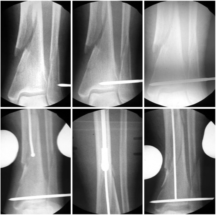
Fig. 2The fluoroscopic X-rays (A~D) reveal using the poller screw for accurate insertion nail. Postoperative X-ray, (E) anteroposterior, and (F) lateral view.

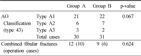
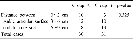
Figure & Data
REFERENCES
Citations
Citations to this article as recorded by 
- Comparative Analysis of Minimally Invasive Plate Osteosynthesis and Intramedullary Nailing in the Treatment of the Distal Tibia Fractures
Ho-Min Lee, Young-Sung Kim, Jong-Pil Kim, Phil-Hyun Chung, Suk Kang, Kaung Suk Jo
Journal of the Korean Fracture Society.2018; 31(3): 94. CrossRef - A Comparison of the Results between Intramedullary Nailing and Minimally Invasive Plate Osteosynthesis in Distal Tibia Fractures
Chul-Hyun Park, Chi-Bum Choi, Bum-Jin Shim, Dong-Chul Lee, Oog-Jin Shon
Journal of the Korean Orthopaedic Association.2014; 49(4): 285. CrossRef
 Cite
CiteComparative Analysis of Minimally Invasive Plate Osteosynthesis Using Periarticular Plate and Intramedullary Nailing in Distal Tibial Metaphyseal Fractures

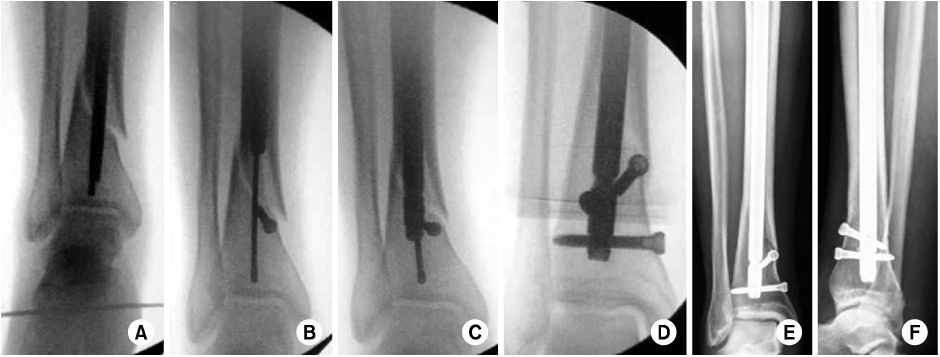
Fig. 1
The fluoroscopic X-rays reveals joy stick method that parallel K-wire fixation on distal tibia for more accurate reduction during insertion of ball-tip guide wire and reaming of the medullary canal (In this case, excute conservative treatment about fibular fracture).
Fig. 2
The fluoroscopic X-rays (A~D) reveal using the poller screw for accurate insertion nail. Postoperative X-ray, (E) anteroposterior, and (F) lateral view.
Fig. 1
Fig. 2
Comparative Analysis of Minimally Invasive Plate Osteosynthesis Using Periarticular Plate and Intramedullary Nailing in Distal Tibial Metaphyseal Fractures
Distal tibial fracture pattern (AO classification) and combined fibular fractures between Group A and B
Distance between Ankle articular surface and fracture site between Group A and B
Table 1
Distal tibial fracture pattern (AO classification) and combined fibular fractures between Group A and B
Table 2
Distance between Ankle articular surface and fracture site between Group A and B

