 E-submission
 TOTA
TOTA TOTS
TOTS
Articles
- Page Path
- HOME > J Musculoskelet Trauma > Volume 29(3); 2016 > Article
-
Case Report
- Medial Plating of Distal Femoral Fracture with Locking Compression Plate-Proximal Lateral Tibia: Cases' Report
- Se-Ang Jang, M.D., Young-Soo Byun, M.D., In-Ho Han, M.D., Dongju Shin, M.D.
-
Journal of the Korean Fracture Society 2016;29(3):206-212.
DOI: https://doi.org/10.12671/jkfs.2016.29.3.206
Published online: July 21, 2016
Department of Orthopaedic Surgery, Daegu Fatima Hospital, Daegu, Korea.
- Address reprint requests to: Dongju Shin, M.D. Department of Orthopaedic Surgery, Daegu Fatima Hospital, 99 Ayang-ro, Dong-gu, Daegu 41199, Korea. Tel: 82-53-940-7324, Fax: 82-53-954-7417, aabga@hanmail.net
• Received: April 15, 2016 • Revised: May 20, 2016 • Accepted: June 9, 2016
Copyright © 2016 The Korean Fracture Society. All rights reserved.
This is an Open Access article distributed under the terms of the Creative Commons Attribution Non-Commercial License (http://creativecommons.org/licenses/by-nc/4.0) which permits unrestricted non-commercial use, distribution, and reproduction in any medium, provided the original work is properly cited.
- 2,593 Views
- 125 Download
- 7 Crossref
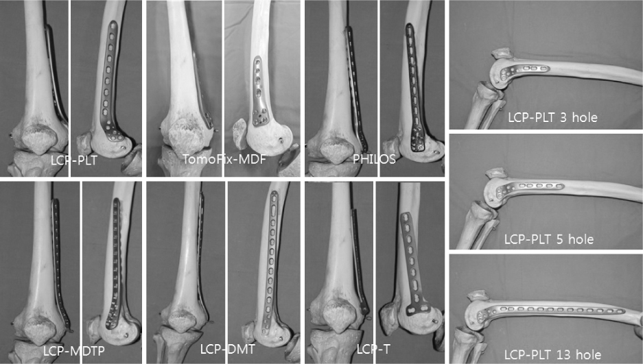
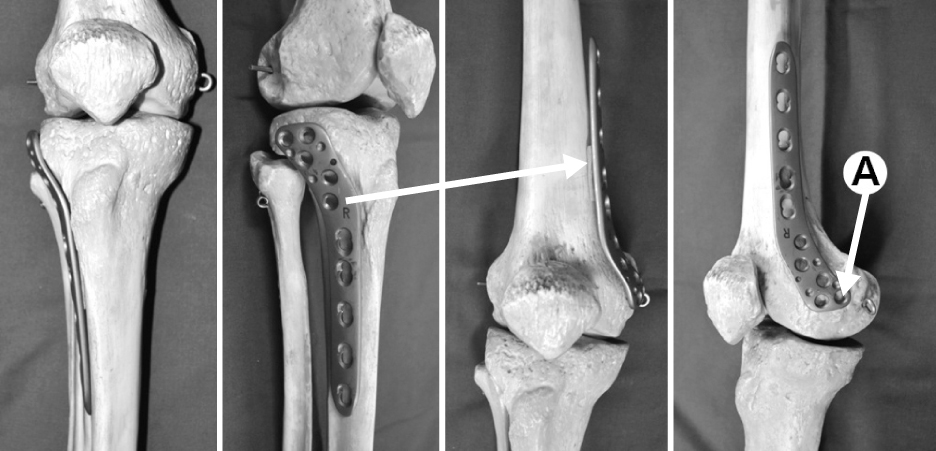
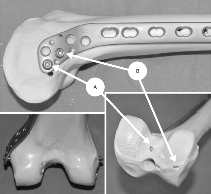
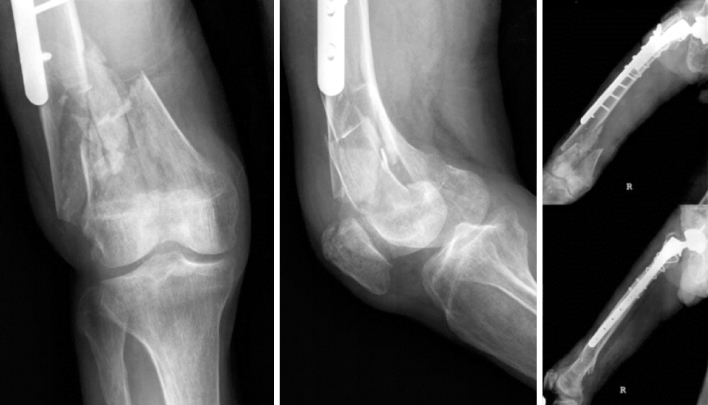
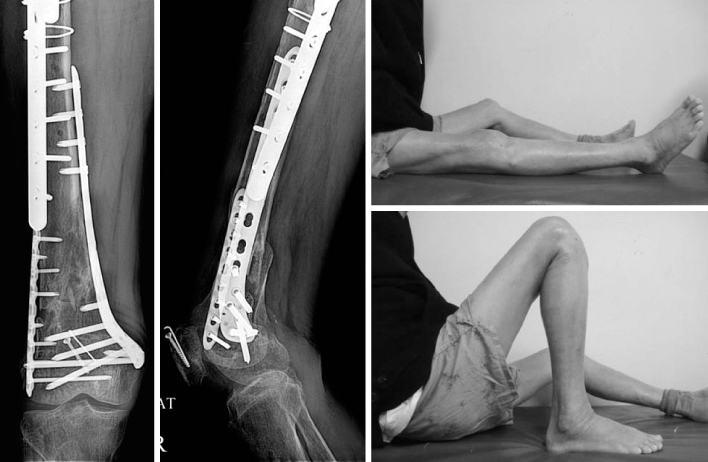


Figure & Data
REFERENCES
Citations
Citations to this article as recorded by 
- A novel anatomical locked medial femoral condyle plate: a biomechanical study
M. A. Ozer, S. Keser, D. Barıs, O. Yazoglu
European Journal of Orthopaedic Surgery & Traumatology.2024; 34(5): 2767. CrossRef - Medial plating of distal femur: which pre-contoured angular stable plate fits best?
Shaam Achudan, Rex Premchand Antony Xavier, Sze Ern Tan
European Journal of Orthopaedic Surgery & Traumatology.2024; 34(6): 3297. CrossRef - Medial augmentation of distal femur fractures using the contralateral distal femur locking plate: A technical note
Jaime Andrés Leal
OTA International.2024;[Epub] CrossRef - The missing piece of the trauma armoury-medial femoral condyle plate
Piyush Upadhyay, Farhan Syed, Darryl N Ramoutar, Jayne Ward
Injury.2022; 53(3): 1237. CrossRef - Surgical Tips and Tricks for Distal Femur Plating
Christopher Lee, Dane Brodke, Ajay Gurbani
Journal of the American Academy of Orthopaedic Surgeons.2021; 29(18): 770. CrossRef - Medial minimally invasive helical plate osteosynthesis of the distal femur – a new technique
G.M. Hohenberger, A.M. Schwarz, P. Grechenig, B. Clement, Mario Staresinic, Bore Bakota
Injury.2021; 52: S27. CrossRef - Feature-Based Design of Personalized Anatomical Plates for the Treatment of Femoral Fractures
Xiaozhong Chen, Zhijian Mao, Xi Jiang
IEEE Access.2021; 9: 43824. CrossRef
 Cite
CiteMedial Plating of Distal Femoral Fracture with Locking Compression Plate-Proximal Lateral Tibia: Cases' Report


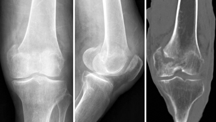
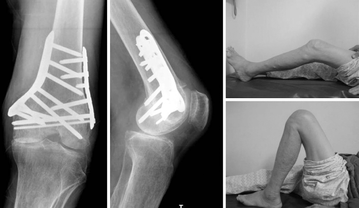
Fig. 1
Photographs of the femur bone model (3B Scientific, Hamburg, Germany) with various plates on medial condyle, locking compression plate-proximal lateral tibia (LCP-PLT), tomoFix-medial distal femur plate (TomoFix-MDF), proximal humerus internal locking plate system (PHILOS), LCP-medial distal tibia plate (LCP-MDTP), LCP-distal metaphyseal tibia (LCP-DMT), and LCP-distal tibia T (LCP-T) plate, in order from upper left corner.
Fig. 2
Photographs of the femur bone model (3B Scientific, Hamburg, Germany) showing the application of locking compression plate- proximal lateral tibia (LCP-PLT) on appropriate position. A: Distal posterior hole.
Fig. 3
Photographs of the femur bone model (Synbone, Malans, Switzerland) with the application of locking compression plate-proximal lateral tibia (LCP- PLT) on appropriate position. A: Distal posterior screw directed to the intercondylar notch, B: 2nd row screw reached the lateral femoral condyle without penetration into the intercondylar notch.
Fig. 4
Radiographs of the right knee and the right femur showing preexisting lateral plate and comminuted fracture of the right distal femur.
Fig. 5
Radiographs and photographs of the right femur at 12 months after the surgery showing good fracture healing and good range of motion of the right knee.
Fig. 6
Radiographs of the left knee and the coronal section of computed tomography scan of the left knee showing severely impacted medial condyle fracture.
Fig. 7
Radiographs and photographs of the left knee at 14 weeks after surgery showing good fracture healing and good range of motion of the left knee.
Fig. 1
Fig. 2
Fig. 3
Fig. 4
Fig. 5
Fig. 6
Fig. 7
Medial Plating of Distal Femoral Fracture with Locking Compression Plate-Proximal Lateral Tibia: Cases' Report

