 E-submission
 TOTA
TOTA TOTS
TOTS
Articles
- Page Path
- HOME > J Musculoskelet Trauma > Volume 29(3); 2016 > Article
-
Original Article
- Radiologic Assessment of Postoperative Stability in Unstable Intertrochanteric Fracture Using Lateral Radiograph
- Suc Hyun Kweon, M.D., Ph.D., Jin Yeong Park, M.D., Seng Hwan Kook, M.D., Byung Min Yoo, M.D.
-
Journal of the Korean Fracture Society 2016;29(3):171-177.
DOI: https://doi.org/10.12671/jkfs.2016.29.3.171
Published online: July 21, 2016
Department of Orthopedic Surgery, Wonkwang University School of Medicine, Iksan, Korea.
*Department of Orthopedic Surgery, St. Carollo Hospital, Suncheon, Korea.
- Address reprint requests to: Suc Hyun Kweon, M.D., Ph.D. Department of Orthopedic Surgery, Wonkwang University Hospital, 895 Muwang-ro, Iksan 54538, Korea. Tel: 82-63-859-1360·Fax: 82-63-852-9329, osksh@wonkwang.ac.kr
• Received: April 5, 2016 • Revised: May 23, 2016 • Accepted: May 23, 2016
Copyright © 2016 The Korean Fracture Society. All rights reserved.
This is an Open Access article distributed under the terms of the Creative Commons Attribution Non-Commercial License (http://creativecommons.org/licenses/by-nc/4.0) which permits unrestricted non-commercial use, distribution, and reproduction in any medium, provided the original work is properly cited.
- 818 Views
- 7 Download
Abstract
-
Purpose
- The purpose of this study was to compare the sliding distance of lag screw in patients with unstable femoral intertrochanteric fractures treated with intramedullary fixation using a cephalomedullary nail with a fixed angle between the neck and shaft of the femur in relation to reduction type by lateral radiographs.
-
Materials and Methods
- Between January 2009 to October 2013, 86 cases (86 patients) with unstable femoral intertrochanteric fractures were treated with intramedullary fixation using a metal nail with a fixed neck-shaft angle and followed for at least 6 months. We used AO/OTA classification, and all cases were unstable fractures. Twenty cases were 31-A22, 54 cases were 31-A23, and 12 cases were 31-A3. There were 30 men and 56 women. Average patient age was 73.7 years (range, 47-97 years). We classified reduction types into three groups as postoperative lateral radiologic findings. Group 1 showed no displacement, group 2 showed anterior displacement of the femur neck, and group 3 showed posterior displacement of the femur neck. The radiological assessment compared the sliding distance of the lag screw between postoperative X-ray and last follow-up X-ray.
-
Results
- Forty-two cases were in group 1, 22 cases were in group 2, and the other 22 cases were in group 3. There was no significant difference in the patient characteristics of each group. The sliding distances of the lag screw were 4.9±3.2 mm, 4.6±3.6 mm, and 8.5±4.9 mm, respectively, and group 3 showed a significant result (p<0.0001, p=0.024).
-
Conclusion
- In cases treated with intramedullary fixation using a cephalomedullary nail with a fixed neck-shaft angle, appropriate reduction with a lateral radiograph before screw fixation is needed to prevent excessive lag screw sliding.
- 1. Chung YK, Hwang JH, Kim HK. The treatment of peritrochanteric fracture of femur with proximal femoral nail: comparative study with dynamic hip screw. J Korean Hip Soc, 2007;19:167-175.Article
- 2. Bridle SH, Patel AD, Bircher M, Calvert PT. Fixation of intertrochanteric fractures of the femur. A randomised prospective comparison of the gamma nail and the dynamdynamic hip screw. J Bone Joint Surg Br, 1991;73:330-334.PubMed
- 3. Chang WS, Zuckerman JD, Kummer FJ, Frankel VH. Biomechanical evaluation of anatomic reduction versus medial displacement osteotomy in unstable intertrochanteric fractures. Clin Orthop Relat Res, 1987;(225):141-146.Article
- 4. Haidukewych GJ, Israel TA, Berry DJ. Reverse obliquity fractures of the intertrochanteric region of the femur. J Bone Joint Surg Am, 2001;83:643-650.Article
- 5. Chang SM, Zhang YQ, Ma Z, Li Q, Dargel J, Eysel P. Fracture reduction with positive medial cortical support: a key element in stability reconstruction for the unstable pertrochanteric hip fractures. Arch Orthop Trauma Surg, 2015;135:811-818.ArticlePDF
- 6. Chevalley F, Gamba D. Gamma nailing of pertrochanteric and subtrochanteric fractures: clinical results of a series of 63 consecutive cases. J Orthop Trauma, 1997;11:412-415.Article
- 7. Hardy DC, Descamps PY, Krallis P, et al. Use of an intramedullary hip-screw compared with a compression hip-screw with a plate for intertrochanteric femoral fractures A prospective, randomized study of one hundred patients. J Bone Joint Surg Am, 1998;80:618-630.Article
- 8. Harrington P, Nihal A, Singhania AK, Howell FR. Intramedullary hip screw versus sliding hip screw for unstable intertrochanteric femoral fractures in the elderly. Injury, 2002;33:23-28.Article
- 9. Kwun KW, Kim SK, Lee SW, Youn KH. Treatment of intertrochanteric fractures of the femur: Comparison of the gamma nail and the dynamic hip screw. J Korean Orthop Assoc, 1993;28:1666-1673.ArticlePDF
- 10. Park MS, Kim GN. Intertrochanteric fractures of the femur treated with sliding hip compression screw and gamma nail: mechanical failure after internal fixation. J Korean Hip Soc, 2000;12:102-111.
- 11. Radford PJ, Needoff M, Webb JK. A prospective randomised comparison of the dynamic hip screw and the gamma locking nail. J Bone Joint Surg Br, 1993;75:789-793.ArticlePDF
- 12. Fracture and dislocation compendium. Orthopaedic trauma association committee for coding and classification. J Orthop Trauma, 1996;10:Suppl 1. v-ix. 1-154.
- 13. Tsukada S, Okumura G, Matsueda M. Postoperative stability on lateral radiographs in the surgical treatment of pertrochanteric hip fractures. Arch Orthop Trauma Surg, 2012;132:839-846.ArticlePDF
- 14. Evans EM. Trochanteric fractures: a review of 110 cases treated by nail-plate fixation. J Bone Joint Surg Br, 1951;33:192-204.
- 15. Evans EM. Trochanteric fractures: a review of 110 cases treated by nail-plate fixation. J Bone Joint Surg Br, 1951;33:192-204.
- 16. Clawson DK. Trochanteric fractures treated by the sliding screw plate fixation method. J Trauma, 1964;4:737-752.Article
- 17. Kyle RF, Gustilo RB, Premer RF. Analysis of six hundred and twenty-two intertrochanteric hip fractures. J Bone Joint Surg Am, 1979;61:216-221.Article
- 18. Davis TR, Sher JL, Horsman A, Simpson M, Porter BB, Checketts RG. Intertrochanteric femoral fractures Mechanical failure after internal fixation. J Bone Joint Surg Br, 1990;72:26-31.ArticlePDF
- 19. Baumgaertner MR, Curtin SL, Lindskog DM, Keggi JM. The value of the tip-apex distance in predicting failure of fixation of peritrochanteric fractures of the hip. J Bone Joint Surg Am, 1995;77:1058-1064.Article
- 20. Ebbinghaus S, Mjöberg B. Posterior angulation in trochanteric fractures detected with roentgen stereophotogrammetry. Ups J Med Sci, 1991;96:235-237.Article
REFERENCES
Fig. 1

Classification of quality of fracture reduction based on postoperative lateral radiographs. (A) Group 1 (reducted). (B) Group 2 (anteriorly displaced proximal fragment). (C) Group 3 (posteriorly displaced proximal fragment).
Fig. 2
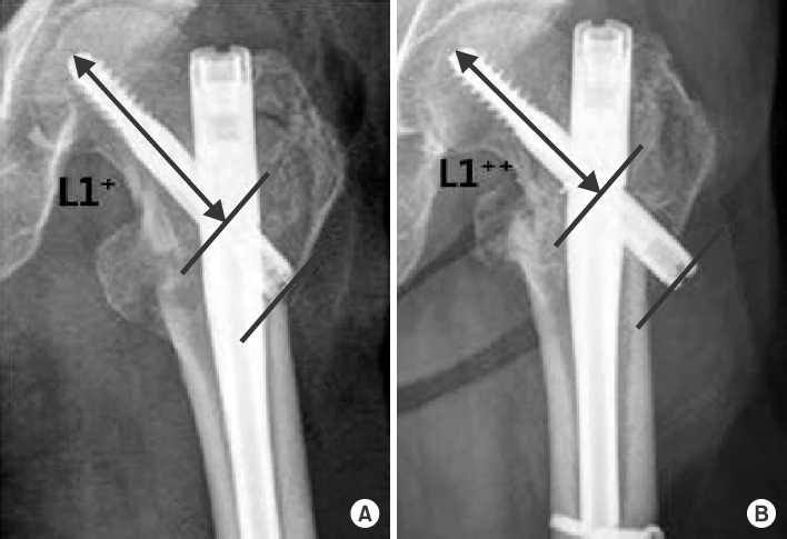
Extent of sliding distance. (A) Postoperative X-ray. (B) Last follow-up X-ray. L1 + : Postoperative screw length from the end of a lag screw to the center of a nail, L1 ++ : Last follow-up length from the end of a lag screw to the center of a nail.
Table 1
![jkfs-29-171-i001.jpg]()
Baseline and Demographic Characteristics of Patients
Table 2
![jkfs-29-171-i002.jpg]()
Sliding Distance of Lag Screw
| Variable | Group 1 | Group 2 | Group 3 |
|---|---|---|---|
| L1 (postoperative)+ | 79.7 | 77.7 | 82.9 |
| L1 (last follow-up)++ | 74.8 | 73.1 | 74.4 |
| L1+-L1++* (mm) | 4.9±3.2 | 4.6±3.6 | 8.5±4.9 |
Group 1 showed no displacement, group 2 showed anterior displacement of the femur neck, and group 3 showed posterior displacement of the femur neck. *Values are presented as mean± standard deviation. L1: Length from the end of a lag screw to the center of a nail, L1 + : Postoperative L1, L1 ++ : Last follow-up L1.
Figure & Data
REFERENCES
Citations
Citations to this article as recorded by 
 Cite
CiteRadiologic Assessment of Postoperative Stability in Unstable Intertrochanteric Fracture Using Lateral Radiograph
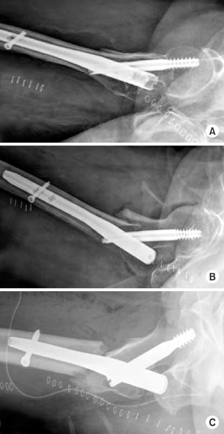

Fig. 1
Classification of quality of fracture reduction based on postoperative lateral radiographs. (A) Group 1 (reducted). (B) Group 2 (anteriorly displaced proximal fragment). (C) Group 3 (posteriorly displaced proximal fragment).
Fig. 2
Extent of sliding distance. (A) Postoperative X-ray. (B) Last follow-up X-ray. L1 + : Postoperative screw length from the end of a lag screw to the center of a nail, L1 ++ : Last follow-up length from the end of a lag screw to the center of a nail.
Fig. 1
Fig. 2
Radiologic Assessment of Postoperative Stability in Unstable Intertrochanteric Fracture Using Lateral Radiograph
Baseline and Demographic Characteristics of Patients
| Characteristic | Group 1 | Group 2 | Group 3 |
|---|---|---|---|
| Mean age (yr) | 74.0 | 72.1 | 74.9 |
| Sex (female:male) | 27:15 | 13:9 | 16:6 |
| Mean height (cm) | 155.6 | 158.4 | 159.8 |
| Mean weight (kg) | 53.6 | 54.2 | 58.1 |
| Mean body mass index (kg/m2) | 21.9 | 21.5 | 22.6 |
| ASA classification (1:2:3:4:5) | 29:11:2:0:0 | 11:11:0:0:0 | 14:8:0:0:0 |
| Gait ability preinjury (0:2:4:6:8:10) | 3:5:8:10:3:13 | 2:1:2:6:3:7 | 1:1:5:12:1:2 |
| AO/OTA classification (A22:A23:A3) | 12:26:4 | 6:13:3 | 2:15:5 |
| Mean tip-apex distance (mm) | 18.3 | 20.6 | 19.4 |
| Mean postoperative follow-up (mo) | 12.5 | 11.9 | 10.7 |
Group 1 showed no displacement, group 2 showed anterior displacement of the femur neck, and group 3 showed posterior displacement of the femur neck. ASA: American Society of Anesthesiologists classification of physical status.
Sliding Distance of Lag Screw
| Variable | Group 1 | Group 2 | Group 3 |
|---|---|---|---|
| L1 (postoperative)+ | 79.7 | 77.7 | 82.9 |
| L1 (last follow-up)++ | 74.8 | 73.1 | 74.4 |
| L1+-L1++* (mm) | 4.9±3.2 | 4.6±3.6 | 8.5±4.9 |
Group 1 showed no displacement, group 2 showed anterior displacement of the femur neck, and group 3 showed posterior displacement of the femur neck. *Values are presented as mean± standard deviation. L1: Length from the end of a lag screw to the center of a nail, L1 + : Postoperative L1, L1 ++ : Last follow-up L1.
Table 1
Baseline and Demographic Characteristics of Patients
Group 1 showed no displacement, group 2 showed anterior displacement of the femur neck, and group 3 showed posterior displacement of the femur neck. ASA: American Society of Anesthesiologists classification of physical status.
Table 2
Sliding Distance of Lag Screw
Group 1 showed no displacement, group 2 showed anterior displacement of the femur neck, and group 3 showed posterior displacement of the femur neck. *Values are presented as mean± standard deviation. L1: Length from the end of a lag screw to the center of a nail, L1 + : Postoperative L1, L1 ++ : Last follow-up L1.

