 E-submission
 TOTA
TOTA TOTS
TOTS
Articles
- Page Path
- HOME > J Musculoskelet Trauma > Volume 27(4); 2014 > Article
-
Original Article
- Treatment of Type IIIb Open Tibial Fractures
- Seong Yeon Lim, M.D., Il Jae Lee, M.D., Jae Ho Joe, M.D., Hyung Keun Song, M.D.
-
Journal of the Korean Fracture Society 2014;27(4):267-273.
DOI: https://doi.org/10.12671/jkfs.2014.27.4.267
Published online: October 20, 2014
Department of Orthopedic Surgery, Ajou University School of Medicine, Suwon, Korea.
*Department of Plastic Surgery, Ajou University School of Medicine, Suwon, Korea.
- Address reprint requests to: Hyung Keun Song, M.D. Department of Orthopedic Surgery, Ajou University Hospital, 164 WorldCup-ro, Yeongtong-gu, Suwon 443-380, Korea. Tel: 82-31-219-5220, Fax: 82-31-219-5229, ostrauma@ajou.ac.kr
• Received: April 4, 2014 • Revised: May 12, 2014 • Accepted: July 10, 2014
Copyright © 2014 The Korean Fracture Society. All rights reserved.
This is an Open Access article distributed under the terms of the Creative Commons Attribution Non-Commercial License (http://creativecommons.org/licenses/by-nc/3.0/) which permits unrestricted non-commercial use, distribution, and reproduction in any medium, provided the original work is properly cited.
- 1,776 Views
- 13 Download
- 1 Crossref
Abstract
-
Purpose
- The purpose of this study is to evaluate the outcome of treatment for patients with Type IIIb open tibial fractures.
-
Materials and Methods
- This study targeted 35 adult patients for whom follow-up was possible over one year after undergoing surgical treatment. There were 29 males and six females with an average age of 45 years.
-
Results
- Fracture location was proximal in 10 cases, midshaft in 13 cases, and the distal part of the tibia in 12 cases. An average of 10 days was observed for definitive fixation with soft tissue coverage of the injury. The mean time to radiographic union was 27 weeks. Sixteen cases (45.7%) of complications were observed. Three cases of superficial infection, two cases of deep infection, four cases of partial flap necrosis, three cases of mal-alignment, three cases of joint stiffness, and one case of hardware breakage were observed. The mean lower extremity functional scale score was 68.5 and the factors influencing the clinical results were severity of open wound (p=0.000) and occurrence of complications (p=0.000) according to results of multiple regression analysis.
-
Conclusion
- In treatment of Type IIIb open tibial fractures, good clinical results can be expected provided that complications are prevented through proper reduction, firm fixation, early soft tissue reconstruction, and early rehabilitation.
- 1. Court-Brown CM, Rimmer S, Prakash U, McQueen MM. The epidemiology of open long bone fractures. Injury, 1998;29:529-534.Article
- 2. DeFranzo AJ, Argenta LC, Marks MW, et al. The use of vacuum-assisted closure therapy for the treatment of lower-extremity wounds with exposed bone. Plast Reconstr Surg, 2001;108:1184-1191.Article
- 3. Melvin JS, Dombroski DG, Torbert JT, Kovach SJ, Esterhai JL, Mehta S. Open tibial shaft fractures: I. Evaluation and initial wound management. J Am Acad Orthop Surg, 2010;18:10-19.Article
- 4. Melvin JS, Dombroski DG, Torbert JT, Kovach SJ, Esterhai JL, Mehta S. Open tibial shaft fractures: II. Definitive management and limb salvage. J Am Acad Orthop Surg, 2010;18:108-117.Article
- 5. Olson SA, Schemitsch EH. Open fractures of the tibial shaft: an update. Instr Course Lect, 2003;52:623-631.
- 6. Gustilo RB, Anderson JT. Prevention of infection in the treatment of one thousand and twenty-five open fractures of long bones: retrospective and prospective analyses. J Bone Joint Surg Am, 1976;58:453-458.
- 7. Sarmiento A, Sobol PA, Sew Hoy AL, Ross SD, Racette WL, Tarr RR. Prefabricated functional braces for the treatment of fractures of the tibial diaphysis. J Bone Joint Surg Am, 1984;66:1328-1339.Article
- 8. Milner SA. A more accurate method of measurement of angulation after fractures of the tibia. J Bone Joint Surg Br, 1997;79:972-974.ArticlePDF
- 9. Sohn OJ, Kang DH. Staged protocol in treatment of open distal tibia fracture: using lateral MIPO. Clin Orthop Surg, 2011;3:69-76.Article
- 10. Binkley JM, Stratford PW, Lott SA, Riddle DL. North American Orthopaedic Rehabilitation Research Network. The Lower Extremity Functional Scale (LEFS): scale development, measurement properties, and clinical application. Phys Ther, 1999;79:371-383.
- 11. Kang CN, Kim JO, Kim DW, et al. Treatment of type IIIB open tibial shaft fractures. J Korean Soc Fract, 1998;11:560-566.Article
- 12. Oh JK, Oh CW, Roh KJ, Chung DM. Treatment of open tibial shaft fractures using unreamed nailing. J Korean Fract Soc, 2005;18:22-28.Article
- 13. Park KC. Acute management of soft tissue defect in open fracture. J Korean Fract Soc, 2010;23:155-159.Article
- 14. Templeman DC, Gulli B, Tsukayama DT, Gustilo RB. Update on the management of open fractures of the tibial shaft. Clin Orthop Relat Res, 1998;(350):18-25.Article
- 15. Godina M. Early microsurgical reconstruction of complex trauma of the extremities. Plast Reconstr Surg, 1986;78:285-292.Article
- 16. Gopal S, Majumder S, Batchelor AG, Knight SL, De Boer P, Smith RM. Fix and flap: the radical orthopaedic and plastic treatment of severe open fractures of the tibia. J Bone Joint Surg Br, 2000;82:959-966.ArticlePDF
- 17. D'Alleyrand JC, Manson TT, Dancy L, et al. Is time to flap coverage of open tibial fractures an independent predictor of flap-related complications? J Orthop Trauma, 2014;28:288-293.Article
- 18. Dedmond BT, Kortesis B, Punger K, et al. The use of negative-pressure wound therapy (NPWT) in the temporary treatment of soft-tissue injuries associated with high-energy open tibial shaft fractures. J Orthop Trauma, 2007;21:11-17.Article
- 19. Gustilo RB, Mendoza RM, Williams DN. Problems in the management of type III (severe) open fractures: a new classification of type III open fractures. J Trauma, 1984;24:742-746.
- 20. Naique SB, Pearse M, Nanchahal J. Management of severe open tibial fractures: the need for combined orthopaedic and plastic surgical treatment in specialist centres. J Bone Joint Surg Br, 2006;88:351-357.
REFERENCES
Fig. 1
(A) A 65-year-old man sustained a type IIIb open tibia segmented fracture. (B) The vacuum-assisted closure system was applied to the open wound after debridement and temporary external fixation and provisional plate fixation. (C) An unreamed tibial nail was applied with an antero-lateral thigh free flap at 12 days after injury. (D) No visible callus on the proximal segmented area was observed at two months after surgery. (E) Plate augmentation without bone graft. (F) Clinical photograph shows a successful result 14 months after injury and the fracture was healed without complication.

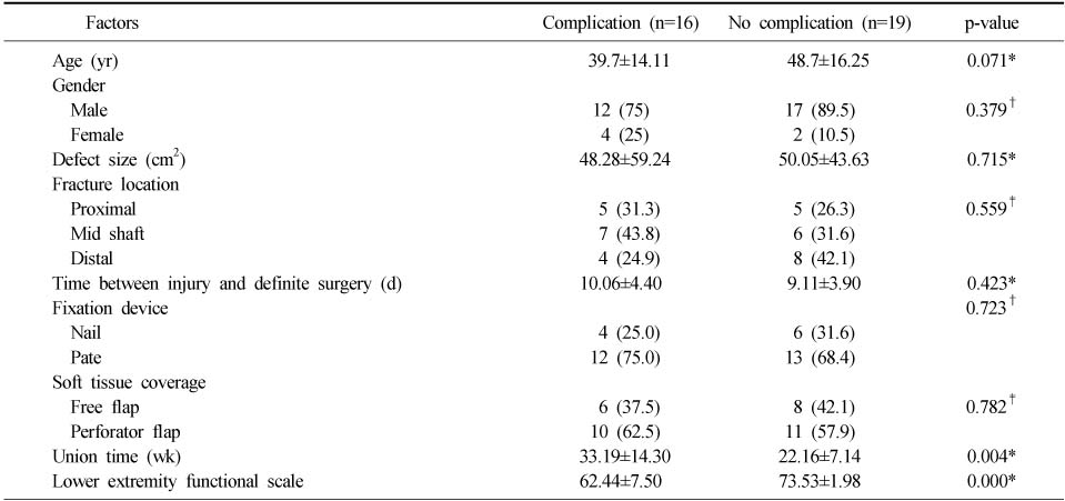
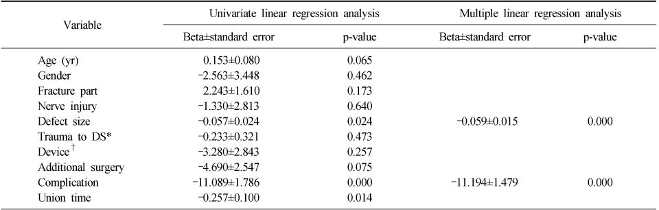
Figure & Data
REFERENCES
Citations
Citations to this article as recorded by 
- Effect of Korean Medicine Treatments in Patients with Proximal Tibia Fracture: A Retrospective Observational Study
Jung Min Lee, Eun-Jung Lee
Journal of Korean Medicine Rehabilitation.2020; 30(3): 141. CrossRef
 Cite
CiteTreatment of Type IIIb Open Tibial Fractures

Fig. 1
(A) A 65-year-old man sustained a type IIIb open tibia segmented fracture. (B) The vacuum-assisted closure system was applied to the open wound after debridement and temporary external fixation and provisional plate fixation. (C) An unreamed tibial nail was applied with an antero-lateral thigh free flap at 12 days after injury. (D) No visible callus on the proximal segmented area was observed at two months after surgery. (E) Plate augmentation without bone graft. (F) Clinical photograph shows a successful result 14 months after injury and the fracture was healed without complication.
Fig. 1
Treatment of Type IIIb Open Tibial Fractures
Factors associated with Complication Following Open Tibial Fractures
Values are presented as mean±standard deviation or number (%). *Mann-Whitney test, †Fisher's exact test, ‡Chi-square test.
Associations of Lower Extremity Functional Scale with Patient Variables
*Time trauma to definitive surgery, †Used on definitive surgery (nail or plate).
Table 1
Factors associated with Complication Following Open Tibial Fractures
Values are presented as mean±standard deviation or number (%). *Mann-Whitney test, †Fisher's exact test, ‡Chi-square test.
Table 2
Associations of Lower Extremity Functional Scale with Patient Variables
*Time trauma to definitive surgery, †Used on definitive surgery (nail or plate).

