 E-submission
 TOTA
TOTA TOTS
TOTS
Search
- Page Path
- HOME > Search
Original Articles
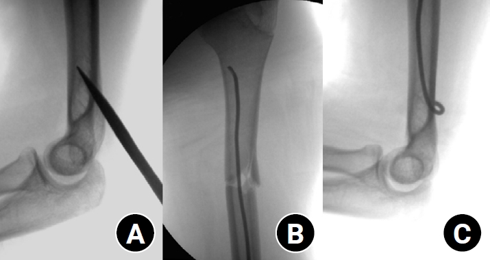
- Clinical and radiographic outcomes of elastic stable intramedullary nailing for pediatric humeral shaft fractures: a retrospective case series
- Kang-San Lee, Dongju Shin, Sang Hee Kim, Il Seo, Tae-Hoon Kim, Sung Jung Kim
- J Musculoskelet Trauma 2026;39(2):156-161. Published online March 10, 2026
- DOI: https://doi.org/10.12671/jmt.2025.00381
-
 Abstract
 PDF
PDF - Background
Pediatric humeral shaft fractures are uncommon and are generally treated conservatively, with satisfactory clinical outcomes reported in most cases. However, conservative management often necessitates prolonged immobilization and frequent outpatient follow-up visits, and it carries an inherent risk of residual angular or translational deformity. Elastic stable intramedullary nailing (ESIN) provides a simple and minimally invasive method of fracture fixation that offers adequate stability without disrupting the periosteal blood supply, thereby permitting early mobilization and promoting rapid bone union. The purpose of this study was to evaluate the clinical and radiological outcomes of ESIN fixation in pediatric patients with humeral shaft fractures.
Methods
The medical records of pediatric patients with humeral shaft fractures who underwent ESIN fixation between January 2015 and November 2025 were retrospectively reviewed. Data collected included patient demographics, mechanism of injury, fracture location, number of elastic nails used, time to union, degree of residual angulation, range of motion (ROM), and postoperative complications.
Results
The mean age of the patients was 10.0 years (range, 7 to 15 years). The mean time to radiographic union was 5.4 weeks (range, 2.4 to 10.4 weeks). The mean coronal angulation was 0.2° (range, −9.1° to 5.8°), while the mean sagittal angulation was −1.3° (range, −6.9° to 5.3°). No cases of infection, nerve injury, or nail migration were observed during the follow-up period. At the final follow-up assessment, all patients demonstrated full shoulder and elbow ROM, with no residual deformity or pain reported.
Conclusions
In this small retrospective case series, ESIN fixation resulted in favorable union rates and excellent functional outcomes in pediatric humeral shaft fractures. Level of evidence: IV.
- 480 View
- 19 Download

- Surgical Treatment of Posterior Wall Fractures of the Acetabulum
- Young Soo Byun, Se Ang Chang, Young Ho Cho, Dae Hee Hwang, Sung Rak Lee, Sang Hee Kim
- J Korean Fract Soc 2007;20(2):123-128. Published online April 30, 2007
- DOI: https://doi.org/10.12671/jkfs.2007.20.2.123
-
Abstract
PDF
- PURPOSE
To evaluate the results of surgical treatment of posterior wall fractures of the acetabulum and to determine the factors affecting the results.
MATERIALS AND METHODS
Thirty-one posterior wall fractures were reviewed; 7 type A1-1, 19 type A1-2 and 5 type A1-3 by AO classification. Postoperatively, the accuracy of the reduction was evaluated. At the final follow-up, clinical and radiographic results were evaluated with medical records and radiographs. The factors affecting the results were determined.
RESULTS
The reduction was graded as anatomical in 22 patients, imperfect in seven and poor in two. The clinical result was excellent in 21 hips, good in six, fair in three and poor in one. The quality of the reduction was strongly associated with the clinical result. The radiographic result was excellent in 22 hips, good in five, fair in two and poor in two. The clinical result was related closely to the radiographic result. Complications were osteoarthritis in three patients, osteonecrosis of the femoral head in one, heterotopic ossification in one, penetration of a screw into the joint in one and iatrogenic sciatic nerve injury in one. The factors affecting the clinical results were fracture patterns, the surgeon's experience, the accuracy of the reduction and late complications.
CONCLUSION
In this present series of posterior wall fractures, as their prognosis depends on the severity of the injury and the accuracy of the reduction, satisfactory result can be obtained by anatomical reduction with thorough preoperative planning and the surgeon's experience.
- 734 View
- 4 Download
- T-Plate Fixation for Two- and Three-Part Fractures of the Proximal Humerus
- Dong Ju Shin, Se Ang Chang, Young Soo Byun, Dae Hee Hwang, Sung Rak Lee, Sang Hee Kim
- J Korean Fract Soc 2005;18(4):426-431. Published online October 31, 2005
- DOI: https://doi.org/10.12671/jkfs.2005.18.4.426
-
Abstract
PDF
- PURPOSE
To evaluate the results and complications of treatment using T-plate fixation for two- and three-part fractures of the proximal humerus.
MATERIALS AND METHODS
Between January 1996 and July 2003, thirty-three patients with two-part and three-part fractures of the proximal humerus were treated by T-plate fixation. There were 21 two-part fractures and 12 three-part fractures including three shoulder dislocations. The reduction was qualified and complications were assessed with final radiographs. The functional outcome was evaluated by Neer's rating system.
RESULTS
Thirty-two cases (96.7%) were united, twenty-nine cases (87.9%) were reduced as good, and twenty-three cases (70%) had excellent or satisfactory results. There were four cases of loss of reduction, three cases of stiff joint, one case of nonunion, and one case of avascular necrosis of the humeral head, but no infection. No correlation was found between the final result and the type of fracture, age, gender, or quality of reduction.
CONCLUSION
T-plate fixation for proximal humeral fractures is a reliable method to obtain good results through satisfactory reduction, rigid fixation, and early movement. Additional tension band wiring can provide stable fixation for osteoporotic or comminuted fractures difficult to obtain stable fixation. -
Citations
Citations to this article as recorded by
- The Surgical Outcomes for Isolated Greater Tuberosity Fracture of Proximal Humerus
Eun-Sun Moon, Myung-Sun Kim, Young-Jin Kim
Journal of the Korean Fracture Society.2007; 20(3): 239. CrossRef
- The Surgical Outcomes for Isolated Greater Tuberosity Fracture of Proximal Humerus
- 805 View
- 3 Download
- 1 Crossref
 First
First Prev
Prev

