 E-submission
 TOTA
TOTA TOTS
TOTS
Search
- Page Path
- HOME > Search
Original Articles
- Risk factors for ankle fractures in older adults based on clinical components of the Fracture Risk Assessment (FRAX) tool and comorbidities in Korea: a retrospective case-control study
- Myeong Jun Song, Se Woong Jang, Jun Young Lee, Seojin Park
- J Musculoskelet Trauma 2025;38(4):193-202. Published online October 24, 2025
- DOI: https://doi.org/10.12671/jmt.2025.00143
-
 Abstract
 PDF
PDF - Background
Ankle fractures are common in older adults; however, their relationship with osteoporotic fractures remains unclear. This study aimed to evaluate potential risk factors for ankle fractures in older adults by analyzing individual clinical components of the Fracture Risk Assessment (FRAX) tool and comorbidities.
Methods
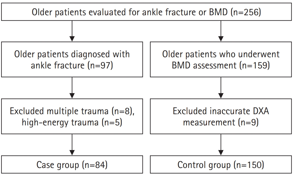
We conducted a retrospective case-control study including 84 patients aged ≥65 years with ankle fractures and 150 controls who underwent bone mineral density (BMD) testing without prior ankle fractures. The variables analyzed included age, sex, body mass index, smoking, alcohol consumption, prior fracture history, and comorbidities such as hypertension, diabetes mellitus, and dementia. BMD was measured at the spine, total hip, and femoral neck.
Results
Univariate analysis showed that alcohol consumption, diabetes mellitus, and total hip T-score categories were significantly associated with ankle fractures. In binary logistic regression, alcohol consumption remained significantly associated with higher ankle fracture risk (odds ratio [OR], 5.302; 95% confidence interval [CI], 1.778–15.811; P=0.003), and both osteopenia and osteoporosis at the total hip were also associated with increased risk (OR, 3.260, P=0.049; OR, 3.561, P=0.031, respectively). Diabetes mellitus did not reach statistical significance in the adjusted model (P=0.074). Model fit was adequate (Hosmer-Lemeshow P=0.377), and post hoc power analysis confirmed sufficient sample size.
Conclusions
These findings suggest that lower total hip BMD and alcohol-related factors may be associated with ankle fracture risk in older adults. The FRAX score itself was not calculated; instead, this study focused on analyzing selected clinical components. Limitations include the retrospective design, lack of fall and medication data, and cross-sectional BMD assessment. Level of evidence: III.
- 1,851 View
- 32 Download

- Demographic and Radiographic Parameters as Predictors of Reduction Loss after Conservative Treatment of Distal Radius Fractures in Adults
- Kyu Jin Kim, Dae Won Shin, Seong Kee Shin
- J Korean Fract Soc 2023;36(2):45-51. Published online April 30, 2023
- DOI: https://doi.org/10.12671/jkfs.2023.36.2.45
-
Abstract
PDF
- Purpose
This study examined the demographic and radiological risk factors for later reduction loss of distal radius fractures treated conservatively. Materials and Methods This study enrolled patients treated for distal radius fractures between January 2017 and December 2019. Seventy-eight patients were included in the analysis and divided into two groups. The patients who showed minimal reduction loss within an acceptable radiologic angle after initial manual reduction were classified as Group A. The patients who showed reduction loss out of an acceptable radiologic angle and finally malunited or converted to surgical treatments were classified as Group B. The patient’s age and bone marrow density were used as demographic data. The initial X-ray images were evaluated to determine the fracture type. Various radiological parameters were measured. Results The 78-patient study cohort consisted of nine men and 69 women with a mean age of 67 years. Forty-eight cases were sorted into Group A, and 30 cases into Group B. On logistic regression analysis, the age of 80 or older was a risk factor for later fracture displacement among the demographic factors (p=0.037, odds ratio=4.937). Among the radiographic factors, the presence of distal ulnar fracture and dorsal cortical comminution were disclosed as risk factors of later displacement (p=0.049, 0.003, odds ratio=3.429, 7.196). Conclusion When conservative management for distal radius fracture is decided in patients more than 80 years of age or accompanied by a distal ulnar fracture or with dorsal cortical comminution, the possibility of later displacement of the distal radius should be considered.
- 826 View
- 4 Download
- Risk Factors for Subsequent Contralateral Hip Fracture following Osteoporotic Hip Fracture Surgery
- Kyung-Jae Lee, Jung-Hoon Choi, Hee-Uk Ye, Young-Hun Kim, Kyung-Hwan Lim
- J Korean Fract Soc 2021;34(2):51-56. Published online April 30, 2021
- DOI: https://doi.org/10.12671/jkfs.2021.34.2.51
-
Abstract
PDF
- Purpose
This study examined the risk factors contributing to subsequent hip fractures in patients with osteoporotic hip fractures.
Materials and Methods
Between March 2008 and February 2016, 68 patients sustained a subsequent contralateral hip fracture after surgery for a primary osteoporotic hip fracture (Study group). The patients were compared with 475 patients who had been followed up for a minimum of one year with a unilateral osteoporotic hip fracture (Control group). The demographic data, bone mineral density (BMD), osteoporosis medication, osteoporotic fracture history, comorbid disease, type of surgery, preoperative, postoperative ambulatory capacity, and postoperative delirium in the two groups were compared.
Results
The demographic data, BMD, osteoporosis medication history, comorbid disease, type of surgery, and postoperative delirium were similar in the two groups. At three months after the primary surgery, the poor ambulatory capacity was significantly higher in the study group than the control group (p<0.001).
Conclusion
The ambulatory capacity after primary surgery is an important risk factor in the occurrence of subsequent hip fractures after osteoporotic hip fracture. Cause analysis regarding the poor ambulatory capacity after surgery will be necessary, and the development of a functional recovery program and careful management of the walking ability recovery will be needed. -
Citations
Citations to this article as recorded by
- Osteoporotic Hip Fracture: How We Make Better Results?
Byung-Chan Choi, Kyung-Jae Lee
Journal of the Korean Fracture Society.2024; 37(1): 52. CrossRef
- Osteoporotic Hip Fracture: How We Make Better Results?
- 1,317 View
- 13 Download
- 1 Crossref
- Risk Factors for Knee Stiffness in Distal Femoral Fractures
- Dong Wook Son, Hyoung Soo Kim, Woo Young Choi
- J Korean Fract Soc 2018;31(4):123-131. Published online October 31, 2018
- DOI: https://doi.org/10.12671/jkfs.2018.31.4.123
-
Abstract
PDF
- PURPOSE
The aims of this study were to evaluate risk factors for knee stiffness after the fixation of distal femoral fractures, and to analyze the clinical and radiologic outcomes.
MATERIALS AND METHODS
This is a retrospective case control study of 104 consecutive patients who have a distal femoral fracture and were treated with a submuscular locking plate. The case group comprised of patients with 12-month postoperative range of motion (ROM) ≤90° or a history of manipulation under anesthesia. The case group was compared with the control group of patients with a 12-month postoperative ROM >90°. The possible risk factors were evaluated by univariate and logistic regression analysis. The postoperative ROM and Knee Society clinical rating system was evaluated for the clinical assessment and the distal femoral angle on a whole-extremity scanogram was measured for radiologic assessments.
RESULTS
Fifty-four patients were included in the study (14 in the case group, 40 in the control group). Univariate analysis showed that comminuted fracture, intra-articular fracture, open fracture, temporary external fixation, severe osteoarthritis, and prolonged immobilization placed patients at an increased risk for knee stiffness. On the other hand, multivariate logistic regression showed that an extensor mechanism injury was the only significant predictor (p=0.001; odds ratio, 42.0; 95% confidence interval, 5.0–350.7). The ROM and Knee Society score were significantly lower in the case group; however, the coronal alignment was similar in the case and control group.
CONCLUSION
Various factors that delay postoperative knee motion place patients at increased risk of knee stiffness. Understanding these risk factors may help surgeons prevent postoperative knee stiffness after distal femoral fractures. In particular, extensor mechanism injury, such as patella fracture or open quadriceps injury, was found to be an independent predictable factor associated with knee stiffness. -
Citations
Citations to this article as recorded by- Post operative knee stiffness after surgical fixation of knee osseous injuries
Oitangor Arthur, Sekamatte Yasin, Mulepo Phillip
Adesh University Journal of Medical Sciences & Research.2026; 8: 56. CrossRef - The outcomes of patients with segmental long bone fractures treated with SIGN nail at Addis Ababa Burn, Emergency and Trauma Hospital
Cheru B. Tesso, Lelisa Merga, Samuel Kebede
OTA International.2026;[Epub] CrossRef - A Comprehensive Approach to Stiffness in Total Knee Arthroplasty
Brian P. Chalmers, Linda I. Suleiman, Peter K. Sculco, Matthew P. Abdel
The Journal of Arthroplasty.2025; 40(9): S59. CrossRef - Staged Management for Distal Femur Fractures: Impacts on Reoperation, Stiffness, and Overall Outcomes
Matthew T. Yeager, Robert W. Rutz, Alex Roszman, Gerald McGwin, James E. Darnley, Joseph P. Johnson, Clay A. Spitler
Journal of Orthopaedic Trauma.2024; 38(11): 577. CrossRef - Outcome of the Masquelet Technique for Complex Bilateral Distal Femoral Bone Defects
Ziad A Aljaafri, Abdullah Alzahrani, Ali Alshehri, Ahmed AlHussain, Faisal Alzahrani, Khalid Alsheikh
Cureus.2023;[Epub] CrossRef - Efficacy of non-operative treatment of patients with knee arthrofibrosis using high-intensity home mechanical therapy: a retrospective review of 11,000+ patients
Shaun K. Stinton, Samantha J. Beckley, Thomas P. Branch
Journal of Orthopaedic Surgery and Research.2022;[Epub] CrossRef - Distal Femoral Replacement and Extensor Mechanism Repair Reinforced With Synthetic Mesh for Distal Femur Fracture With Patellar Ligament Avulsion
Charles Powell, Kristopher Sanders, Neal Huang, Luis Felipe Colón, Colton Norton
Arthroplasty Today.2022; 16: 31. CrossRef - The fragility of statistical significance in distal femur fractures: systematic review of randomized controlled trials
Michael Megafu, Hassan Mian, Emmanuel Megafu, Sulabh Singhal, Alexander Lee, Richawna Cassie, Paul Tornetta, Robert Parisien
European Journal of Orthopaedic Surgery & Traumatology.2022; 33(6): 2411. CrossRef - Association Between Femoral “Spike” Size After Intramedullary Nailing and Subsequent Knee Motion Surgery
Michael G. Schloss, Nathan N. O'Hara, Syed M. R. Zaidi, Zachary D. Hannan, Dimitrius Marinos, Jared Atchison, Alexandra Mulliken, Jason W. Nascone, Robert V. O'Toole
Journal of Orthopaedic Trauma.2021; 35(2): 100. CrossRef - Distal Femur Replacement Versus Surgical Fixation for the Treatment of Geriatric Distal Femur Fractures: A Systematic Review
Brett P. Salazar, Aaron R. Babian, Malcolm R. DeBaun, Michael F. Githens, Gustavo A. Chavez, L. Henry Goodnough, Michael J. Gardner, Julius A. Bishop
Journal of Orthopaedic Trauma.2021; 35(1): 2. CrossRef
- Post operative knee stiffness after surgical fixation of knee osseous injuries
- 1,102 View
- 19 Download
- 10 Crossref
- Perioperative Blood Loss in Intramedullary Hip Screw for Intertrochanteric Fracture: Analysis of Risk Factors
- Jai Hyung Park, Hwa Jae Jung, Hun Kyu Shin, Eugene Kim, Se Jin Park, Taeg Su Ko, Jong Hyon Park
- J Korean Fract Soc 2015;28(1):53-58. Published online January 31, 2015
- DOI: https://doi.org/10.12671/jkfs.2015.28.1.53
-
Abstract
PDF
- PURPOSE
We compared visible blood loss and calculated blood loss after intramedullary fixation in intertrochanteric fracture, and evaluated correlation between blood loss and its risk factors.
MATERIALS AND METHODS
A total of 256 patients who underwent closed reduction and intramedullary fixation in femoral intertrochanteric fracture between 2004 and 2013 were enrolled in this study. The total blood loss was calculated using the formula reported by Mercuiali and Brecher. We analyzed several factors, including fracture pattern (according to Evans classification), gender, age, body mass index (BMI), anesthesia method, cardiovascular and cerebrovascular disease, preoperative anemia, American Society of Anesthesiologists (ASA) score and use of antithrombotic agents.
RESULTS
Total calculated blood loss (2,100+/-1,632 ml) differed significantly from visible blood loss (564+/-319 ml). In addition, the blood loss of unstable fracture patient was 2,496+/-1,395 ml and multivariate analysis showed a significant relationship between blood loss and fracture pattern (p<0.01). However, other factors showed no statistically significant difference.
CONCLUSION
Total calculated blood loss was much greater than visible blood loss. Patients with unstable intertrochanteric fracture should be treated with care in order to reduce blood loss.
- 744 View
- 1 Download
- Neurologic Injury within Pelvic Ring Injuries
- Ji Wan Kim, Dong Hoon Baek, Jae Hyun Kim, Young Chang Kim
- J Korean Fract Soc 2014;27(1):17-22. Published online January 31, 2014
- DOI: https://doi.org/10.12671/jkfs.2014.27.1.17
-
Abstract
PDF
- PURPOSE
To evaluate the incidence of neurologic injury in pelvic ring injuries and to assess the risk factors for neurologic injury related to pelvic fractures.
MATERIALS AND METHODS
Sixty-two patients with the pelvic ring injury were enrolled in the study from March 2010 to May 2013. When the neurologic injury was suspected clinically, the electro-diagnostic tests were performed. Combined injuries, fracture types, and longitudinal displacements were examined for correlations with the neurologic injury.
RESULTS
There were 7 cases of AO/OTA type A, 37 cases of type B, and 18 cases of type C. Among them, 25 patients (40%) had combined spine fractures, and the average of longitudinal displacement was 7 mm (1-50 mm). Of the 62 patients, 13 (21%) had neurologic injury related with pelvic fractures; 5 with lumbosacral plexus injury, 5 with L5 or S1 nerve injury, 2 with obturator nerve injury, and 1 case of lateral femoral cutaneous nerve injury. There were no relationships between the neurologic injuries and fracture types (p=0.192), but the longitudinal displacements of posterior ring and combined spine fractures were related to the neurologic injury within pelvic ring injury (p=0.006, p=0.048).
CONCLUSION
The incidence of neurologic injury in pelvis fracture was 21%. In this study, the longitudinal displacements of posterior ring and combined spine fractures were risk factors for neurological injury in pelvic ring injury. -
Citations
Citations to this article as recorded by- Surgical Outcome of Posterior Pelvic Fixation Using S1, S2 Screws in Vertically Unstable Pelvic Ring Injury
Kwang Hee Yeo, Nam Hoon Moon, Jae Min Ahn, Jae Yoon Jeong, Jae Hoon Jang
Journal of the Korean Fracture Society.2018; 31(1): 9. CrossRef
- Surgical Outcome of Posterior Pelvic Fixation Using S1, S2 Screws in Vertically Unstable Pelvic Ring Injury
- 1,152 View
- 8 Download
- 1 Crossref
- Risk Factors of Periprosthetic Fracture after Total Knee Arthroplasty
- Ju Young Shin, Hyung Jun Kim, Seung Han Cha, Dong Heon Kim
- J Korean Fract Soc 2012;25(1):1-7. Published online January 31, 2012
- DOI: https://doi.org/10.12671/jkfs.2012.25.1.1
-
Abstract
PDF
- PURPOSE
To evaluate the incidence rate and risk factors for periprosthetic fracture after total knee replacement (TKR).
MATERIALS AND METHODS
We carried out a retrospective case-control study of 596 patients (951 knees) who underwent TKR between 1999 and 2006 and who were followed up over 36 months. We classified patients into group I (study group) and group II (control group). We subdivided risk factors as pre-operative, intra-operative, and post-operative factors. Age, osteoporosis, revision arthroplasty, CVA, and alcohol dependence were categorized as pre-operative factors; anterior femoral notching and prosthetic types (mobile, fixed, and load-bearing) were considered intra-operative factors; and post-operative activity level was classified as a post-operative factor. We obtained information from the patients' charts, X-ray film, and telephone interviews.
RESULTS
The overall incidence rate was 2.25%; 3 patients were male, and 18 were female (14.28% and 85.72%, respectively). Old age (p<0.01, odds ratio=1.14), osteoporosis (p=0.01, odds ratio=4.74), revision arthroplasty (p=0.01, odds ratio=7.46), CVA (p=0.02, odds ratio=8.55), and alcohol dependence (p=0.03, odds ratio=44.54) were statistically significant among the pre-operative factors. Among the intra-operative factors, anterior femoral notching (p<0.01, odds ratio=11.74) was significant, and continued heavy labor (p<0.01, odds ratio=8.14) was significant among the post-operative factors.
CONCLUSION
We concluded that old age, osteoporosis, revision arthroplasty, comorbidity related with falling down, anterior femoral notching, and continued heavy labor were associated with periprosthetic fracture after TKR. -
Citations
Citations to this article as recorded by- Periprosthetic Distal Femoral Fracture Following Total Knee Arthroplasty Associated With Anterior Femoral Notching: A Case Report
Nay Aung Zin, Thiha Zaw, Sandar Shune Let Aung, Min Maung Maung, Htoo Aung Naing Shwe, Tun Tun
Cureus.2026;[Epub] CrossRef
- Periprosthetic Distal Femoral Fracture Following Total Knee Arthroplasty Associated With Anterior Femoral Notching: A Case Report
- 922 View
- 2 Download
- 1 Crossref
- Delirium after Intertrochanteric Fractures of Femur in Elderly Patients
- Kyu Bok Kang, Dong Hun Suh, Seong Rok Oh
- J Korean Fract Soc 2011;24(2):131-137. Published online April 30, 2011
- DOI: https://doi.org/10.12671/jkfs.2011.24.2.131
-
Abstract
PDF
- PURPOSE
To evaluate the incidence, risk factors and prognosis of delirium in elderly patients with intertrochanteric fractures of femur.
MATERIALS AND METHODS
162 patients who underwent operation for intertrochanteric fracture of femur from July 2005 to January 2007 were reviewed retrospectively. Delirium was diagnosed by using Confusion Assessment Method (CAM). Medical records were reviewed for the information of the patients, Gross motor function classification of Palisano et al. was used for the evaluation of ambulatory status. Univariate analysis and multivariate analysis were done to find out the risk factors.
RESULTS
2 cases out of 162 (1.2%) met the criteria of delirium at admission, and 39 cases (24.1%) after surgery. Univariate analysis and multivariate analysis identified age, hematocrit, dementia, the duration of opiate use, and pulmonary complication as risk factors. Hospital stay was longer and postoperative ambulatory status was worse in the patients with delirium.
CONCLUSION
Delirium is a frequent complication of intertrochanteric fractures of old age and associated with worse results. Cognitive function as well as physical status should be evaluated before and after surgery. Delirium needs more active prevention and treatment for better results. -
Citations
Citations to this article as recorded by- Incidence and Associated Factors of Delirium after Orthopedic Surgery
Si-Wook Lee, Chul-Hyun Cho, Ki-Cheor Bae, Kyung-Jae Lee, Eun-Seok Son, Sang-Hyun Um
Journal of the Korean Orthopaedic Association.2019; 54(2): 157. CrossRef - Laozi. De la figure du maître mythique à la divinité taoïque
Kyong-Kon Kim
Archimède. Archéologie et histoire ancienne.2019; 6: 16. CrossRef - Outcomes of Patients With Delirium in Long-Term Care Facilities: A Prospective Cohort Study
Kyoung Ja Moon, Heeok Park
Journal of Gerontological Nursing.2018; 44(9): 41. CrossRef - Relationship between Delirium and Clinical Prognosis among Older Patients underwent Femur Fracture Surgery
Jae-Lan Shim, Seon-Young Hwang
Journal of the Korea Academia-Industrial cooperation Society.2016; 17(2): 649. CrossRef - The effects of a tailored intensive care unit delirium prevention protocol: A randomized controlled trial
Kyoung-Ja Moon, Sun-Mi Lee
International Journal of Nursing Studies.2015; 52(9): 1423. CrossRef - Automatic Delirium Prediction System and Nursing-Sensitive Outcomes in the Medical Intensive Care Unit
Ha-young Cho, Xianghua Song, Jinshi Piao, Yinji Jin, Sun-Mi Lee
Clinical Nursing Research.2015; 24(1): 29. CrossRef - Postoperative Delirium in Elderly Patients with Osteoarthritis Surgery: Incidence and Risk Factors
Eun A Park, Min Young Kim
Journal of muscle and joint health.2015; 22(2): 57. CrossRef - Is Delirium an Unrecognized Threat to Patient Safety in Korean Intensive Care Units?
Kyoung-Ja Moon, Jinshi Piao, Yinji Jin, Sun-Mi Lee
Journal of Nursing Care Quality.2014; 29(1): 91. CrossRef
- Incidence and Associated Factors of Delirium after Orthopedic Surgery
- 1,437 View
- 11 Download
- 8 Crossref
- Risk Factors of Postoperative Delirium in Elderly Patients with Hip Fractures
- Ki Hwan Kim, Duk Hwan Kho, Ju Yong Shin, Jin Yong Choi, Eung Sik Kim, Dong Heon Kim
- J Korean Fract Soc 2008;21(3):189-194. Published online July 31, 2008
- DOI: https://doi.org/10.12671/jkfs.2008.21.3.189
-
Abstract
PDF
- PURPOSE
To find out the relationship between various risk factors and post-operative delirium in elderly patients with hip fractures.
MATERIALS AND METHODS
Out of 135 patients older than 65 years old who underwent the surgery for hip fracture in our department, between the periods of March 2003 to March 2005, 14 patients (10.4%) developed post-operative delirium and 121 patients (89.6%) did not. We studied risk factors of post-operative delirium in two groups.
RESULTS
In chi-square test between delirium group and non-delirium group, the patients were more likely to develop post-operative delirium if they had previous episodes of delirium, abnormal cognitive function, low walking ability before admission, high dependency on ADL (Activities of Daily Living), other medical accompanying diseases, history of dementia, post-operative hypoxia, post-operative electrolyte imbalance, low post-operative hemoglobin and hematocrit, low post-operative albumin and were older than 75 years old (p<0.05). Sex, type of fracture, anesthesia and the time between admission and operation did not show much difference between the two groups.
CONCLUSION
The risk factors of postoperative delirium in elderly patients with hip fracture have a tendency to be multifactorial. Therefore, we conclude that being prepared by thorough understanding of the risk factors and their relationships will help prevent post-operative delirium and result in good postoperative prognosis. -
Citations
Citations to this article as recorded by- Increased Serum Neuropeptide Galanin Level Is a Predictor of Cognitive Dysfunction in Patients with Hip Fracture
Zichao Xue, Ke Zhang, Biao Luo, Long Fan, Ruizhe Zhao, Guangliang Hu, Yuzhen Xu
Disease Markers.2021; 2021: 1. CrossRef - Sleep Disturbance Strongly Related to the Development of Postoperative Delirium in Proximal Femoral Fracture Patients Aged 60 or Older
Myung-Rae Cho, Suk-Kyoon Song, Cheol-Hwan Ryu
Hip & Pelvis.2020; 32(2): 93. CrossRef - Incidence and Associated Factors of Delirium after Orthopedic Surgery
Si-Wook Lee, Chul-Hyun Cho, Ki-Cheor Bae, Kyung-Jae Lee, Eun-Seok Son, Sang-Hyun Um
Journal of the Korean Orthopaedic Association.2019; 54(2): 157. CrossRef - Relationship between Delirium and Clinical Prognosis among Older Patients underwent Femur Fracture Surgery
Jae-Lan Shim, Seon-Young Hwang
Journal of the Korea Academia-Industrial cooperation Society.2016; 17(2): 649. CrossRef - Relationship between Knowledge, Stress, and Nursing Performance about Care for Delirium in Geriatric Hospital Nurses
Eun-Hee Kim
Journal of Korean Clinical Health Science.2016; 4(2): 593. CrossRef - The effects of a tailored intensive care unit delirium prevention protocol: A randomized controlled trial
Kyoung-Ja Moon, Sun-Mi Lee
International Journal of Nursing Studies.2015; 52(9): 1423. CrossRef - Is Delirium an Unrecognized Threat to Patient Safety in Korean Intensive Care Units?
Kyoung-Ja Moon, Jinshi Piao, Yinji Jin, Sun-Mi Lee
Journal of Nursing Care Quality.2014; 29(1): 91. CrossRef - The Effects of Delirium Care Training Program for Nurses in Hospital Nursing Units
Moonja Kim, Haejung Lee
Korean Journal of Adult Nursing.2014; 26(5): 489. CrossRef - Knowledge, Performance and Stress about Care for Delirium in Orthopedic Hospital Nurses
Mi Young Kim, Young Eun
Journal of muscle and joint health.2013; 20(1): 72. CrossRef - The Experience of Delirium Care and Clinical Feasibility of the CAM-ICU in a Korean ICU
Joo-Hee Jung, Jung-Hye Lim, Eun-Jung Kim, Hyo-Chan An, Min-Kyung Kang, Jin Lee, Yu-Kyung Min, Eun-Zoo Park, Xiang-Hwa Song, Hye-Ryoung Kim, Sun-Mi Lee
Clinical Nursing Research.2013; 22(1): 95. CrossRef - Development and validation of the Korean Nursing Delirium Scale
Kyoung-Nam Kim, Cheol-Ho Kim, Kwang-Il Kim, Hyun-Jung Yoo, Si-Young Park, Yeon-Hwan Park
Journal of Korean Academy of Nursing.2012; 42(3): 414. CrossRef - Influencing Factors of the Incidence of Delirium in Elderly Patients with Arthroplasty
Young-Whee Lee, Hye-Bin Im, Eun-Jeong Jeong, Hee-Sun Ma
Korean Journal of Adult Nursing.2012; 24(4): 348. CrossRef - Delirium After Spinal Surgery in Korean Population
Jin Kyu Lee, Ye-Soo Park
Spine.2010; 35(18): 1729. CrossRef - The Incidence and Related Factors of Delirium in Elderly Patients with Hip Fracture after Surgery
Bo-Kyung Sohn, Yerl-Bo Sung, Eun-Jin Park, Dong-Woo Lee
Journal of the Korean Geriatrics Society.2010; 14(3): 162. CrossRef
- Increased Serum Neuropeptide Galanin Level Is a Predictor of Cognitive Dysfunction in Patients with Hip Fracture
- 2,945 View
- 6 Download
- 14 Crossref
- Risk Group of Lateral Meniscus Injury in Fracture of Lateral Tibial Condyle
- Jung Man Kim, Cheong Ho Chang, Doo Hoon Sun, Jeong Tae Seo, Seok Joong Kim
- J Korean Soc Fract 1999;12(3):601-606. Published online July 31, 1999
- DOI: https://doi.org/10.12671/jksf.1999.12.3.601
-
Abstract
PDF
- PURPOSE
Untreated meniscus injuries accompanied with fractures of lateral tibial condyle may cause prolonged pain and post-traumatic arthritis. Lateral displacement of fractured fragment of lateral tibial condyle was found to be related to the incidence of lateral meniscus tear. We established the risk groups for lateral meniscus injuries on the basis of the degree of the lateral displacement of lateral tibial condyle.
MATERIALS and METHODS
Risk groups for lateral meniscus injury in fractures of lateral tibial condyle were evaluated in 39 knees, retrospectively, using plain roentgenograms and arthroscopic findings. On the knee anteroposterior radiography, displacement of lateral tibia condyle were classified into three groups according to the probability of lateral meniscus INJURY: high risk group for above 8mm of lateral displacement; moderate risk group for 4-8mm; low risk group for less than 4mm.
RESULTS
High risk group has ten meniscus injuries among 13 knees(76.9%) and moderate risk group had the seven meniscus injuries of 18 knees(38.9%). Low risk group of eight knees had no meniscus injury(0%). These different incidences among groups were statistically significant(p<0.05).
CONCLUSIONS
The authors suggest that the incidence of lateral meniscus injury was related to the degree of lateral displacement of lateral tibial condyle, and our definition of risk groups are useful for prediction of lateral meniscus injury in fracture of lateral tibial condyle.
- 714 View
- 0 Download
- Analysis of Rick Fractors of HiP fractures after the eighth Decade of Life
- Seung Ho Yune, Sang Rho Ahn, Chan Hee Park, Jun Young Yang
- J Korean Soc Fract 1994;7(1):131-136. Published online May 31, 1994
- DOI: https://doi.org/10.12671/jksf.1994.7.1.131
-
Abstract
PDF
- In Order to evaluate to the risk factors of hip fracture, we reviewed 34 patients, who were over 70 years old, from Jan. 1991 to Dec. 1993. The patients who have preexisting desease such as chronic illness, or in case of traffic accident, and fall down injury were excluded. We measured the singhs index and femoral neck-shaft angle on simple X-ray, bone mineral density for quantitative measurement of mineral on dual photon absorptiometry, and calcium, phosphate, and alkaline phosphatase on serologic study. We compared the results with control group who have not orthopaedic problems in 20 old aged person, and the results obtained were as follows. 1. The male to female ratio is 11:23, a high rate in female, and the mean age of patients is 74.3 years old, ragne from 70 to 89 years. 2. Femoral neck-shaft angle of patient group is 141.67±5.3 compared to 143.40±4.8 of control, showed statistical significance. 3. Bony trabeculae index of proximal femur of patient group is 1.83±0.83 compared to 2.91 ± 0.86 of control, showed statistical significance. 4. In patient group, about 30% decreased in bone mineral density compared control. This result must be requisite to reinvestigation and statlstical analysis were not carried due to numbers of members of control. 5. There is no significant difference between two groups regard to serologic study. In summary, risk factors of hip fractures are aged female with decreased femoral neck-shaft angle, below third degree in singhs index and lower bone mineral density compared with same age. According to the our data, in case of hip fractures in old aged, for prevent the postoperative complication and refracture, prophylactic theraphy to collectible causes are required, in addllion to operative internal fixation.
- 622 View
- 0 Download
 First
First Prev
Prev

