 E-submission
 TOTA
TOTA TOTS
TOTS
Articles
- Page Path
- HOME > J Musculoskelet Trauma > Volume 28(4); 2015 > Article
-
Review Article
- Reconstruction of a Traumatic Soft Tissue Defect
- Jong Woong Park, M.D., Ph.D.
-
Journal of the Korean Fracture Society 2015;28(4):256-265.
DOI: https://doi.org/10.12671/jkfs.2015.28.4.256
Published online: October 19, 2015
Department of Orthopaedic Surgery, Division of Hand Surgery & Reconstructive Microsurgery, Korea University College of Medicine, Seoul, Korea.
- Address reprint requests to: Jong Woong Park, M.D., Ph.D. Department of Orthopaedic Surgery, Korea University Anam Hospital, 73 Inchon-ro, Seongbuk-gu, Seoul 02841, Korea. Tel: 82-2-920-5320, Fax: 82-2-920-5320, ospark@korea.ac.kr
Copyright © 2015 The Korean Fracture Society. All rights reserved.
This is an Open Access article distributed under the terms of the Creative Commons Attribution Non-Commercial License (http://creativecommons.org/licenses/by-nc/4.0) which permits unrestricted non-commercial use, distribution, and reproduction in any medium, provided the original work is properly cited.
- 1,447 Views
- 7 Download
- 2 Crossref
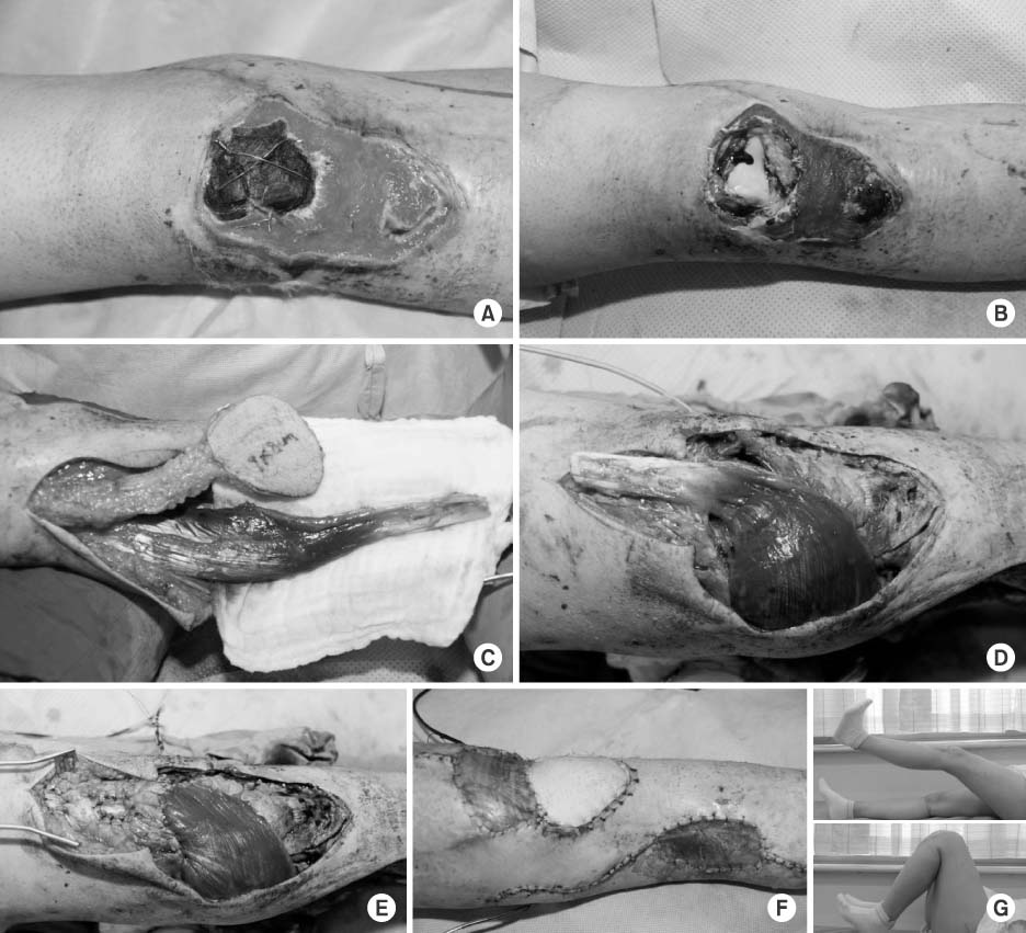

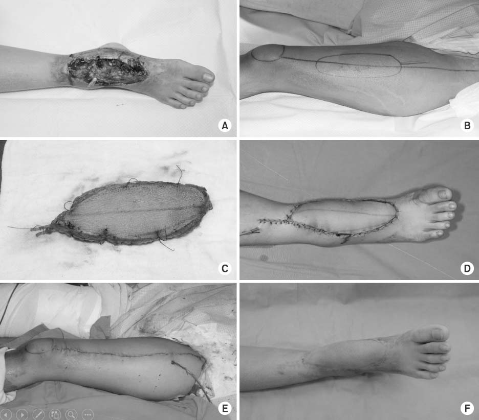
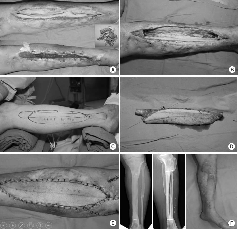
Figure & Data
REFERENCES
Citations
Citations to this article as recorded by 
 Cite
CiteReconstruction of a Traumatic Soft Tissue Defect

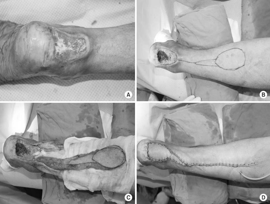


Fig. 1
Open fracture around the knee joint. (A) Necrosis of patella and loss of patella tendon. (B) After debridement, the knee joint is opened and reconstruction of quadriceps mechanism is required. (C) Medial gastrocnemius muscle flap and saphenous neurocutaneous island flap are harvested. (D, E) The gastrocnemius muscle is turned to be sutured to the quadriceps muscle. (F) The remaining defect is covered with the saphenous neurocutaneous island flap. (G) Two years later, full range of motion of the knee joint is recovered and the motor grade reaches grade 4.
Fig. 2
Reverse sural artery flap. (A) Soft tissue defect at the posterior aspect of the ankle. (B) Reverse sural artery flap is designed. (C) Pedicled flap is dissected. (D) After completion of flap insetting.
Fig. 3
Anterolateral thigh (ALT) flap. (A) Contaminated soft tissue defect on the anterior ankle. (B) ALT flap is designed. (C) Dissected ALT flap. (D) Flap insetting. (E) Donor site is primarily closed. (F) Two months after the operation.
Fig. 4
Free vascularized fibular osteocutaneous flap. (A) Large tibia and soft tissue defect after infected open fracture. (B) Sequestrum is removed. (C, D) Free vascularized fibular graft is harvested from the ipsilateral leg. (E) Flap is fixed and repaired. (F) Two years after bone and soft tissue reconstruction.
Fig. 1
Fig. 2
Fig. 3
Fig. 4
Reconstruction of a Traumatic Soft Tissue Defect

