 E-submission
 TOTA
TOTA TOTS
TOTS
Articles
- Page Path
- HOME > J Musculoskelet Trauma > Volume 28(4); 2015 > Article
-
Original Article
- The Result of Open Reduction and Mini-Plate Fixation for Displaced Talar Neck Fracture
- Woong Chae Na, M.D., Sang Hong Lee, M.D., Jun Young Lee, M.D., Ph.D, Sang Jun Lee, M.D., Boseon Kim, M.D.
-
Journal of the Korean Fracture Society 2015;28(4):215-222.
DOI: https://doi.org/10.12671/jkfs.2015.28.4.215
Published online: October 19, 2015
Department of Orthopaedic Surgery, Chosun University School of Medicine, Gwangju, Korea.
- Address reprint requests to: Jun Young Lee, M.D., Ph.D. Department of Orthopaedic Surgery, Chosun University Hospital, 365 Pilmun-daero, Dong-gu, Gwangju 61453, Korea. Tel: 82-62-220-3147, Fax: 82-62-226-3379, leejy88@chosun.ac.kr
• Received: May 22, 2015 • Revised: July 8, 2015 • Accepted: July 26, 2015
Copyright © 2015 The Korean Fracture Society. All rights reserved.
This is an Open Access article distributed under the terms of the Creative Commons Attribution Non-Commercial License (http://creativecommons.org/licenses/by-nc/4.0) which permits unrestricted non-commercial use, distribution, and reproduction in any medium, provided the original work is properly cited.
- 942 Views
- 2 Download
- 2 Crossref
Abstract
-
Purpose
- We evaluated the complications, radiological and clinical results after operative treatment using a mini-plate for fixation of displaced talar neck fractures.
-
Materials and Methods
- There were 20 cases of displaced talar neck fractures from May 2006 to December 2011; we performed a retrospective chart review of 15 patients treated by open reduction and internal fixation using a mini-plate who had more than 2 years of follow-up. According to Hawkin's classification, there were 7 cases of type II fractures and 8 cases of type III fractures. During postoperative 12-16 weeks we checked magnetic resonance imaging. The assessment of clinical results was based on the American Orthopaedic Foot and Ankle Society (AOFAS) ankle-hindfoot scale.
-
Results
- Mean union period was 11.6 weeks (10-15 weeks). Nonunion and malunion did not occur in all cases. The mean AOFAS score was 88.2 points (80-97 points). There were 5 cases of avascular necrosis. Of these, there were 3 cases of body collapse and 4 cases of post-traumatic arthritis. In the statistical analysis, there was no correlation between the elements including gender, Hawkin's classification and union rates and clinical results.
-
Conclusion
- Mini-plate fixation of a displaced talar neck fracture is thought to be a good technique, with a low rate of malunion and also showed satisfactory results in radiological and clinical assessment.
- 1. Baumhauer JF, Alvarez RG. Controversies in treating talus fractures. Orthop Clin North Am, 1995;26:335-351.Article
- 2. Halvorson JJ, Winter SB, Teasdall RD, Scott AT. Talar neck fractures: a systematic review of the literature. J Foot Ankle Surg, 2013;52:56-61.Article
- 3. Hawkins LG. Fractures of the neck of the talus. J Bone Joint Surg Am, 1970;52:991-1002.Article
- 4. Kenwright J, Taylor RG. Major injuries of the talus. J Bone Joint Surg Br, 1970;52:36-48.ArticlePDF
- 5. Canale ST. Fractures of the neck of the talus. Orthopedics, 1990;13:1105-1115.Article
- 6. Grob D, Simpson LA, Weber BG, Bray T. Operative treatment of displaced talus fractures. Clin Orthop Relat Res, 1985;(199):88-96.Article
- 7. Lorentzen JE, Christensen SB, Krogsoe O, Sneppen O. Fractures of the neck of the talus. Acta Orthop Scand, 1977;48:115-120.Article
- 8. Cronier P, Talha A, Massin P. Central talar fractures--therapeutic considerations. Injury, 2004;35:Suppl 2. SB10-SB22.Article
- 9. Sangeorzan BJ, Wagner UA, Harrington RM, Tencer AF. Contact characteristics of the subtalar joint: the effect of talar neck misalignment. J Orthop Res, 1992;10:544-551.Article
- 10. Ohl X, Harisboure A, Hemery X, Dehoux E. Long-term follow-up after surgical treatment of talar fractures: twenty cases with an average follow-up of 7.5 years. Int Orthop, 2011;35:93-99.
- 11. Penny JN, Davis LA. Fractures and fracture-dislocations of the neck of the talus. J Trauma, 1980;20:1029-1037.Article
- 12. Gelberman RH, Mortensen WW. The arterial anatomy of the talus. Foot Ankle, 1983;4:64-72.
- 13. Peterson L, Goldie IF. The arterial supply of the talus. A study on the relationship to experimental talar fractures. Acta Orthop Scand, 1975;46:1026-1034.Article
- 14. Canale ST, Kelly FB Jr. Fractures of the neck of the talus. Long-term evaluation of seventy-one cases. J Bone Joint Surg Am, 1978;60:143-156.Article
- 15. Daniels TR, Smith JW. Talar neck fractures. Foot Ankle, 1993;14:225-234.
- 16. Fleuriau Chateau PB, Brokaw DS, Jelen BA, Scheid DK, Weber TG. Plate fixation of talar neck fractures: preliminary review of a new technique in twenty-three patients. J Orthop Trauma, 2002;16:213-219.Article
- 17. Frawley PA, Hart JA, Young DA. Treatment outcome of major fractures of the talus. Foot Ankle Int, 1995;16:339-345.ArticlePDF
- 18. Sanders DW, Busam M, Hattwick E, Edwards JR, McAndrew MP, Johnson KD. Functional outcomes following displaced talar neck fractures. J Orthop Trauma, 2004;18:265-270.
- 19. Vallier HA, Nork SE, Barei DP, Benirschke SK, Sangeorzan BJ. Talar neck fractures: results and outcomes. J Bone Joint Surg Am, 2004;86:1616-1624.Article
- 20. Fortin PT, Balazsy JE. Talus fractures: evaluation and treatment. J Am Acad Orthop Surg, 2001;9:114-127.Article
- 21. Attiah M, Sanders DW, Valdivia G, et al. Comminuted talar neck fractures: a mechanical comparison of fixation techniques. J Orthop Trauma, 2007;21:47-51.
- 22. Rammelt S, Zwipp H. Talar neck and body fractures. Injury, 2009;40:120-135.Article
- 23. Swanson TV, Bray TJ, Holmes GB Jr. Fractures of the talar neck. A mechanical study of fixation. J Bone Joint Surg Am, 1992;74:544-551.Article
- 24. Juliano PJ, Dabbah M, Harris TG. Talar neck fractures. Foot Ankle Clin, 2004;9:723-736. vi.
- 25. Xue Y, Zhang H, Pei F, et al. Treatment of displaced talar neck fractures using delayed procedures of plate fixation through dual approaches. Int Orthop, 2014;38:149-154.
- 26. Daniels TR, Smith JW, Ross TI. Varus malalignment of the talar neck. Its effect on the position of the foot and on subtalar motion. J Bone Joint Surg Am, 1996;78:1559-1567.Article
REFERENCES
Fig. 1

Intraoperative photograph shows fracture site exposure by medial malleolar osteotomy and fixation using a mini-plate on the superior portion of the deltoid ligament attachment site.
Fig. 2
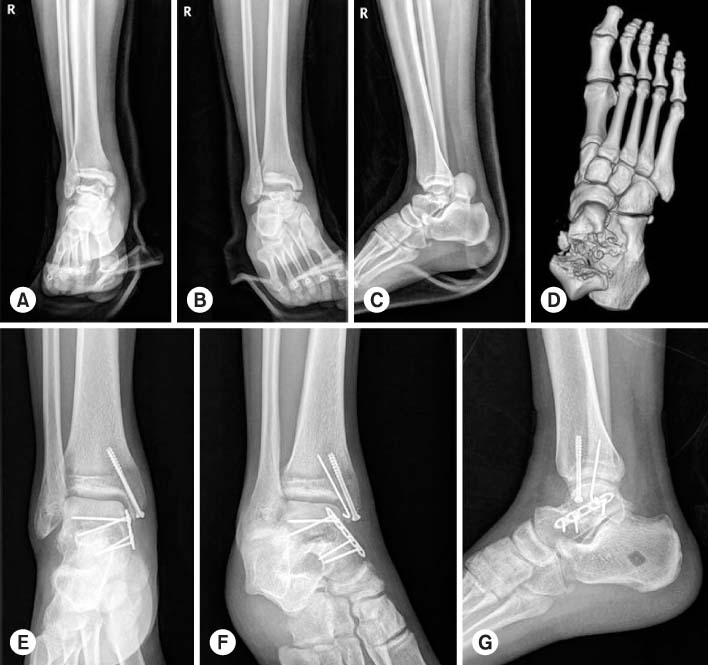
(A-D) The preoperative simple radiographs and 3-dimensional computed tomography show a Hawkin's type III comminuted talar neck fracture. (E-G) Comminuted talar neck fracture was fixed by a miniplate and medial malleolar osteotomy was fixed by a 4.0 cannulated screw.
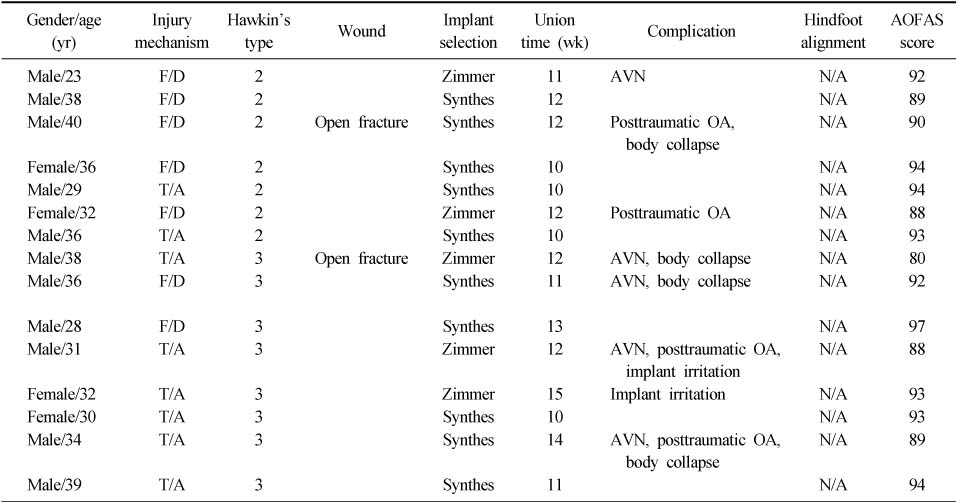
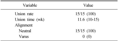
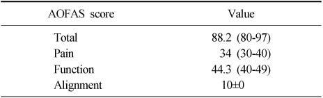
Figure & Data
REFERENCES
Citations
Citations to this article as recorded by 
- Outcome of Type 3 Talar Neck Fractures by Means of Medial Malleolar Osteotomy and Large Distractor
Sung Hae Park, Jun Young Lee, Jung Woo Lee
Journal of the Korean Orthopaedic Association.2019; 54(1): 45. CrossRef - The Measurement of Normal Talus in Korean Cadaver
Dong-Jun Ha, Heui-Chul Gwak, Jeon-Gyo Kim, Jung-Han Kim, Chang-Rak Lee, Young-Jun Kim, Jeong-Han Lee, Byung-Ho Ha, Ui-Cheol Kim
Journal of Korean Foot and Ankle Society.2016; 20(4): 163. CrossRef
 Cite
CiteThe Result of Open Reduction and Mini-Plate Fixation for Displaced Talar Neck Fracture
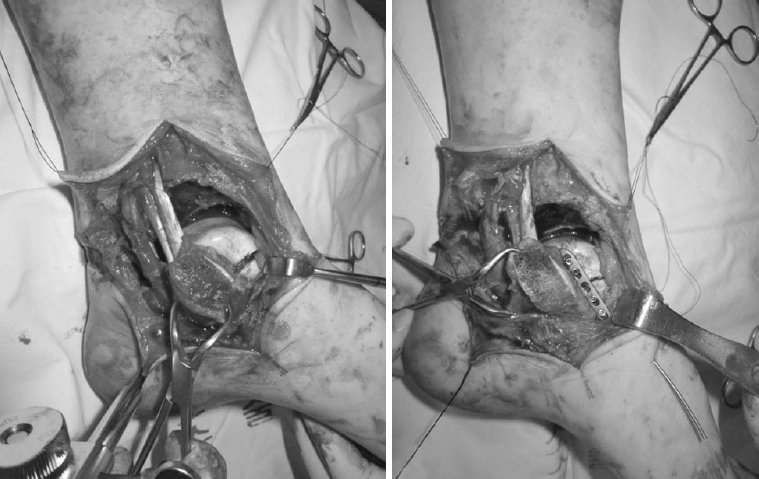

Fig. 1
Intraoperative photograph shows fracture site exposure by medial malleolar osteotomy and fixation using a mini-plate on the superior portion of the deltoid ligament attachment site.
Fig. 2
(A-D) The preoperative simple radiographs and 3-dimensional computed tomography show a Hawkin's type III comminuted talar neck fracture. (E-G) Comminuted talar neck fracture was fixed by a miniplate and medial malleolar osteotomy was fixed by a 4.0 cannulated screw.
Fig. 1
Fig. 2
The Result of Open Reduction and Mini-Plate Fixation for Displaced Talar Neck Fracture
Demographic Data
AOFAS: American Orthopaedic Foot and Ankle Society, F/D: Fall down, T/A: Traffic accident, AVN: Avascular necrosis, OA: Osteoarthritis, N/A: Normal alignment.
Radiographic Results
Values are presented as number/total number (%) or median (range). p-value: age, 0.981; gender, 0.327; Hawkin's type, 0.482.
Clinical Results
Values are presented as median (range) or number±standard deviation. p-value: age, 0.221; gender, 0.584; Hawkin's type, 0.823.
Table 1
Demographic Data
AOFAS: American Orthopaedic Foot and Ankle Society, F/D: Fall down, T/A: Traffic accident, AVN: Avascular necrosis, OA: Osteoarthritis, N/A: Normal alignment.
Table 2
Radiographic Results
Values are presented as number/total number (%) or median (range). p-value: age, 0.981; gender, 0.327; Hawkin's type, 0.482.
Table 3
Clinical Results
Values are presented as median (range) or number±standard deviation. p-value: age, 0.221; gender, 0.584; Hawkin's type, 0.823.

