 E-submission
 TOTA
TOTA TOTS
TOTS
Articles
- Page Path
- HOME > J Musculoskelet Trauma > Volume 20(1); 2007 > Article
-
Case Report
- Costoclavicular Syndrome: A Case Report
- Sung Keun Sohn, M.D., Sung Soo Kim, M.D., Chang Geun Song, M.D., Jong Ill Kwak, M.D.
-
Journal of the Korean Fracture Society 2007;20(1):86-89.
DOI: https://doi.org/10.12671/jkfs.2007.20.1.86
Published online: June 14, 2016
Department of Orthopedic Surgery, School of Medicine, Dong-A University, Busan, Korea.
- Address reprint requests to: Sung Soo Kim, M.D. Department of Orthopaedic Surgery, Dong-A University Hospital, 1, Dongdaesin-dong 3-ga, Seo-gu, Busan 602-715, Korea. Tel: 82-51-240-5166, Fax: 82-51-254-6757, sskim2@dau.ac.kr
Copyright © The Korean Fracture Society. All rights reserved
- 2,443 Views
- 10 Download
- 2 Crossref
Abstract
- Costoclavicular syndrome is one of the four syndromes of thoracic outlet syndrome in which have similiar symptoms, and may result from cervical and thoracic scoliosis, formation of excessive callus or nonunion after fractures of clavicle or first rib. Conservative treatment may be offered. Surgical treatment includes scalenectomy with supraclavicular approach, transaxillary first rib resection with scalenectomy and correction of clavicular abnormality. The purpose of this paper is to evaluate the result of surgical intervention in costoclavicular syndrome of a 38-year old man with clavicular nonunion after an operation.
- 1. Caldwell JW, Crane CR, Krusen EM. Nerve conduction studies: an aid in the dignosis of thoracic outlet syndrome. South Med J, 1971;64:210-212.
- 2. Chung HY. Surgery for entrapments of the thoracic outlet. J Korean Microsurg Soc, 1999;8:1-9.
- 3. Hahn SB, Park BM, Lim YJ. Thoracic outlet syndrome. J Korean Orthop Assoc, 1990;25:919-926.ArticlePDF
- 4. Lee SE, Nam IL, Lee SS, Lee DH, Lee KJ. Nonunion of the clavicular fracture. J Korean Soc Fract, 1999;12:741-748.Article
- 5. Mulder DS, Greenwood FA, Brooks CE. Posttraumatic thoracic outlet syndrome. J Trauma, 1973;13:706-715.Article
- 6. Neer CS 2nd. Nonunion of the clavicle. J Am Med Assoc, 1960;172:1006-1011.Article
- 7. Novak CB, Mackinnon SE, Patterson GA. Evaluation of patients with thoracic outlet syndrom. J Hand Surg Am, 1993;18:292-299.
- 8. Patterson RH. Cervical rib and the scalenus muscle syndrome. Ann Surg, 1940;111:531-545.
- 9. Smith KF. The thoracic outlet syndrome: a protocol of treatments. J Orthop Sports Physical Therapy, 1979;1:89-99.
- 10. Tindall SC. Chronic injuries of peripheral nerves by entrapment. In: Youmans JR, editor. Neurological surgery. 4th ed. Philadelphia: WB Saunders Co; 1996. p. 2182-2187.
REFERENCES


Figure & Data
REFERENCES
Citations
Citations to this article as recorded by 
- The Need to Expand the Concept of Thoracic Outlet Syndrome: A Proposal
Jaeseok Kim
Journal of Korean Medical Society of Acupotomology.2022; 6(1): 1. CrossRef - Costoclavicular Syndrome Secondary to Nonunion of a Displaced Fracture of the Clavicle, Misdiagnosed as a Simple Muscle Strain - A Case Report -
Ho-Seung Jeon, Haeng-Kee Noh, Seo-Goo Kang, Jong-Min Kim, Seung-Ju Jeon
Journal of the Korean Fracture Society.2013; 26(1): 60. CrossRef
 Cite
CiteCostoclavicular Syndrome: A Case Report
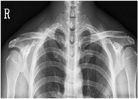
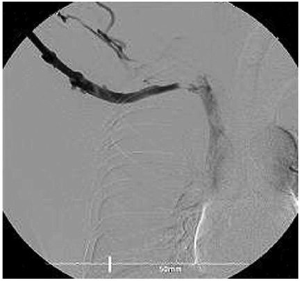
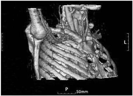
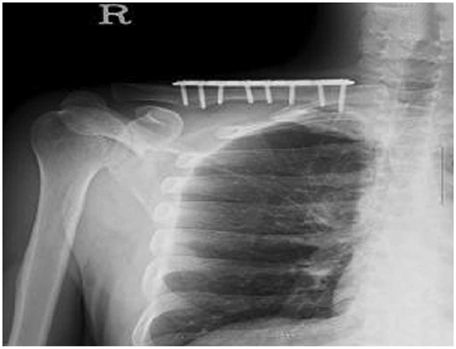
Fig. 1
The preoperative radiograph shows inferior displacement of distal fragment at fracture site without callus.
Fig. 2
The venogram showing the stenosis is aggravated by hyperabduction of the right shoulder.
Fig. 3
The three dimensional computed tomogram showing subclavian vein is compressed by distal fragment at fracture site.
Fig. 4
The radiograph taken 5 months after surgery shows well maintained bony alignment.
Fig. 1
Fig. 2
Fig. 3
Fig. 4
Costoclavicular Syndrome: A Case Report

