 E-submission
 TOTA
TOTA TOTS
TOTS
Articles
- Page Path
- HOME > J Musculoskelet Trauma > Volume 28(4); 2015 > Article
-
Original Article
- The Usefulness of Poller Screw with Antegrade Nailing in the Initial Treatment of Infraisthmal Femur Shaft Fracture
- Jeong-Hyun Yoo, M.D., Ph.D., Hyoung-Soo Kim, M.D., Ph.D., Chang-Geun Kim, M.D., Ho-Il Kwak, M.D., Sang-Heon Song, M.D., Ph.D.
-
Journal of the Korean Fracture Society 2015;28(4):230-236.
DOI: https://doi.org/10.12671/jkfs.2015.28.4.230
Published online: October 19, 2015
Department of Orthopedic Surgery, Myongji Hospital, Seonam University College of Medicine, Goyang, Korea.
- Address reprint requests to: Sang-Heon Song, M.D., Ph.D. Department of Orthopedic Surgery, Myongji Hospital, Seonam University College of Medicine, 55 Hwasu-ro 14beon-gil, Deogyang-gu, Goyang 10475, Korea. Tel: 82-31-810-5424, Fax: 82-31-969-0500, sh.gabriel.song@gmail.com
• Received: June 15, 2015 • Revised: July 6, 2015 • Accepted: July 24, 2015
Copyright © 2015 The Korean Fracture Society. All rights reserved.
This is an Open Access article distributed under the terms of the Creative Commons Attribution Non-Commercial License (http://creativecommons.org/licenses/by-nc/4.0) which permits unrestricted non-commercial use, distribution, and reproduction in any medium, provided the original work is properly cited.
- 736 Views
- 7 Download
Abstract
-
Purpose
- The purpose of this study was to evaluate the radiologic and clinical outcomes after intramedullary nailing with Poller screw insertion at initial stage in infraisthmal femur shaft fractures.
-
Materials and Methods
- Seven consecutive patients (7 femurs) treated with antegrade intramedullary nailing with Poller screw insertion for the infraisthmal femur shaft fracture were reviewed retrospectively. There were 4 male and 3 female patients. Mean age was 46.1 years (20-72 years). Operative time including Poller screw insertion, time for union, malalignment, and range of motion were evaluated.
-
Results
- All 7 cases had primarily healed successfully. Mean time for radiologic union was 19.1 weeks (16-24 weeks) postoperatively. One case had 5 degree valgus malalignment. One case of 15 mm shortening was reported and he required shoe lift orthosis. All cases had a full range of motion in hip and knee joint.
-
Conclusion
- Antegrade intramedullary nailing with Poller screw insertion is useful in the initial treatment of infraisthmal femur shaft fracture, because it could provide additional stability. An additional 20 minutes were required but a Poller screw should be considered according to the anatomic location of a femur shaft fracture.
- 1. Bucholz RW, Jones A. Fractures of the shaft of the femur. J Bone Joint Surg Am, 1991;73:1561-1566.Article
- 2. Weiss RJ, Montgomery SM, Al Dabbagh Z, Jansson KA. National data of 6409 Swedish inpatients with femoral shaft fractures: stable incidence between 1998 and 2004. Injury, 2009;40:304-308.Article
- 3. Wolinsky PR, McCarty E, Shyr Y, Johnson K. Reamed intramedullary nailing of the femur: 551 cases. J Trauma, 1999;46:392-399.
- 4. Pihlajamäki HK, Salminen ST, Böstman OM. The treatment of nonunions following intramedullary nailing of femoral shaft fractures. J Orthop Trauma, 2002;16:394-402.Article
- 5. Brumback RJ. The rationales of interlocking nailing of the femur, tibia, and humerus. Clin Orthop Relat Res, 1996;(324):292-320.Article
- 6. Kessler SB, Hallfeldt KK, Perren SM, Schweiberer L. The effects of reaming and intramedullary nailing on fracture healing. Clin Orthop Relat Res, 1986;(212):18-25.Article
- 7. Yang KH, Kim JR, Park J. Nonisthmal femoral shaft nonunion as a risk factor for exchange nailing failure. J Trauma Acute Care Surg, 2012;72:E60-E64.Article
- 8. Krettek C, Rudolf J, Schandelmaier P, Guy P, Könemann B, Tscherne H. Unreamed intramedullary nailing of femoral shaft fractures: operative technique and early clinical experience with the standard locking option. Injury, 1996;27:233-254.Article
- 9. Stedtfeld HW, Mittlmeier T, Landgraf P, Ewert A. The logic and clinical applications of blocking screws. J Bone Joint Surg Am, 2004;86:Suppl 2. 17-25.Article
- 10. Park J, Kim SG, Yoon HK, Yang KH. The treatment of nonisthmal femoral shaft nonunions with im nail exchange versus augmentation plating. J Orthop Trauma, 2010;24:89-94.Article
- 11. Kim JJ, Kim SK, Ahn JH. the use of poller screws in nonunion of femoral shaft following intramedullary nailing of femoral shaft fracture: a case report. J Korean Orthop Assoc, 2005;40:1024-1027.ArticlePDF
REFERENCES
Fig. 1
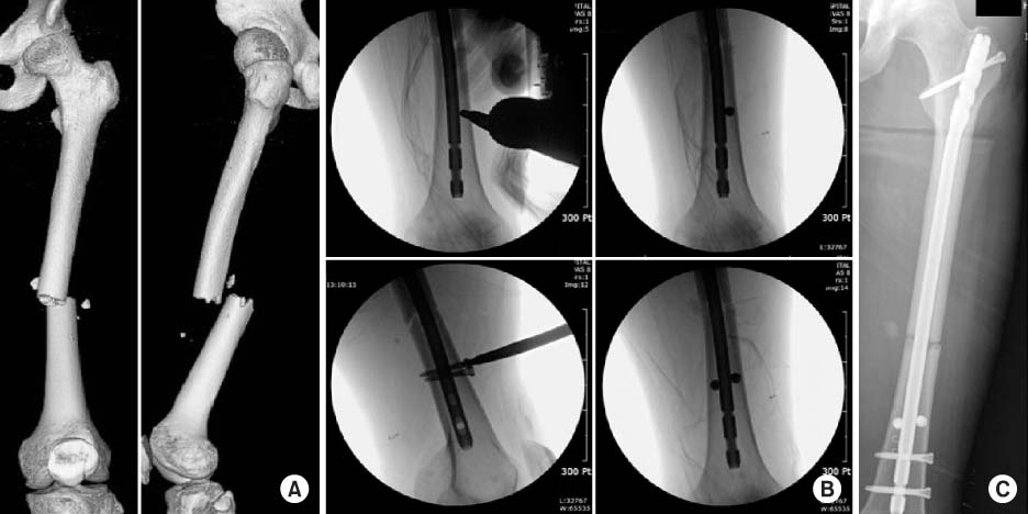
(A) Three-dimensional-reconstructed computed tomography image shows a fragmented wedge fracture at the femur shaft infraisthmal area. (B) Intra-operative C-arm images show the sequence of Poller screw insertion. (C) Postoperative radiograph shows a well reduced femur with good positioned Poller screws.
Fig. 2

A 20-year-old male was poly-traumatized after a traffic accident. (A) Anteroposterior radiograph shows an irregular complex femur shaft fracture with a non-displaced fracture of the ipsilateral femoral neck. (B) He underwent damage control-external fixator application at the time of brain surgery in the neurosurgery department. (C) After 3-weeks, he underwent antegrade nailing with two Poller screws after closed reduction with preservation of fracture site biology. Serial radiographs (D: initial postoperative, E: postoperative 2 month, F: postoperative 4 months) show bridging callus progression until radiologic healing.
Fig. 3
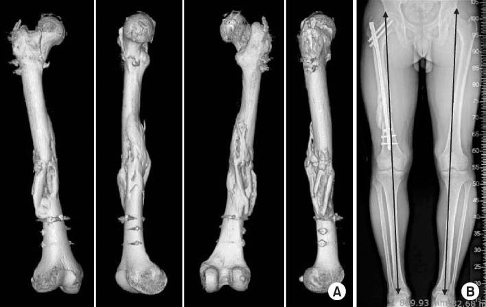
(A) Three-dimensional-reconstructed computed tomography images at postoperative 5 months show full consolidation of a complex femur shaft fracture including neck fracture component. (B) However standing lower extremity scanogram shows a limb length discrepancy of 15 mm.
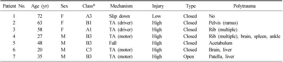
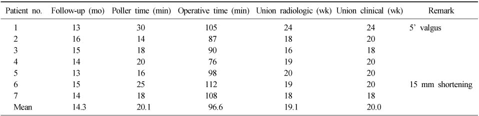
Figure & Data
REFERENCES
Citations
Citations to this article as recorded by 
 Cite
CiteThe Usefulness of Poller Screw with Antegrade Nailing in the Initial Treatment of Infraisthmal Femur Shaft Fracture

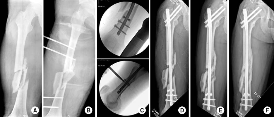

Fig. 1
(A) Three-dimensional-reconstructed computed tomography image shows a fragmented wedge fracture at the femur shaft infraisthmal area. (B) Intra-operative C-arm images show the sequence of Poller screw insertion. (C) Postoperative radiograph shows a well reduced femur with good positioned Poller screws.
Fig. 2
A 20-year-old male was poly-traumatized after a traffic accident. (A) Anteroposterior radiograph shows an irregular complex femur shaft fracture with a non-displaced fracture of the ipsilateral femoral neck. (B) He underwent damage control-external fixator application at the time of brain surgery in the neurosurgery department. (C) After 3-weeks, he underwent antegrade nailing with two Poller screws after closed reduction with preservation of fracture site biology. Serial radiographs (D: initial postoperative, E: postoperative 2 month, F: postoperative 4 months) show bridging callus progression until radiologic healing.
Fig. 3
(A) Three-dimensional-reconstructed computed tomography images at postoperative 5 months show full consolidation of a complex femur shaft fracture including neck fracture component. (B) However standing lower extremity scanogram shows a limb length discrepancy of 15 mm.
Fig. 1
Fig. 2
Fig. 3
The Usefulness of Poller Screw with Antegrade Nailing in the Initial Treatment of Infraisthmal Femur Shaft Fracture
Patient Demographics Related with Injury Type, Mechanism and Fracture Classification
*AO-OTA classification. F: Female, M: Male, TA: Traffic accident.
Clinico-Radiological Results Showing Poller Screw Fixation Time, Whole Operative Time, Time for Union (Radiologic, Clinical) and Clinical Remarks
Table 1
Patient Demographics Related with Injury Type, Mechanism and Fracture Classification
*AO-OTA classification. F: Female, M: Male, TA: Traffic accident.
Table 2
Clinico-Radiological Results Showing Poller Screw Fixation Time, Whole Operative Time, Time for Union (Radiologic, Clinical) and Clinical Remarks

